Embed Size (px)
Citation preview

1
Blood
Chapter 19
Blood
• Blood is a specialized fluid connective tissue that contains cells suspended in a fluid matrix
• Functions of blood include:– Transport of dissolved gases, nutrients, hormonesTransport of dissolved gases, nutrients, hormones
and metabolic wastes– Regulation of the pH and ion composition of
interstitial fluids– Restriction of fluid loss at injury sites– Defense against toxins and pathogens– Stabilization of body temperature
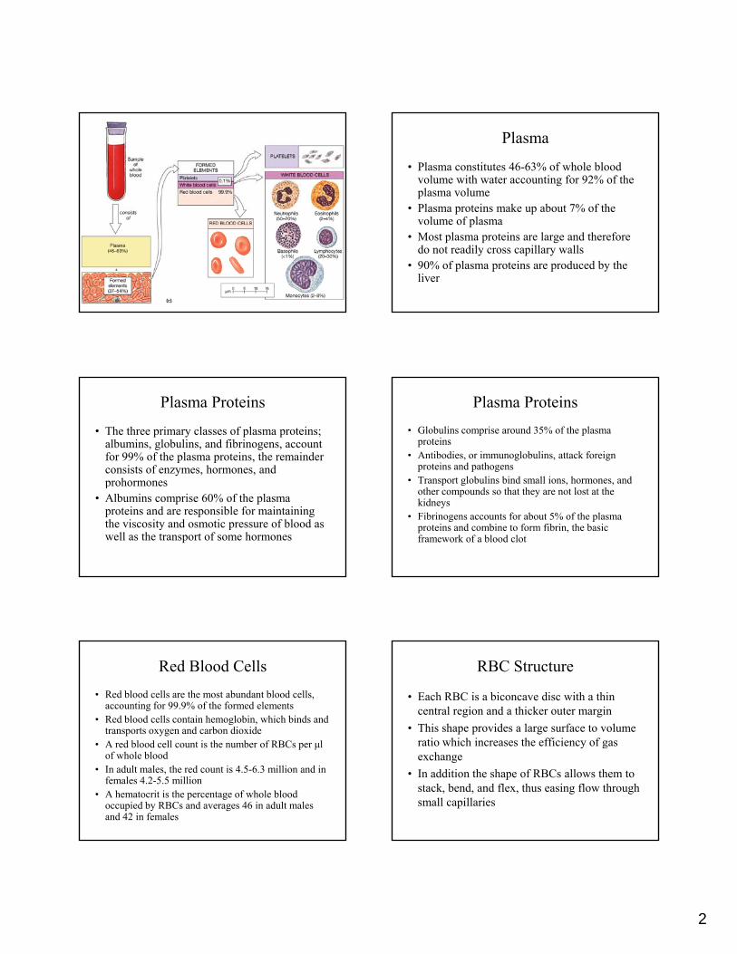
Blood Composition
• Blood consists of plasma and formed elements• Blood plasma is the fluid matrix that contains
plasma proteins in solution• Formed elements are blood cells and cellFormed elements are blood cells and cell
fragments that are suspended in the plasma• The formed elements consist of
– Red Blood Cells (RBCs)– White Blood Cells (WBCs)– Platelets
Blood Composition
• Red blood cells, or erythrocytes, are the most abundant blood cells and are essential for the transport of oxygen
• White blood cells or leukocytes are involve• White blood cells, or leukocytes, are involve with the body’s defense
• Platelets are small, membrane bound, cell fragments that contain enzymes and other substances involve in clotting
Fractionation & Hemopoesis
• Plasma and the formed elements constitute whole blood
• The components of whole blood can be separated or fractionated by centrifugationseparated, or fractionated, by centrifugation, for analytical or clinical purposes
• Hemopoesis is the process of blood cell formation
• Hemocytoblasts are circulating stem cells that divide to form all types of blood cells
2
Plasma
• Plasma constitutes 46-63% of whole blood volume with water accounting for 92% of the plasma volume
• Plasma proteins make up about 7% of the p pvolume of plasma
• Most plasma proteins are large and therefore do not readily cross capillary walls
• 90% of plasma proteins are produced by the liver
Plasma Proteins
• The three primary classes of plasma proteins; albumins, globulins, and fibrinogens, account for 99% of the plasma proteins, the remainder consists of enzymes, hormones, and prohormones
• Albumins comprise 60% of the plasma proteins and are responsible for maintaining the viscosity and osmotic pressure of blood as well as the transport of some hormones
Plasma Proteins• Globulins comprise around 35% of the plasma
proteins• Antibodies, or immunoglobulins, attack foreign
proteins and pathogensT l b li bi d ll i h d• Transport globulins bind small ions, hormones, and other compounds so that they are not lost at the kidneys
• Fibrinogens accounts for about 5% of the plasma proteins and combine to form fibrin, the basic framework of a blood clot
Red Blood Cells• Red blood cells are the most abundant blood cells,
accounting for 99.9% of the formed elements• Red blood cells contain hemoglobin, which binds and
transports oxygen and carbon dioxideA d bl d ll i h b f RBC l• A red blood cell count is the number of RBCs per μl of whole blood
• In adult males, the red count is 4.5-6.3 million and in females 4.2-5.5 million
• A hematocrit is the percentage of whole blood occupied by RBCs and averages 46 in adult males and 42 in females
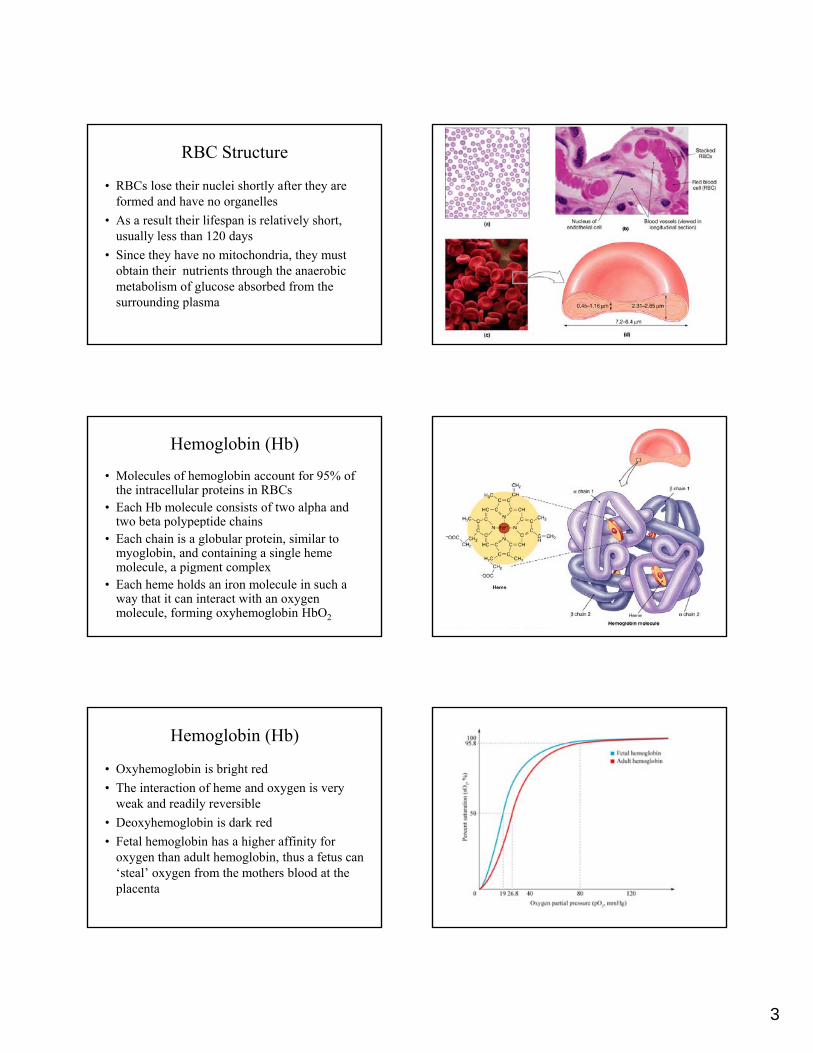
RBC Structure
• Each RBC is a biconcave disc with a thin central region and a thicker outer margin
• This shape provides a large surface to volume ratio which increases the efficiency of gasratio which increases the efficiency of gas exchange
• In addition the shape of RBCs allows them to stack, bend, and flex, thus easing flow through small capillaries
3
RBC Structure
• RBCs lose their nuclei shortly after they are formed and have no organelles
• As a result their lifespan is relatively short, usually less than 120 daysusually less than 120 days
• Since they have no mitochondria, they must obtain their nutrients through the anaerobic metabolism of glucose absorbed from the surrounding plasma
Hemoglobin (Hb)
• Molecules of hemoglobin account for 95% of the intracellular proteins in RBCs
• Each Hb molecule consists of two alpha and two beta polypeptide chainsp yp p
• Each chain is a globular protein, similar to myoglobin, and containing a single heme molecule, a pigment complex
• Each heme holds an iron molecule in such a way that it can interact with an oxygen molecule, forming oxyhemoglobin HbO2
Hemoglobin (Hb)
• Oxyhemoglobin is bright red• The interaction of heme and oxygen is very
weak and readily reversibleh l bi i d k d• Deoxyhemoglobin is dark red
• Fetal hemoglobin has a higher affinity for oxygen than adult hemoglobin, thus a fetus can ‘steal’ oxygen from the mothers blood at the placenta
4
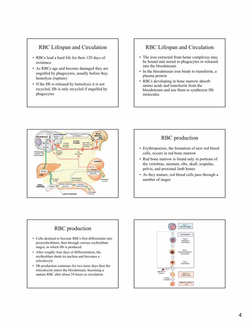
RBC Lifespan and Circulation
• RBCs lead a hard life for their 120 days of existence
• As RBCs age and become damaged they are engulfed by phagocytes usually before theyengulfed by phagocytes, usually before they hemolyze (rupture)
• If the Hb is released by hemolysis it is not recycled, Hb is only recycled if engulfed by phagocytes
RBC Lifespan and Circulation
• The iron extracted from heme complexes may be bound and stored in phagocytes or released into the bloodstream
• In the bloodstream iron binds to transferrin, a ,plasma protein
• RBCs developing in bone marrow absorb amino acids and transferrin from the bloodstream and use them to synthesize Hb molecules
RBC production
• Erythropoeisis, the formation of new red blood cells, occurs in red bone marrow
• Red bone marrow is found only in portions of the vertebrae sternum ribs skull scapulaethe vertebrae, sternum, ribs, skull, scapulae, pelvis, and proximal limb bones
• As they mature, red blood cells pass through a number of stages
RBC production
• Cells destined to become RBCs first differentiate into proerythroblasts, then through various erythroblast stages, in which Hb is produced
• After roughly four days of differentiation, the g y yerythroblast sheds its nucleus and becomes a reticulocyte
• Hb production continues for two more days then the reticulocyte enters the bloodstream, becoming a mature RBC after about 24 hours in circulation
5
Regulation of Erythropoesis
• Erythropoesis is directly stimulated by the peptide hormone erythropoetin (aka EPO, erythropoesis stimulating hormone) and indirectly by thyroxine, androgens, and growth hormone, but not by estrogens
• EPO is a glycoprotein that appears in the plasma when peripheral tissues, especially the kidneys, are exposed to low oxygen levels
• Once in the bloodstream EPO travels to the red bone marrow, stimulates stem cells and speeds the maturation of developing RBCs
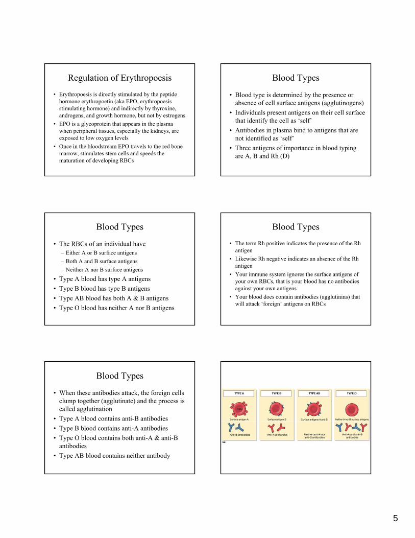
Blood Types
• Blood type is determined by the presence or absence of cell surface antigens (agglutinogens)
• Individuals present antigens on their cell surface that identify the cell as ‘self’that identify the cell as ‘self’
• Antibodies in plasma bind to antigens that are not identified as ‘self’
• Three antigens of importance in blood typing are A, B and Rh (D)
Blood Types
• The RBCs of an individual have– Either A or B surface antigens– Both A and B surface antigens
Neither A nor B s rface antigens– Neither A nor B surface antigens• Type A blood has type A antigens• Type B blood has type B antigens• Type AB blood has both A & B antigens• Type O blood has neither A nor B antigens
Blood Types
• The term Rh positive indicates the presence of the Rh antigen
• Likewise Rh negative indicates an absence of the Rh antigeng
• Your immune system ignores the surface antigens of your own RBCs, that is your blood has no antibodies against your own antigens
• Your blood does contain antibodies (agglutinins) that will attack ‘foreign’ antigens on RBCs
Blood Types
• When these antibodies attack, the foreign cells clump together (agglutinate) and the process is called agglutination
• Type A blood contains anti B antibodies• Type A blood contains anti-B antibodies• Type B blood contains anti-A antibodies• Type O blood contains both anti-A & anti-B
antibodies• Type AB blood contains neither antibody

6
Rh
• In contrast to antigens A & B, Rh negative individuals (no Rh antigen) do not necessarily contain Rh positive antibodies
• These antibodies are present only if the p yindividual is sensitized by previous exposure to Rh positive RBCs
• This exposure can occur during transfusion or during the pregnancy of an Rh negative woman with an Rh positive child
Testing for Compatibility
• At least 50 surface antigens have been identified on RBCs, but standard blood typing considers only those most likely to produce dangerous levels of agglutination; A, B, & Rh
• The test involves taking drops of blood and mixing them separately with solutions containing anti-A, anti-B, & anti-Rh antibodies
• The resulting pattern of agglutination indicates blood type
White Blood Cells
• Unlike RBCs, white blood cells have nuclei and organelles, but no hemoglobin
• WBCs, or leukocytes, help defend the body against invasion by pathogens, and remove g y p g ,toxins, wastes, and abnormal or damaged cells
• All are capable of amoeboid movement, migration out of the bloodstream and positive chemotaxis
• Some are capable of phagocytosis
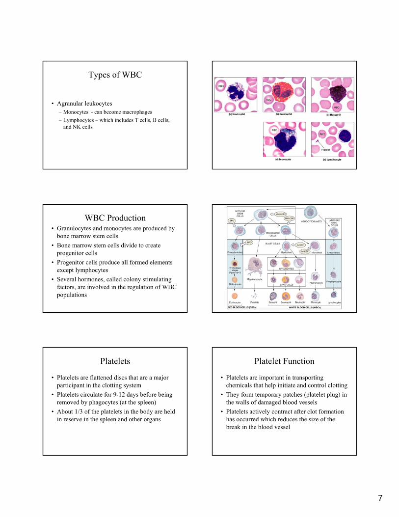
• There are two major groups of WBCs; granular leukocytes and agranular leukocytes
• Granular leukocytes
Types of WBC
– Neutrophils – 50 to 70 % total WBC population– Eosinophils – phagocytes attracted to foreign
compounds that have been marked with antibodies– Basophils – migrate to damaged tissue and release
histamine and heparin
7
• Agranular leukocytes– Monocytes - can become macrophages
Types of WBC
– Lymphocytes – which includes T cells, B cells, and NK cells
• Granulocytes and monocytes are produced by bone marrow stem cells
• Bone marrow stem cells divide to create progenitor cells
WBC Production
• Progenitor cells produce all formed elements except lymphocytes
• Several hormones, called colony stimulating factors, are involved in the regulation of WBC populations
Platelets
• Platelets are flattened discs that are a major participant in the clotting system
• Platelets circulate for 9-12 days before being removed by phagocytes (at the spleen)removed by phagocytes (at the spleen)
• About 1/3 of the platelets in the body are held in reserve in the spleen and other organs
Platelet Function
• Platelets are important in transporting chemicals that help initiate and control clotting
• They form temporary patches (platelet plug) in the walls of damaged blood vesselsthe walls of damaged blood vessels
• Platelets actively contract after clot formation has occurred which reduces the size of the break in the blood vessel

8
Hemostasis
• The process of hemostasis prevents the loss of blood through damaged vessel walls
• At the same time, it establishes a framework for tissue repairfor tissue repair
• Hemostasis consists of three phases:– Vascular phase– Platelet phase– Coagulation phase
Hemostasis
• During the vascular phase blood vessels constrict in a vascular spasm which involves the contraction of smooth muscle fibers in the vessel wall
• During the platelet phase, platelets begin to attach (via platelet adhesion) to the exposed tissue surfaces of the vessel
• As more platelets arrive they stick to one another and form a platelet plug
Hemostasis
• During the coagulation phase factors released by platelets and endothelial cells interact with clotting factors to form a clot
• During the clotting phase the clotting factors g g p g(enzymes and proenzymes) interact in a clotting cascade
• The three cascades involved are called the extrinsic (in the vessel wall), intrinsic (inside the bloodstream) and common (where both converge) pathways
The common pathway
• The common pathway begins when enzymes from either the extrinsic or intrinsic pathway activate factor X, forming the enzyme prothrombinase
• Prothrombinase converts prothrombin into thrombin
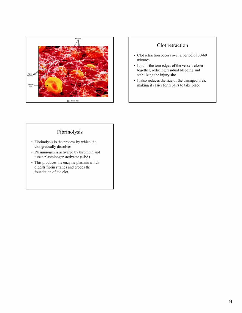
• Thrombin completes the clotting process by converting fibrinogen, a plasma protein, into insoluble strand of fibrin
9
Clot retraction
• Clot retraction occurs over a period of 30-60 minutes
• It pulls the torn edges of the vessels closer together reducing residual bleeding andtogether, reducing residual bleeding and stabilizing the injury site
• It also reduces the size of the damaged area, making it easier for repairs to take place
Fibrinolysis
• Fibrinolysis is the process by which the clot gradually dissolves
• Plasminogen is activated by thrombin and tissue plasminogen activator (t PA)tissue plasminogen activator (t-PA)
• This produces the enzyme plasmin which digests fibrin strands and erodes the foundation of the clot