Embed Size (px)
Citation preview
BIOLOGY OF MELANOCYTESDone by: Mohammed AbduljabbarDone by: Mohammed Abduljabbar
KAUH
• OBJECTIVES:
Definition & function of MelanocyteDefinition & function of Melanocyte
Definition & function of Melanin
E b i d l t f M l tEmbryonic development of Melanocytes
Site‐specific Melanocytes
Melanization steps
MelanosomesMelanosomes
Regulation of Melanocyte function
C it l h l i di dCongenital hypomelanosis disorders
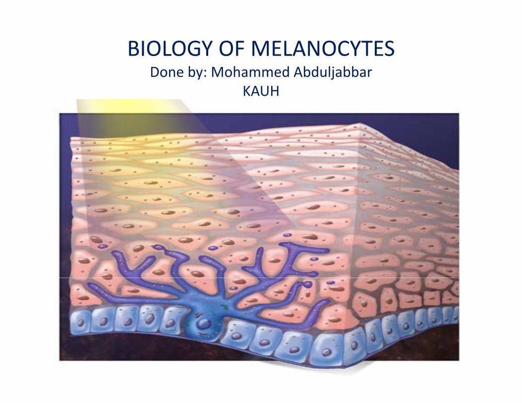
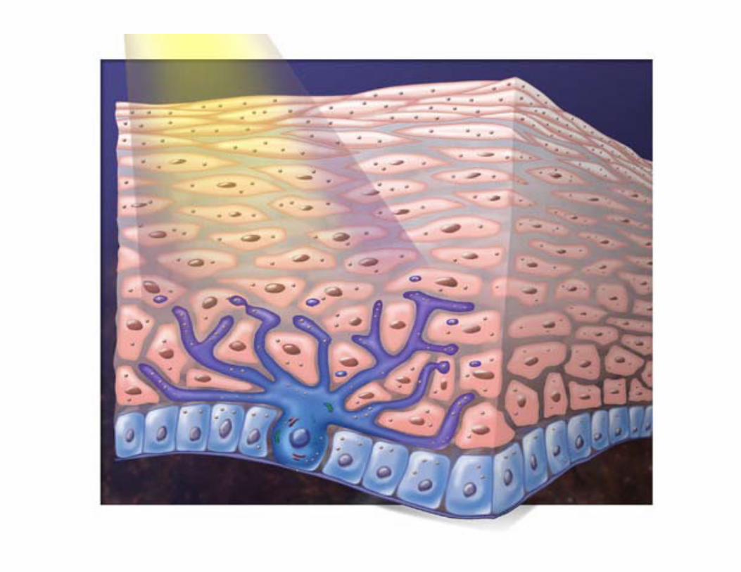
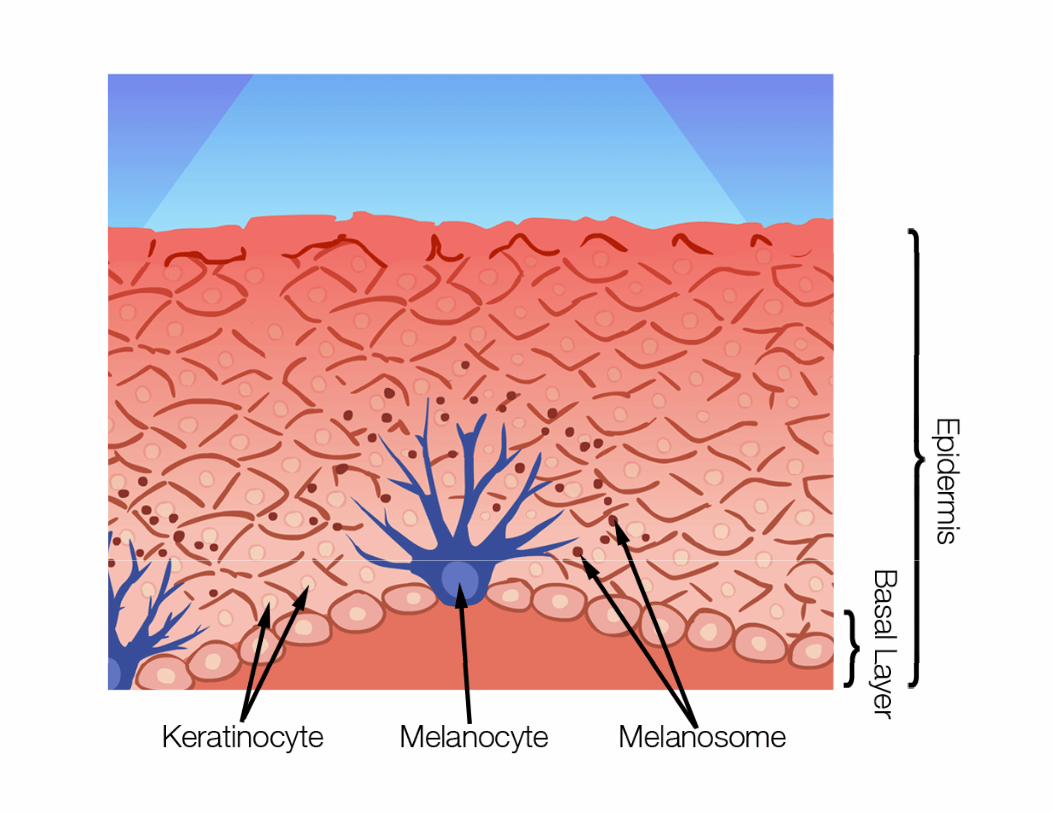
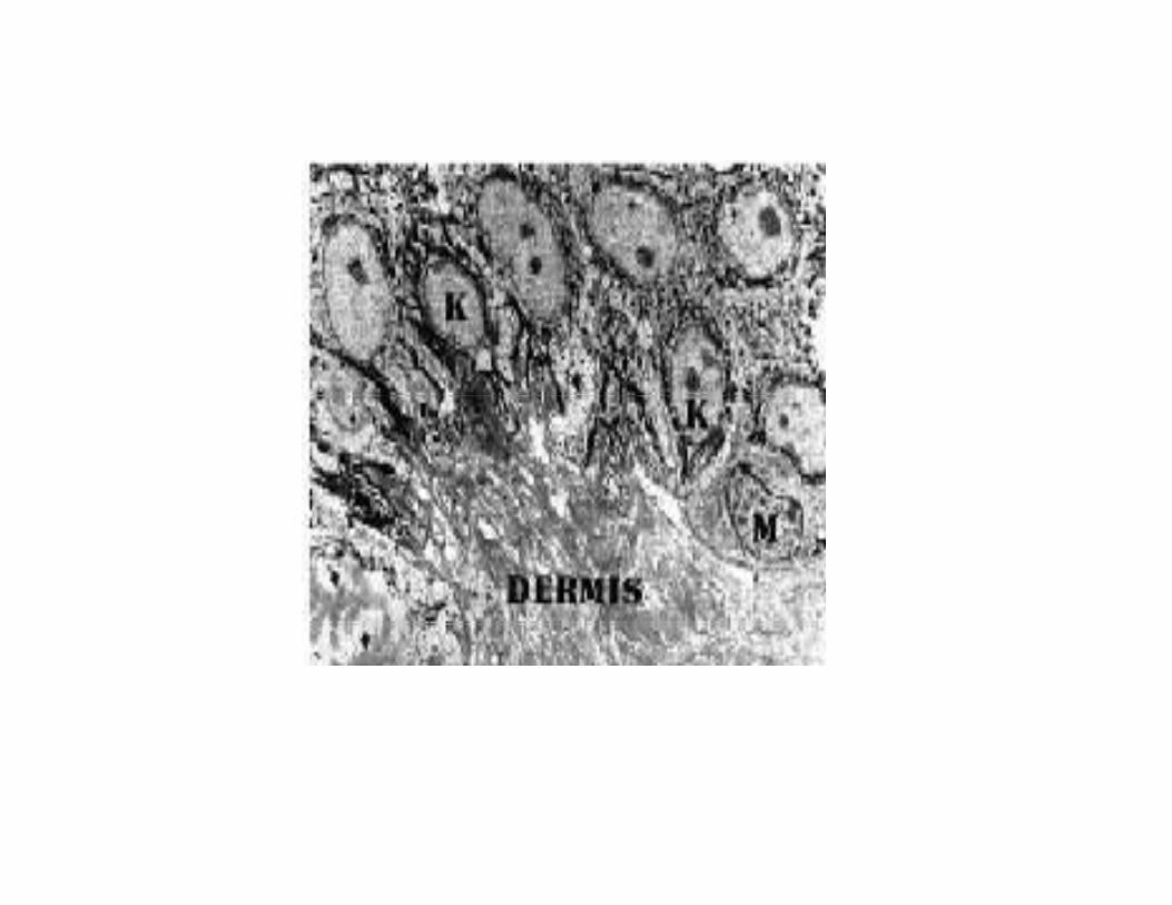
• Definition Melanocyte: are dendritic cells located in the bottom layer(the stratum y (basale layer) of skin’s epidermis.
Other sites: hair follicles,eye(uvea)eye(uvea),
ear(cochlea),
meningesmeninges
• Function of cutaneous melanocytes:Function of cutaneous melanocytes:
is to synthesize melanin in membrane‐bound oragnelles called melanosomes & to transferoragnelles called melanosomes & to transfer melanosomes to neighboring keratinocytes to provide protection from UV irradiationprovide protection from UV irradiation.
• Function of otic , ocular & meningeal melanocytes.
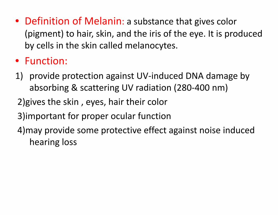
• Definition of Melanin: a substance that gives color (pigment) to hair, skin, and the iris of the eye. It is produced by cells in the skin called melanocytes.
• Function: 1) provide protection against UV‐induced DNA damage by
absorbing & scattering UV radiation (280‐400 nm)
2)gives the skin , eyes, hair their color
3)important for proper ocular function
4)may provide some protective effect against noise induced hearing loss
• Factors causing melanocytes to increase melanin production:p
1)UV irradiation (direct effect)
2)Keratinocyte paracrine factors induced by UV2)Keratinocyte paracrine factors induced by UV irradiation
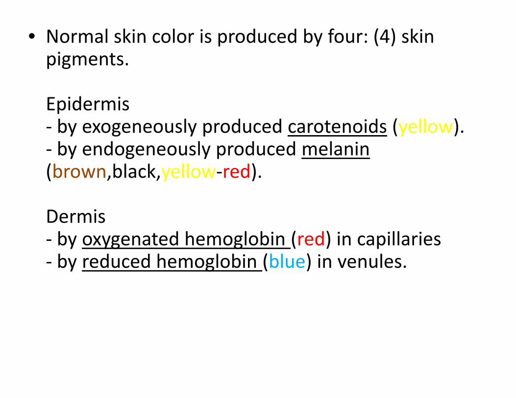
• Normal skin color is produced by four: (4) skin pigments.
Epidermis by exogeneously produced carotenoids (yellow)‐ by exogeneously produced carotenoids (yellow).
‐ by endogeneously produced melanin(brown black yellow‐red)(brown,black,yellow red).
Dermis ‐ by oxygenated hemoglobin (red) in capillaries ‐ by reduced hemoglobin (blue) in venules.
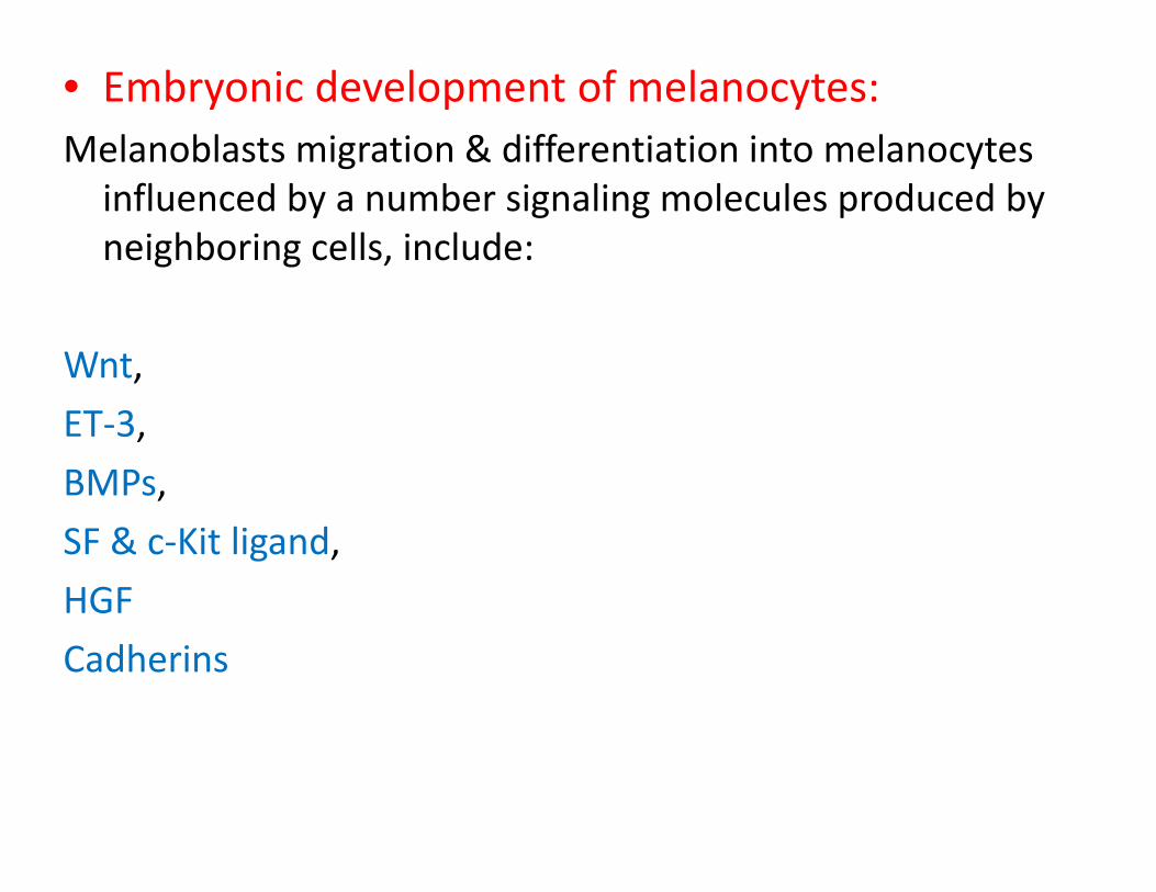
• Embryonic development of melanocytes:Melanoblasts migration & differentiation into melanocytes g yinfluenced by a number signaling molecules produced by neighboring cells, include:
Wnt,
ET‐3,
BMPs,
SF & c‐Kit ligand,
HGF
Cadherins
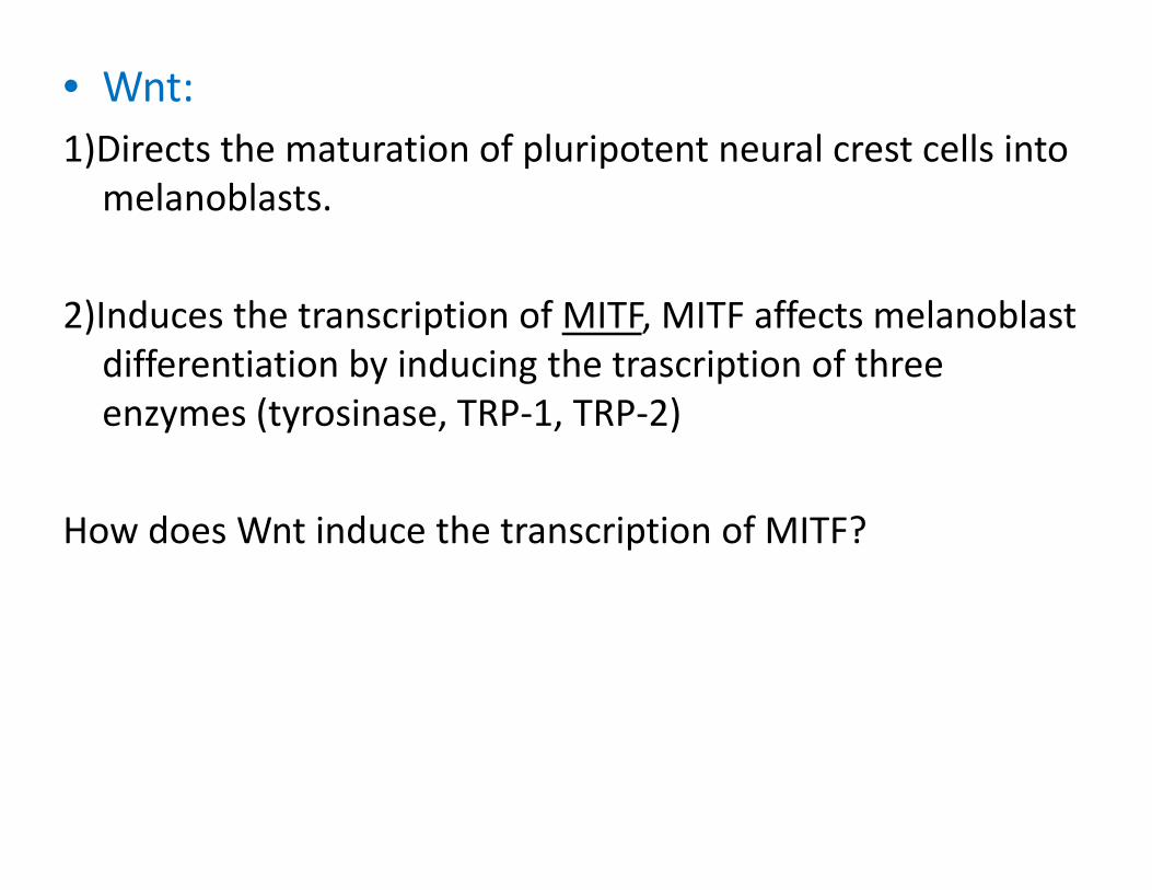
• Wnt:1)Directs the maturation of pluripotent neural crest cells into ) p pmelanoblasts.
2)Induces the transcription of MITF, MITF affects melanoblast differentiation by inducing the trascription of three enzymes (tyrosinase, TRP‐1, TRP‐2)
How does Wnt induce the transcription of MITF?
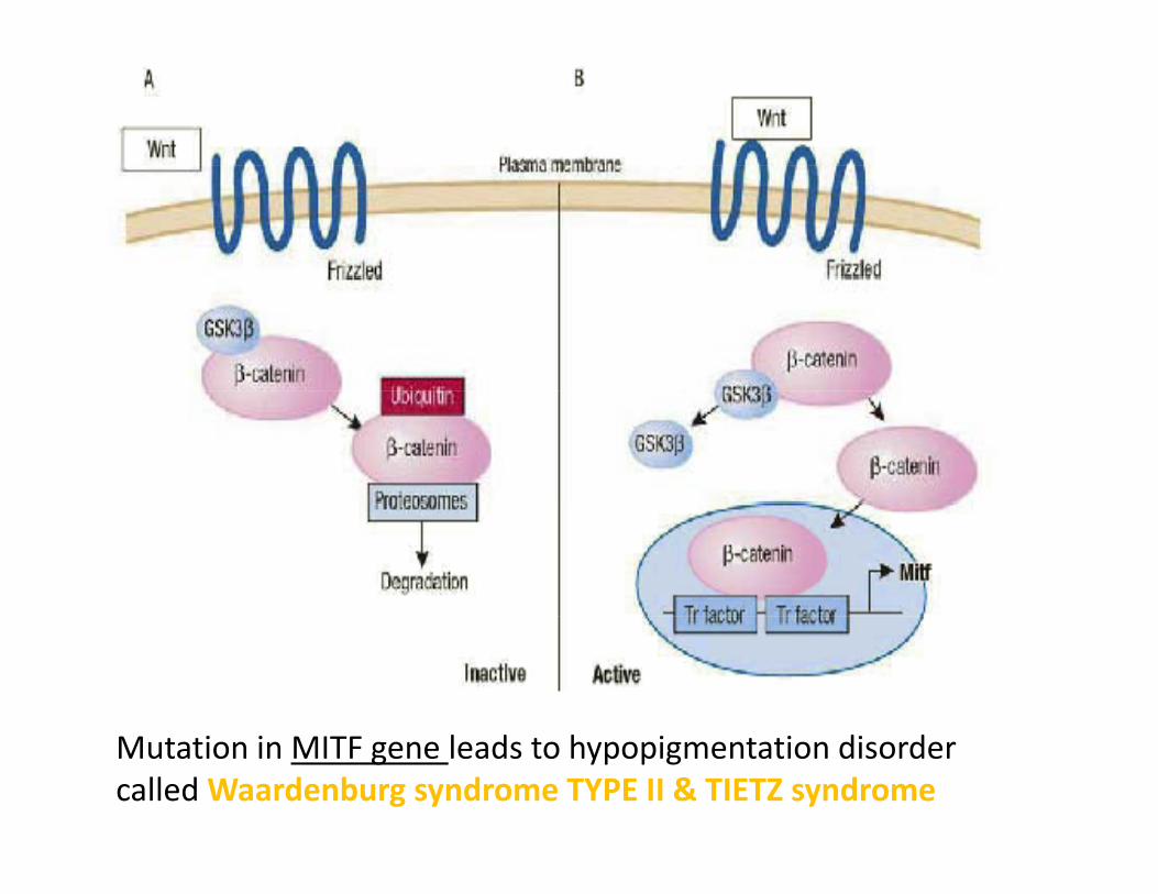
Mutation in MITF gene leads to hypopigmentation disorder called Waardenburg syndrome TYPE II & TIETZ syndrome
• Bone Morphogenetic proteins (BMPs)
• Endothelins (ETs):Include three members ET1 ET2 ET3Include three members ET1,ET2,ET3
They bind either EdnrA or EdnrB receptors
ET3 & its receptor EdnrB receptor are important duringET3 & its receptor EdnrB receptor are important during melanoblast migration & their proper expression is required fo survival, prolifration.required fo survival, prolifration.
Defect in ET3 or its receptor EdnrB leads loss of melanocytesDefect in ET3 or its receptor EdnrB leads loss of melanocytes “Waardenburg syndrome TYPE IV”
• Steel factor:
is expressed by epidermal keratinocytes & its receptor c‐Kit isis expressed by epidermal keratinocytes & its receptor c Kit is expressed on melanoblasts.
Both SF & c‐Kit are important in melanoblast development & migration.g
mutations of c‐Kit or SF & leads to melanoblast failure tomutations of c Kit or SF & leads to melanoblast failure to migrate to the skin “Piebaldism”
• Cadherins: family of transmembrane glycoproteins (E,P,N) that promote y g y p ( , , ) pcalcium dependent cell to cell adhesion.
It is thought that coordinate expression of E‐cadherins by epidermal keratinocytes & melanocytes plays a role in suppressing melanocyte prolifration in the epidermis. At
d l l h ftimes, some epidermal melanocytes switch from E‐cadherin to N‐cadherin & this switch allows them to escape the keratinocye mediated growth suppression &escape the keratinocye‐mediated growth suppression & prolifrate/aggregate in nests to form nevocellular nevi
• HGF:HGF:
the role of it in melanocyte development still unclear
• Site specific melanocytes:
Cutaneous melanocytes:Cutaneous melanocytes:
There is approximately one melanocyte per five to six keratinocyteskeratinocytes
Melanocytes synthesize & store melanin in cytosolic organellsMelanocytes synthesize & store melanin in cytosolic organells called melanosomes that are transferred to keratinocytes.
As keratinocytes are continuously being desquemated, there is a constant need for synthesis & transfer of melanosomes from melanocytes to keratinocytes to maintain cutaneous i ipigmentation
Melanocyte density/square mm ranges from 550 to 1200, i h h hi h i i hi f & liwith the highest concentration within face & genetalia
Melanocyte density is almost the same in all individuals of different ethnic background & thus cutaneous pigmentation doesn’t depend on melanocyte number.
Q‐ it depends on what?
1)Melanogenic activity within the melanocyte
2)The proportion of mature melanosomes
3)Type of melanin (eumelanin, or pheomelanin)
4)melanosomes transfer & distribution within the keratinocytes
5)Size of melanosomes
• Hair follicle melanocytes:
In contrast to interfollicular melanocytes, the follicular melanin unit undergoes cyclic modifications in coordinationmelanin unit undergoes cyclic modifications in coordination with hair cycle
Hair color is determined by amount of melanin transferred toHair color is determined by amount of melanin transferred to keratinocytes forming the hair shaft as well as by the ratio of eumelanin(black‐brown) to pheomelanin
• Ocular melanocytes:
Unlike cutaneous melanocytes, ocular melanocytes are in contact only with each other & don’t transfercontact only with each other & don t transfer melanosomes.
Q‐what are the functions of melanin in the eye?
Albinosmay have visual abnormalitis due to absence of melanin
• Otic melanocytes:
Reside in cochlea & are important for hearing, as loss of otic melanocytes may leads to deafness as inWaardenburgmelanocytes may leads to deafness as in Waardenburg syndrome TYPE II.
Q‐What about albinos? Are they deaf?y
A‐No
• Cephalic melanocytes
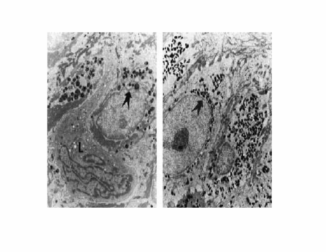
• Melanosome:
Is membrane‐bound organelle in which melanin biosynthesis & storage take placebiosynthesis & storage take place
And depending on the type of melanin(eumelanin orAnd depending on the type of melanin(eumelanin or pheomelanin) synthesized, melanosomes can be divided into eumelanosome & pheomelanosome
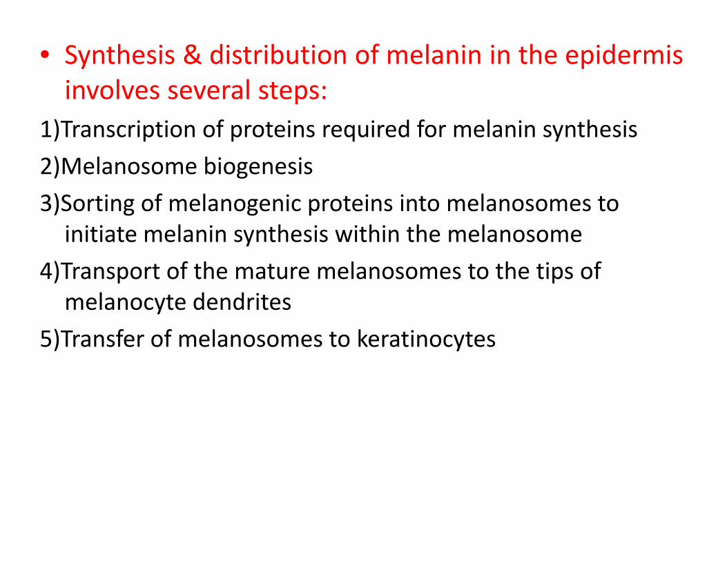
• Synthesis & distribution of melanin in the epidermis involves several steps:p
1)Transcription of proteins required for melanin synthesis
2)Melanosome biogenesis) g
3)Sorting of melanogenic proteins into melanosomes to initiate melanin synthesis within the melanosomey
4)Transport of the mature melanosomes to the tips of melanocyte dendrites
5)Transfer of melanosomes to keratinocytes
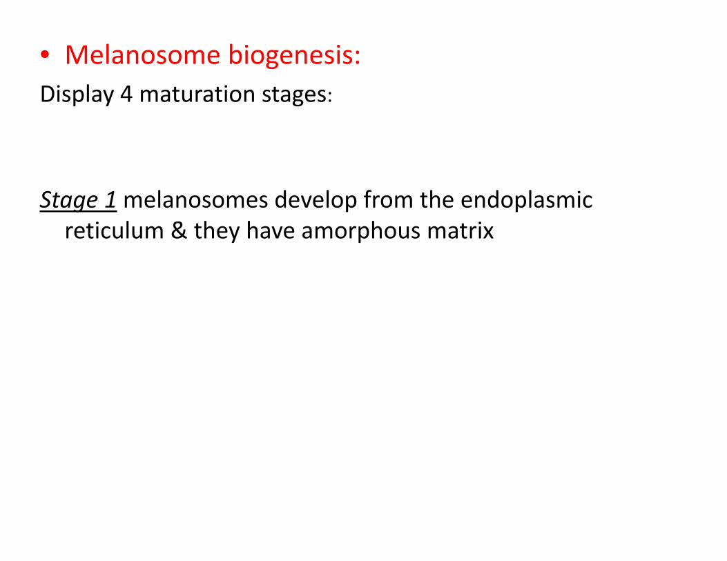
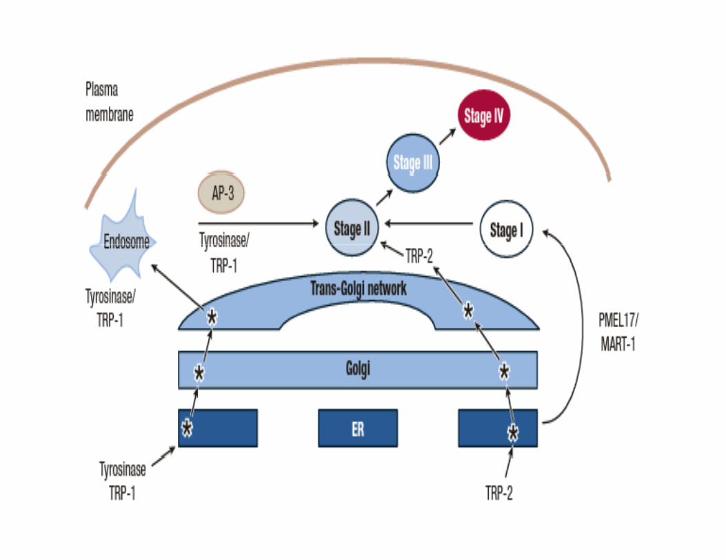
• Melanosome biogenesis:Display 4 maturation stages:p y g
Stage 1melanosomes develop from the endoplasmic reticulum & they have amorphous matrix
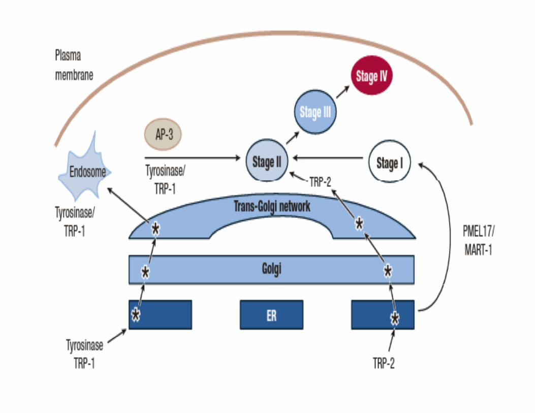
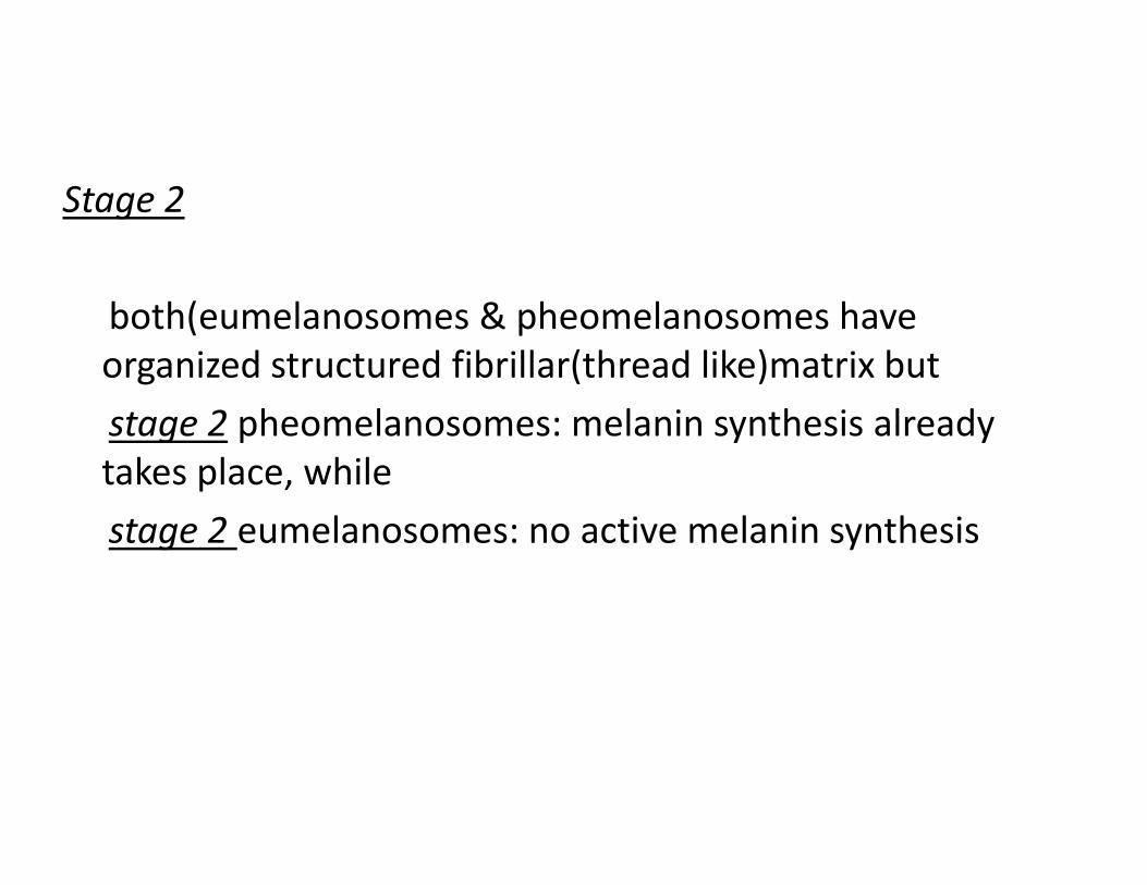
Stage 2
both(eumelanosomes & pheomelanosomes have organized structured fibrillar(thread like)matrix but
stage 2 pheomelanosomes: melanin synthesis already takes place, while
stage 2 eumelanosomes: no active melanin synthesis
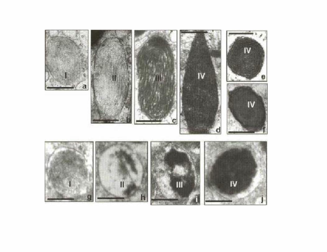
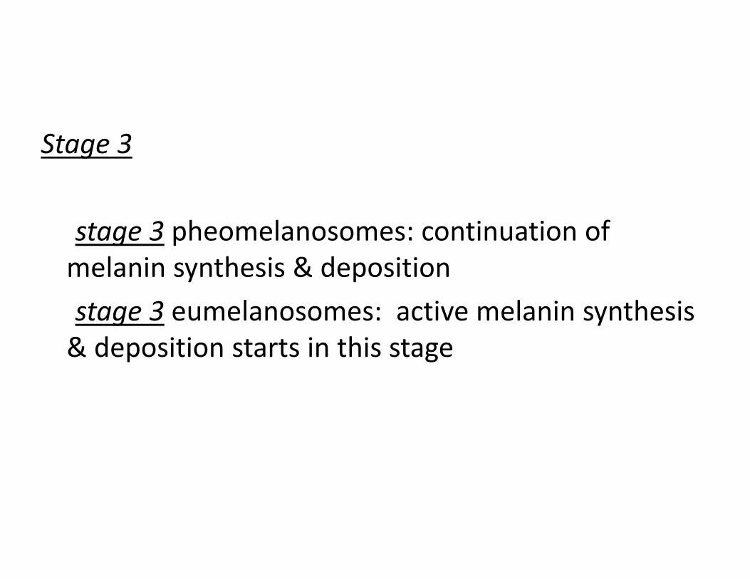
Stage 3
stage 3 pheomelanosomes: continuation of melanin synthesis & deposition
stage 3 eumelanosomes: active melanin synthesis & deposition starts in this stage
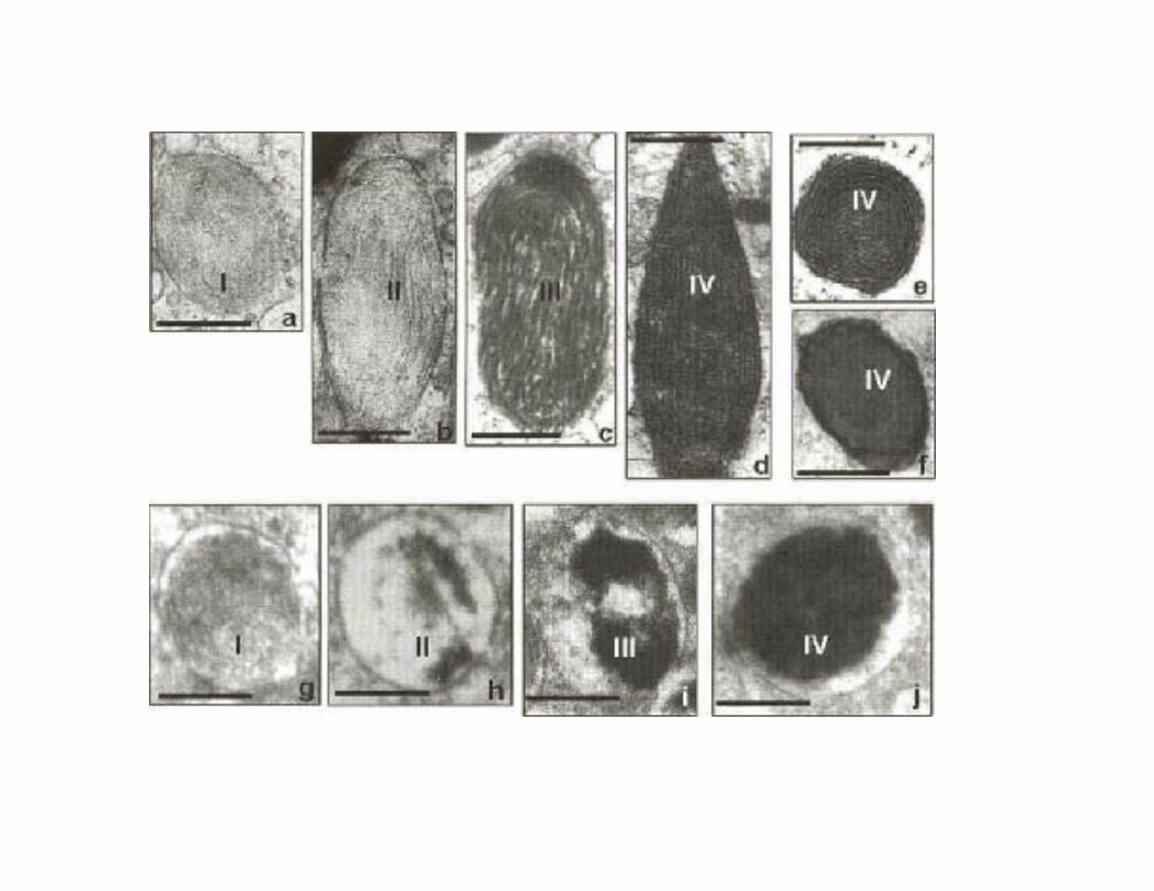
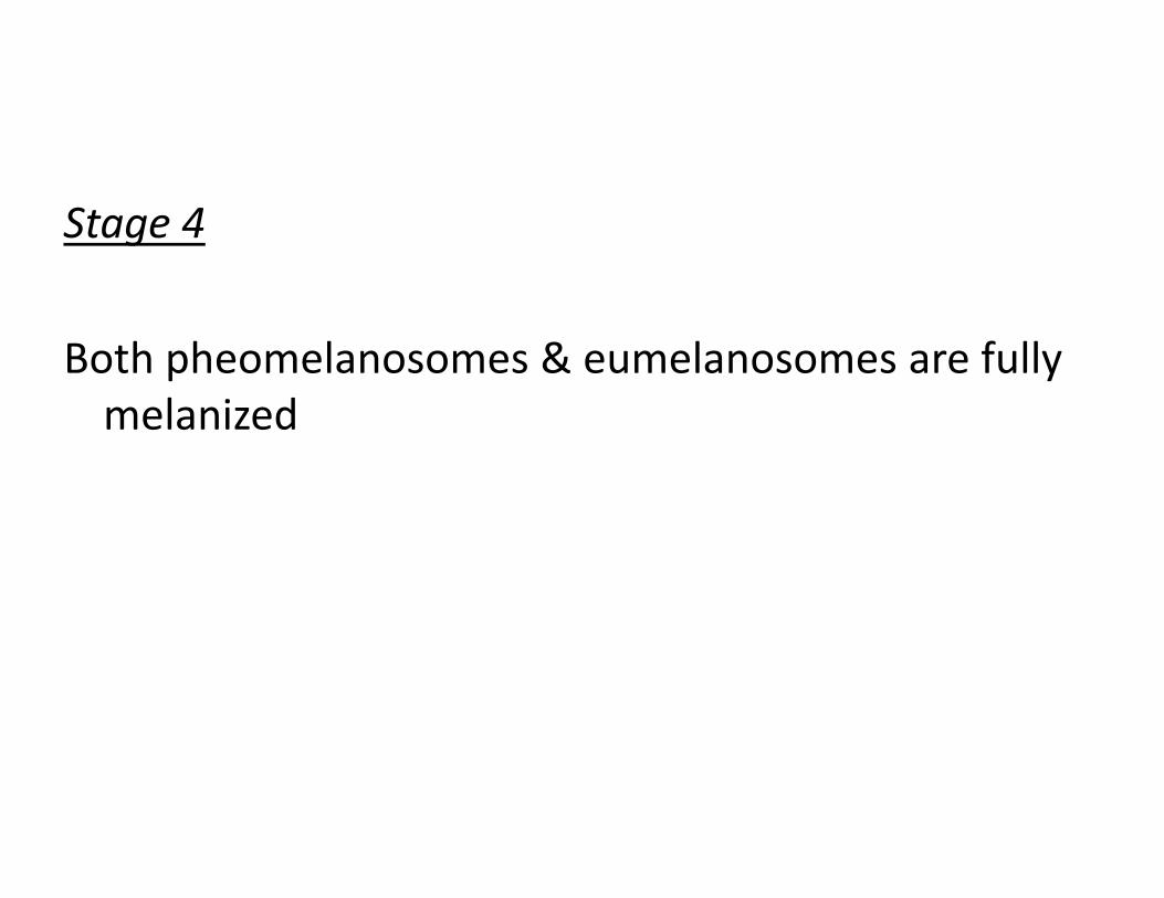
Stage 4
Both pheomelanosomes & eumelanosomes are fully melanized

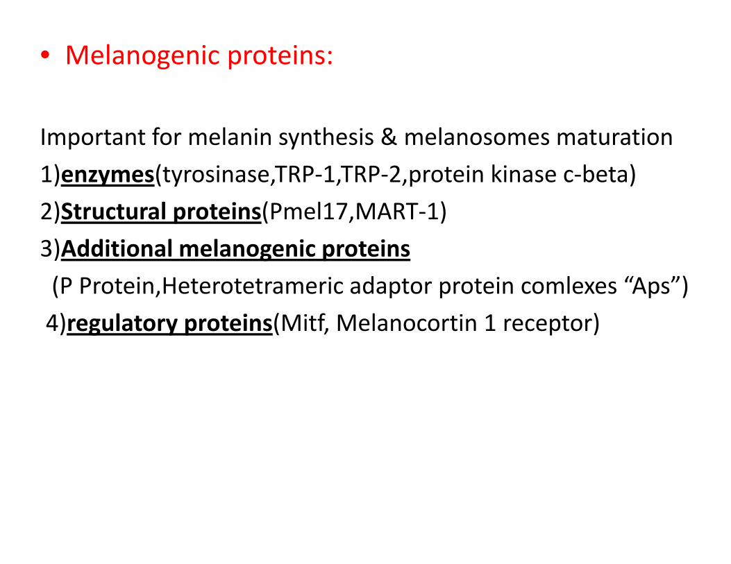
• Melanogenic proteins:
Important for melanin synthesis & melanosomes maturation
1)enzymes(tyrosinase TRP‐1 TRP‐2 protein kinase c‐beta)1)enzymes(tyrosinase,TRP‐1,TRP‐2,protein kinase c‐beta)
2)Structural proteins(Pmel17,MART‐1)
3)Additional melanogenic proteins3)Additional melanogenic proteins
(P Protein,Heterotetrameric adaptor protein comlexes “Aps”)
4) l t t i (Mitf M l ti 1 t )4)regulatory proteins(Mitf, Melanocortin 1 receptor)
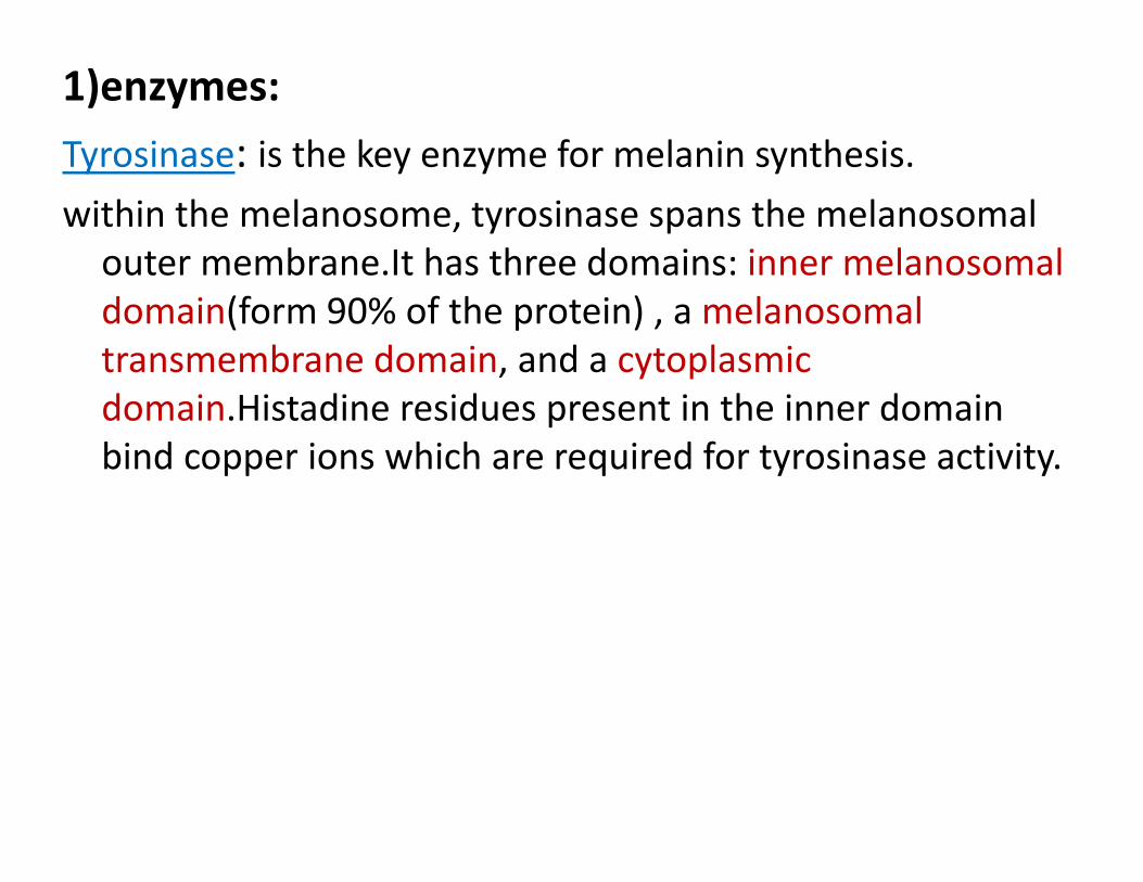
1)enzymes:
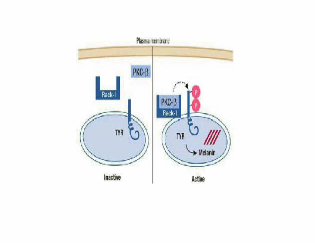
Tyrosinase: is the key enzyme for melanin synthesis.Tyrosinase: is the key enzyme for melanin synthesis.
within the melanosome, tyrosinase spans the melanosomal outer membrane.It has three domains: inner melanosomalouter membrane.It has three domains: inner melanosomal domain(form 90% of the protein) , a melanosomal transmembrane domain, and a cytoplasmic domain.Histadine residues present in the inner domain bind copper ions which are required for tyrosinase activity.

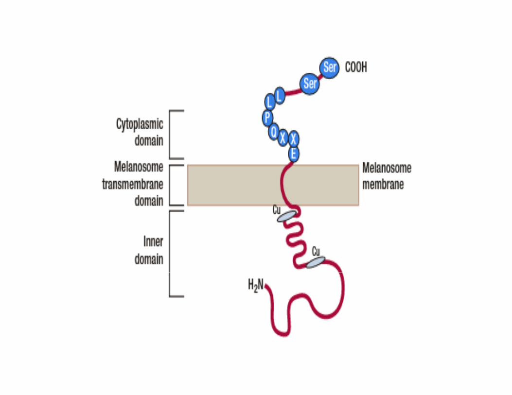
In addition to that,two serine residues on theIn addition to that,two serine residues on the cytoplasmic domain must be phospholyrated by protein kinase C‐beta to activate tyrosinase, in theprotein kinase C beta to activate tyrosinase, in the absence of this phosphorylation, pigmentation does not occure.not occure.
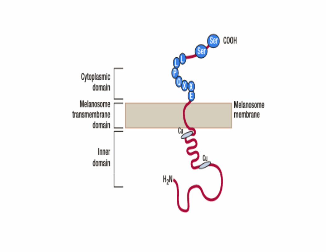
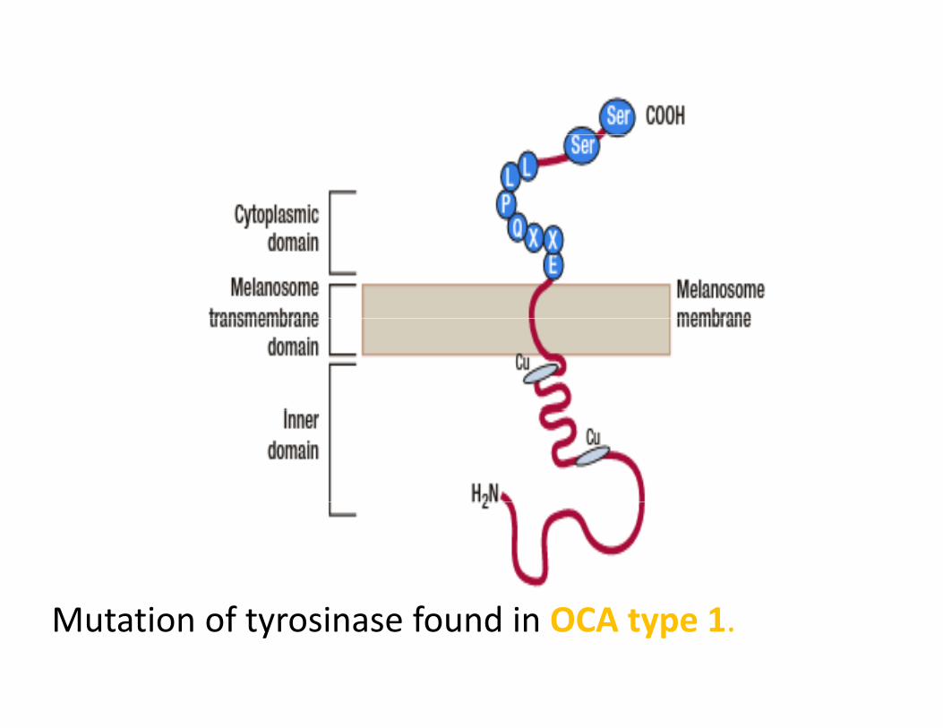
Mutation of tyrosinase found in OCA type 1.
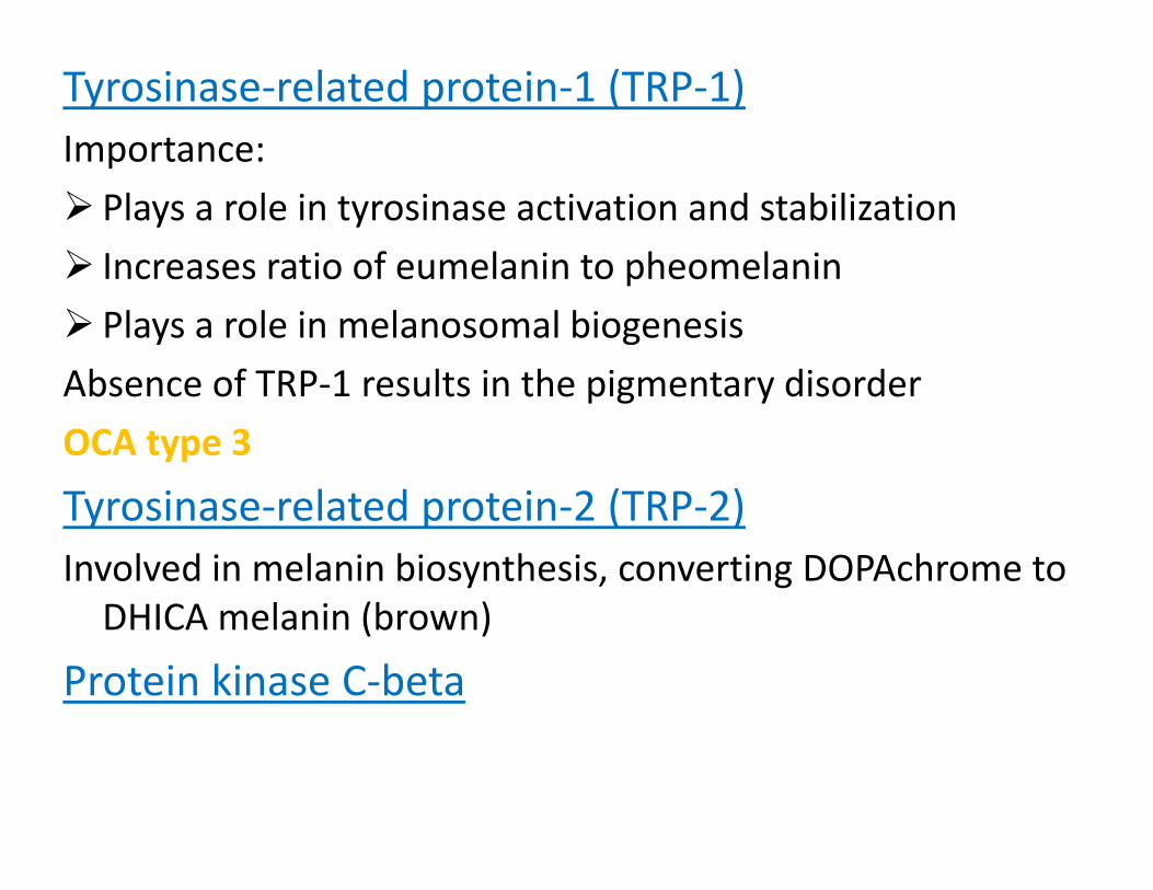
Tyrosinase‐related protein‐1 (TRP‐1)Importance: p
Plays a role in tyrosinase activation and stabilization
Increases ratio of eumelanin to pheomelaninIncreases ratio of eumelanin to pheomelanin
Plays a role in melanosomal biogenesis
Absence of TRP‐1 results in the pigmentary disorderAbsence of TRP 1 results in the pigmentary disorder
OCA type 3
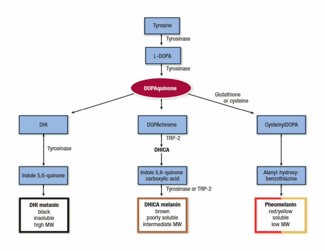
Tyrosinase related protein 2 (TRP 2)Tyrosinase‐related protein‐2 (TRP‐2)Involved in melanin biosynthesis, converting DOPAchrome to DHICA melanin (brown)DHICA melanin (brown)
Protein kinase C‐beta
2)Structural proteins(Pmel17 & MART‐1)
Both are important for proper deposition of melaninBoth are important for proper deposition of melanin within the melanosomes.
Upto date no haypopigmentary disorders associatedUpto date, no haypopigmentary disorders associated with nonfunctioning MART‐1 or Pmel17 have been identifiedidentified
3)Additional melanogenic proteins
P ProteinP ProteinProposed Functions:
• Maintains acidic enviroment within the melanosomes• Maintains acidic enviroment within the melanosomes
• Transporting tyrosinase into the melanosomes
Individuals lacking P Protein display OCA type 2 largely dueIndividuals lacking P Protein display OCA type 2, largely due to improper melanosomal PH.

Heterotetrameric adaptor protein complexes(Aps)AP‐3 & possibly AP‐1 facilitate tyrosinase transport from p y y pendosomes to melanosomes.
Individuals with defects in AP‐3 display
Hermansky‐Pudlak syndrome 2(OCA+ platelets dysfunction+ pulmonary disease)

4)Regulatory proteins:
Microphthalmia‐associated transcription factorMicrophthalmia associated transcription factorMitf gene is the master gene for melanocyte survival & is a key factor regulating the transcription of the majorkey factor regulating the transcription of the major melanogenic proteins(tyrosinase, TRP‐1 , TRP‐2 ,PKC‐beta & MART1)
Mutations in Mitf found in the pigmentary disorders
waardenburg syndrome type 2 & tietz syndrome.
Mitf expression is under the control of several transcription factors including Sox10 & Pax3.
Sox10(is mutated in WS type 4)
Pax3(is mutated in WS type 3 & 1)
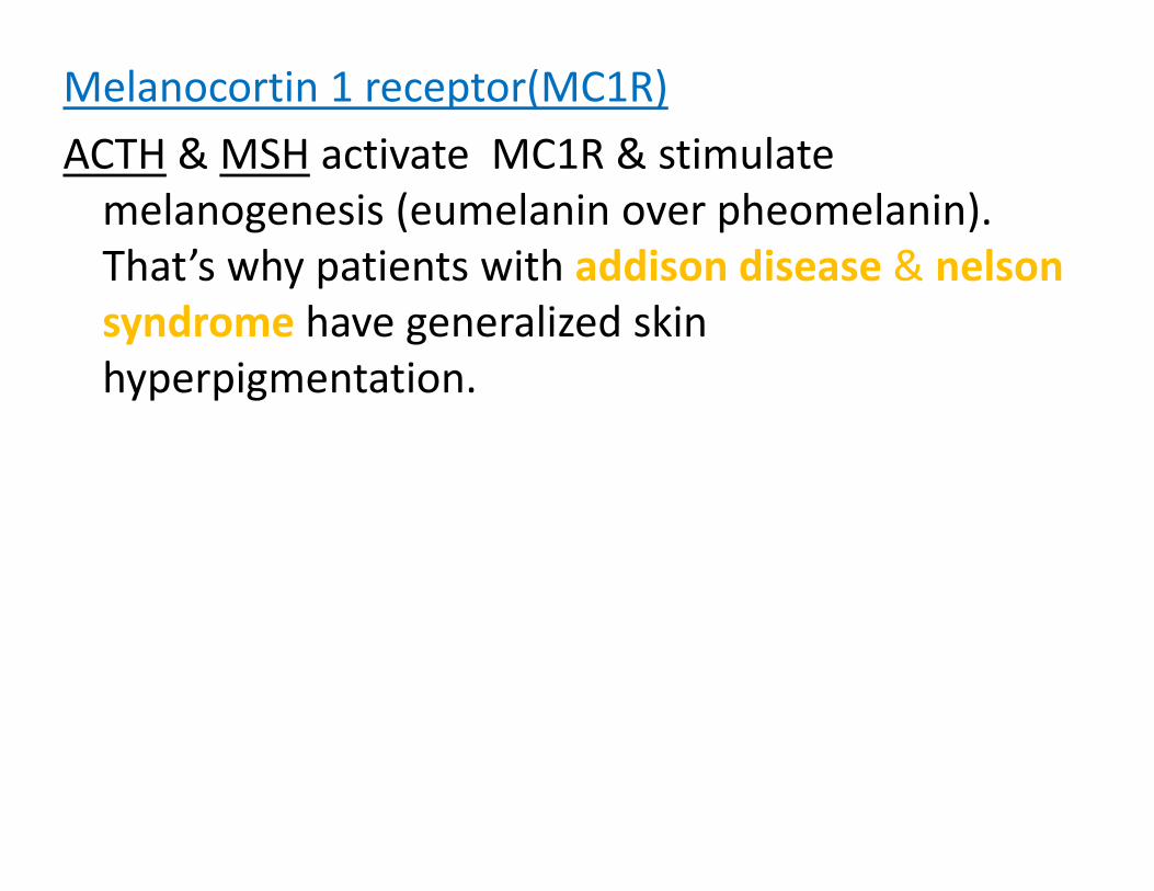
Melanocortin 1 receptor(MC1R)
ACTH & MSH activate MC1R & stimulateACTH & MSH activate MC1R & stimulate melanogenesis (eumelanin over pheomelanin). That’s why patients with addison disease & nelsonThat s why patients with addison disease & nelsonsyndrome have generalized skin hyperpigmentation.hyperpigmentation.

• Melanin biosynthesis:
Site:melanocytes(melanosomes)Site:melanocytes(melanosomes)
Types:
E l i (b bl k)Eumelanin(brown,black)
Pheomelanin(red‐yellow)
The main function of melanin:
Protection against UV induced DNA damage.Protection against UV induced DNA damage.
Q‐why is light skinned‐red haired individuals are more prone to UV induced melanoma?prone to UV induced melanoma?
• Melanocyte dendrites:
Are branching processes that interact withAre branching processes that interact with keratinocytes
The major structural component of melanocyteThe major structural component of melanocyte dendrites is actin
Q h t d i l t d d i it ?Q‐what does increase melanocyte dendricity?
‐UV irradiation
‐keratinocyte derived factors(ET1,nerve growth factor,ACTH,MSH,PGE2,PGF2)
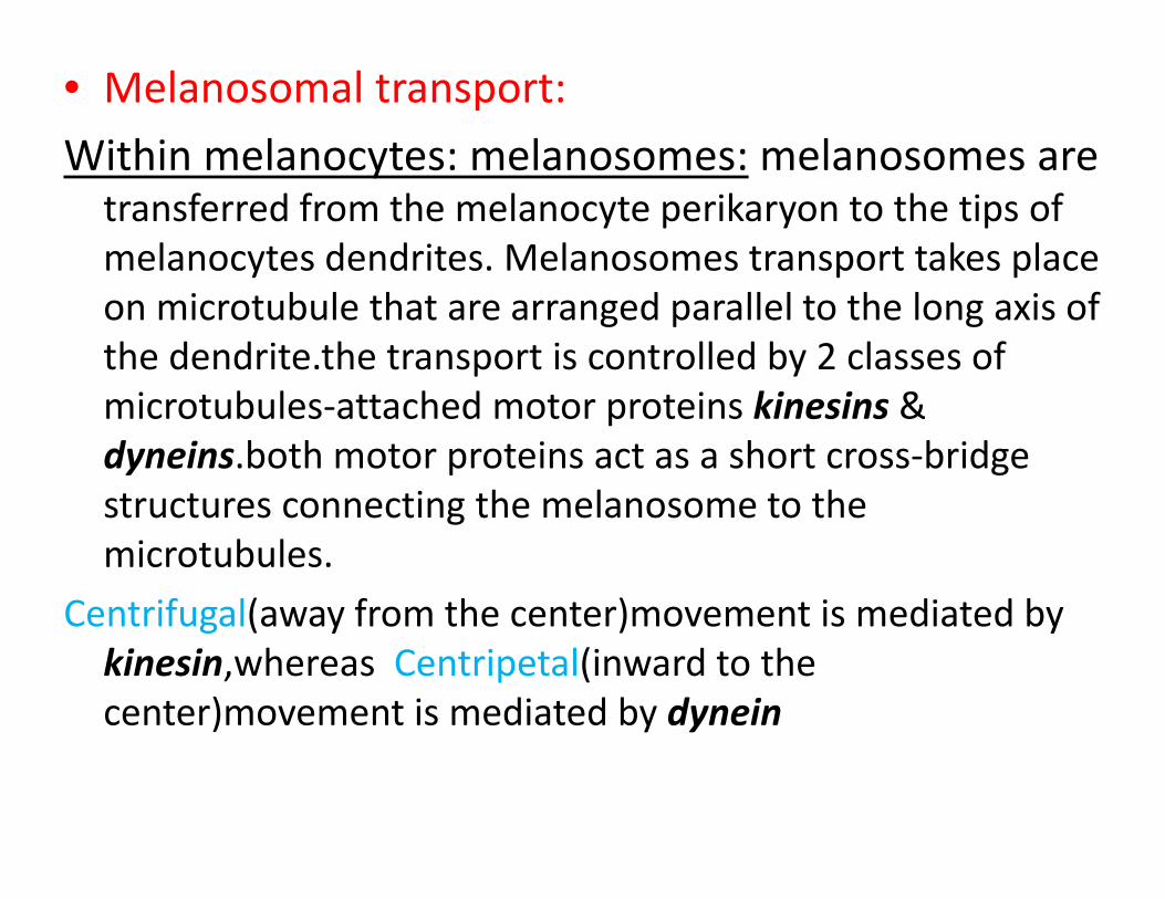
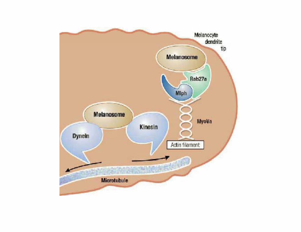
• Melanosomal transport:
Within melanocytes: melanosomes: melanosomes areWithin melanocytes: melanosomes: melanosomes are transferred from the melanocyte perikaryon to the tips of melanocytes dendrites. Melanosomes transport takes place on microtubule that are arranged parallel to the long axis of the dendrite.the transport is controlled by 2 classes of i b l h d i ki i µtubules‐attached motor proteins kinesins &
dyneins.both motor proteins act as a short cross‐bridge structures connecting the melanosome to thestructures connecting the melanosome to the microtubules.
Centrifugal(away from the center)movement is mediated byCentrifugal(away from the center)movement is mediated by kinesin,whereas Centripetal(inward to the center)movement is mediated by dynein
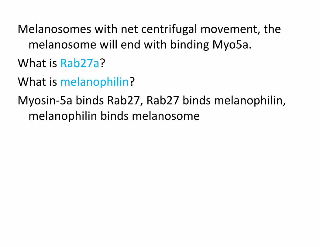
Melanosomes with net centrifugal movement, the melanosome will end with binding Myo5amelanosome will end with binding Myo5a.
What is Rab27a?
What is melanophilin?
Myosin‐5a binds Rab27, Rab27 binds melanophilin, melanophilin binds melanosome
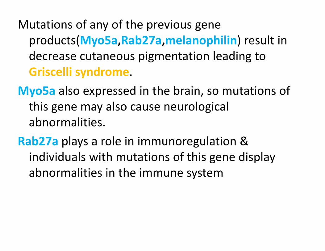
Mutations of any of the previous gene products(Myo5a,Rab27a,melanophilin) result in p ( y p )decrease cutaneous pigmentation leading to Griscelli syndrome.y
Myo5a also expressed in the brain, so mutations of this gene may also cause neurologicalthis gene may also cause neurological abnormalities.
Rab27a plays a role in immunoregulation &Rab27a plays a role in immunoregulation & individuals with mutations of this gene display abnormalities in the immune systemabnormalities in the immune system
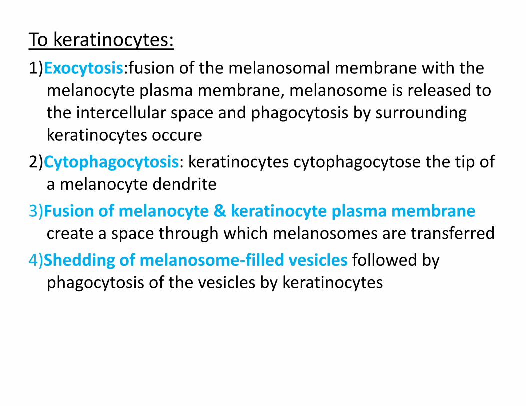
To keratinocytes:1)Exocytosis:fusion of the melanosomal membrane with the ) ymelanocyte plasma membrane, melanosome is released to the intercellular space and phagocytosis by surrounding keratinocytes occure
2)Cytophagocytosis: keratinocytes cytophagocytose the tip of l d da melanocyte dendrite
3)Fusion of melanocyte & keratinocyte plasma membrane t th h hi h l t f dcreate a space through which melanosomes are transferred
4)Shedding of melanosome‐filled vesicles followed by phagocytosis of the vesicles by keratinocytesphagocytosis of the vesicles by keratinocytes
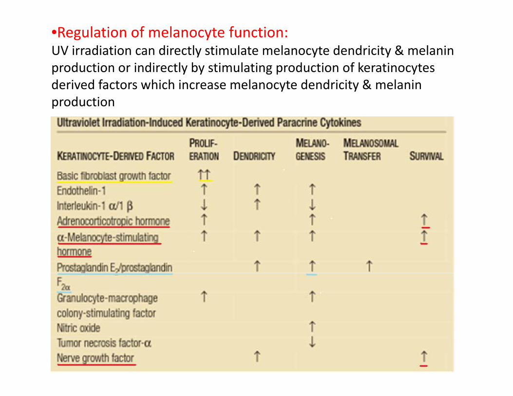
•Regulation of melanocyte function:UV irradiation can directly stimulate melanocyte dendricity & melanin production or indirectly by stimulating production of keratinocytesp y y g p yderived factors which increase melanocyte dendricity & melanin production
• Congenital hypomelanosis disorders
Arise due to defect in:
1)Melanocyte embryonic development1)Melanocyte embryonic development
WS1‐4, Tietz syndrome, Piebaldism
2)Disorder of melanin synthesis2)Disorder of melanin synthesis
OCA1‐4
3)M l f ti t t & t f3)Melanosome formation, transport & transfer
HPS, Chediak‐Higashi syndrome, Gracelli syndrome

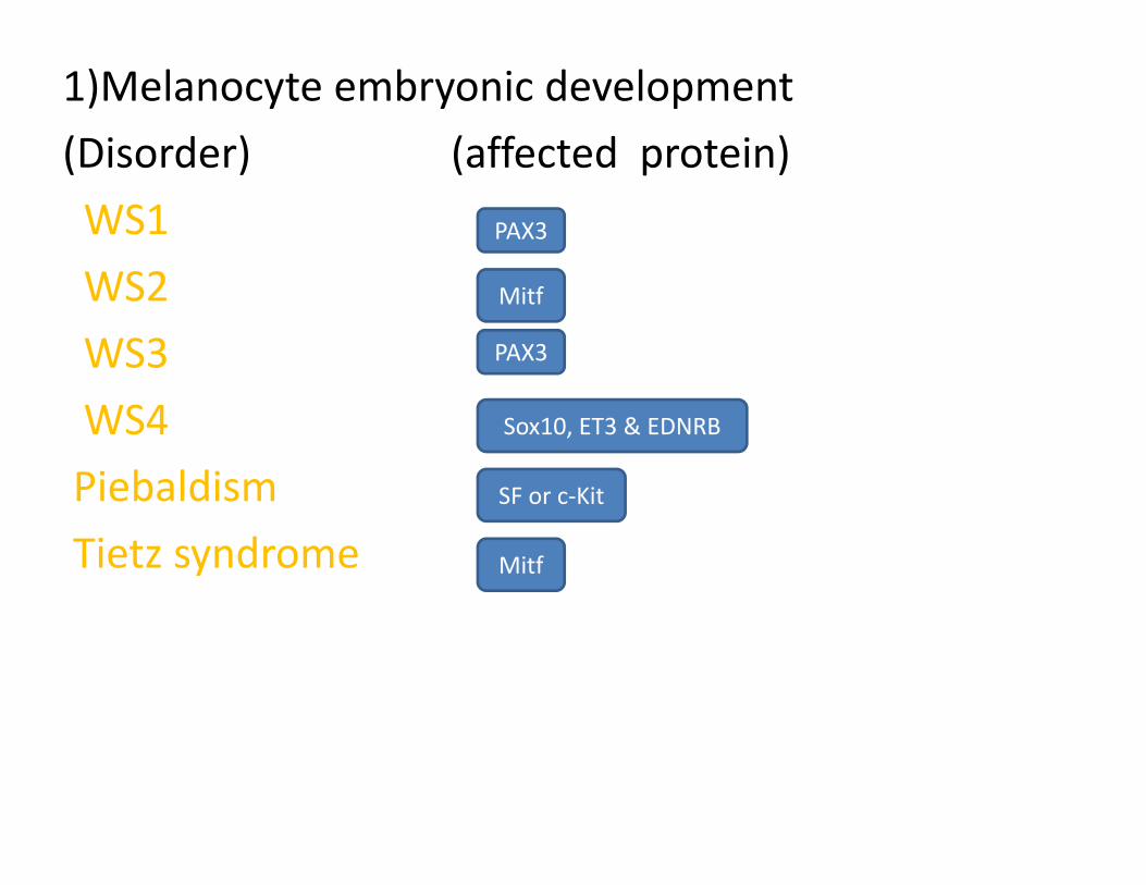
1)Melanocyte embryonic development
(Disorder) (affected protein)(Disorder) (affected protein)
WS1
WS2PAX3
WS2
WS3Mitf
PAX3
WS4
Piebaldism
Sox10, ET3 & EDNRB
SF or c‐KitPiebaldism
Tietz syndromeSF or c Kit
Mitf
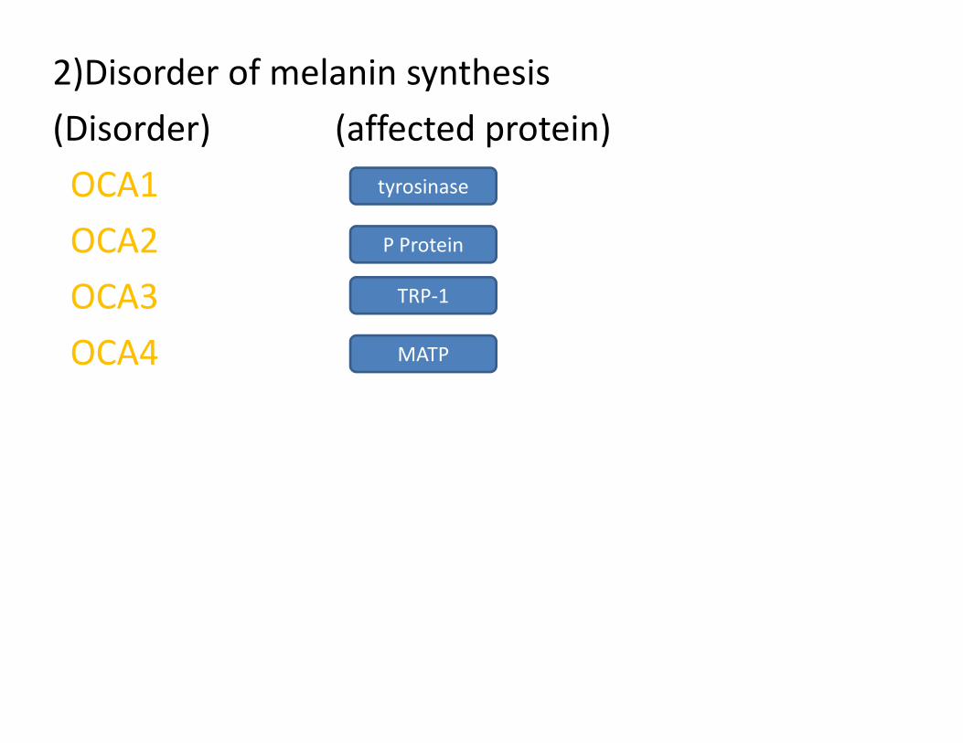
2)Disorder of melanin synthesis
(Disorder) (affected protein)(Disorder) (affected protein)
OCA1
OCA2
tyrosinase
OCA2
OCA3 TRP‐1
P Protein
OCA4 MATP
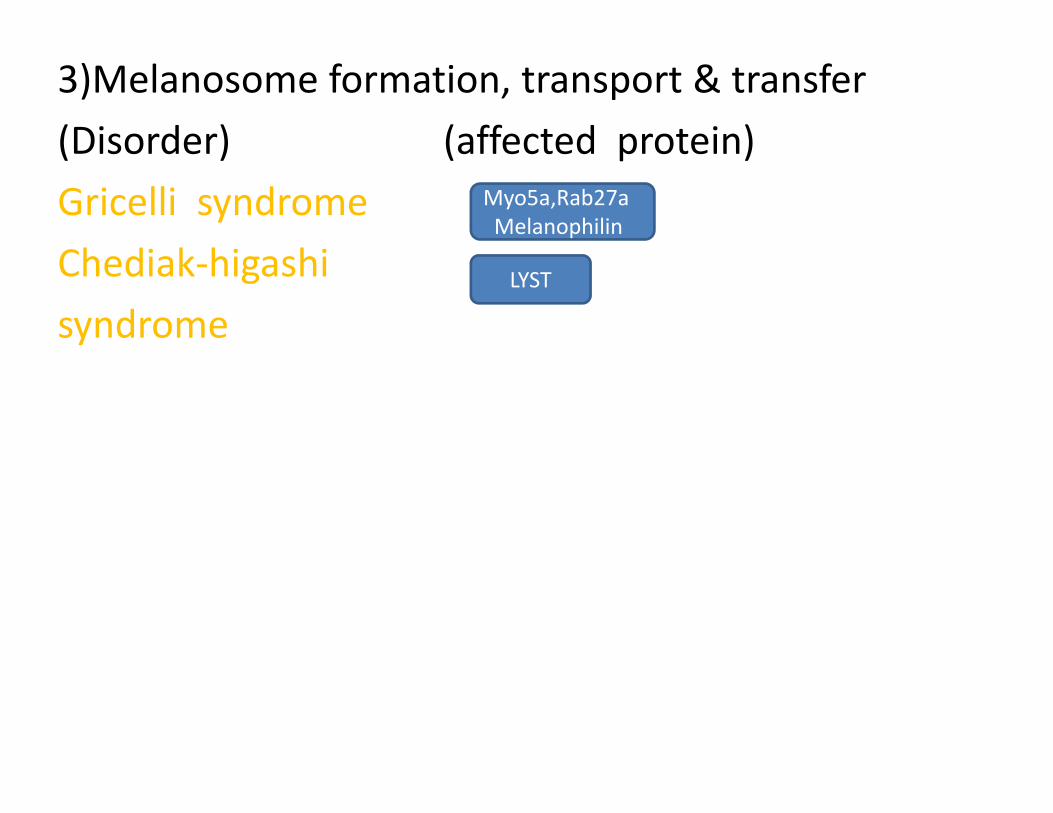
3)Melanosome formation, transport & transfer
(Disorder) (affected protein)(Disorder) (affected protein)
Gricelli syndrome
Ch di k hi hi
Myo5a,Rab27aMelanophilin
Chediak‐higashi
syndromeLYST