Embed Size (px)
Citation preview

CALIFORNIA STATE UNIVERSITY, NORTHRIDGE
The Roles of CXCR4 and CXCR7 in Melanocyte and Melanoma Motility
A thesis submitted in partial fulfillment of the requirements
for the degree of Master of Science in Biology
By:
Samantha Hain
August 2019

ii
The thesis of Samantha Hain is approved:
__________________________________________ ______________________
Dr. Ravinder Abrol Date
__________________________________________ ______________________
Dr. Cindy Malone Date
__________________________________________ ______________________
Dr. Maria Elena de Bellard, Chair Date
California State University, Northridge

iii
Acknowledgments
This thesis project is financially supported by the Association of Retired Faculty
Graduate Thesis Award, the Peter Bellinger Student Research Award, and the Center on
Disabilities Award, all provided through California State University, Northridge (CSUN).
Funding was also provided by the CSUN’s 2019 Thesis Support Program, and the
Department of Rehabilitation. I would like to express deep gratitude to my trainees for their
assistance and participation. First, I thank Natalie Dahan, Roland Lacap and Tyler Tran for
their hard work in assisting with both movement and wound assays, as well as qPCR, and
data analysis in cell tracking. Furthermore, funding was also provided by these
undergraduate students: Natalie with BUILD/PODER, and Roland with MARC.
Additionally, I express great appreciation to Ronelle Caguioa and Anil Chaganti for all
their work in data analysis of the movement and wound assays.
In developing the ideas presented here, I have received helpful input,
encouragement, and critique from my research supervisors, Dr. Maria Elena de Bellard and
Dr. Ravinder Abrol. I thank the Abrol Lab for their collaboration and access to the modified
receptors and Figure 1. I also thank my thesis committee: Dr. Maria Elena de Bellard, Dr.
Ravinder Abrol, and Dr. Cindy Malone for their feedback and support. I would like to give
one last acknowledgment to show my personal appreciation to Martin Van der Plas and my
parents, Rich and Sheryl Hain, for all their advice and support throughout this Thesis
Project.
iv
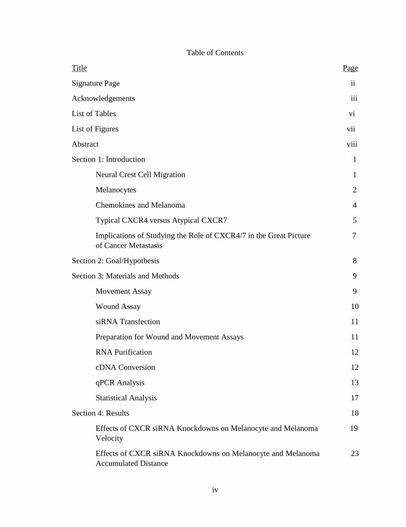
Table of Contents
Title Page
Signature Page ii
Acknowledgements iii
List of Tables vi
List of Figures vii
Abstract viii
Section 1: Introduction 1
Neural Crest Cell Migration 1
Melanocytes 2
Chemokines and Melanoma 4
Typical CXCR4 versus Atypical CXCR7 5
Implications of Studying the Role of CXCR4/7 in the Great Picture 7
of Cancer Metastasis
Section 2: Goal/Hypothesis 8
Section 3: Materials and Methods 9
Movement Assay 9
Wound Assay 10
siRNA Transfection 11
Preparation for Wound and Movement Assays 11
RNA Purification 12
cDNA Conversion 12
qPCR Analysis 13
Statistical Analysis 17
Section 4: Results 18
Effects of CXCR siRNA Knockdowns on Melanocyte and Melanoma 19
Velocity
Effects of CXCR siRNA Knockdowns on Melanocyte and Melanoma 23
Accumulated Distance

v
Effects of CXCR siRNA Knockdowns on Melanocyte and Melanoma 26
Persistence and Chemotaxis Index
Effects of CXCR siRNA Knockdowns on Melanocyte Wound Assays 29
Effects of CXCR siRNA Knockdowns on Melanoma Wound Assays 32
qPCR and GFP Expression of Each Transfection in Melanocyte 34
and Melanoma
Section 5: Discussion 37
Future Directions 45
Conclusion 46
References 47
vi
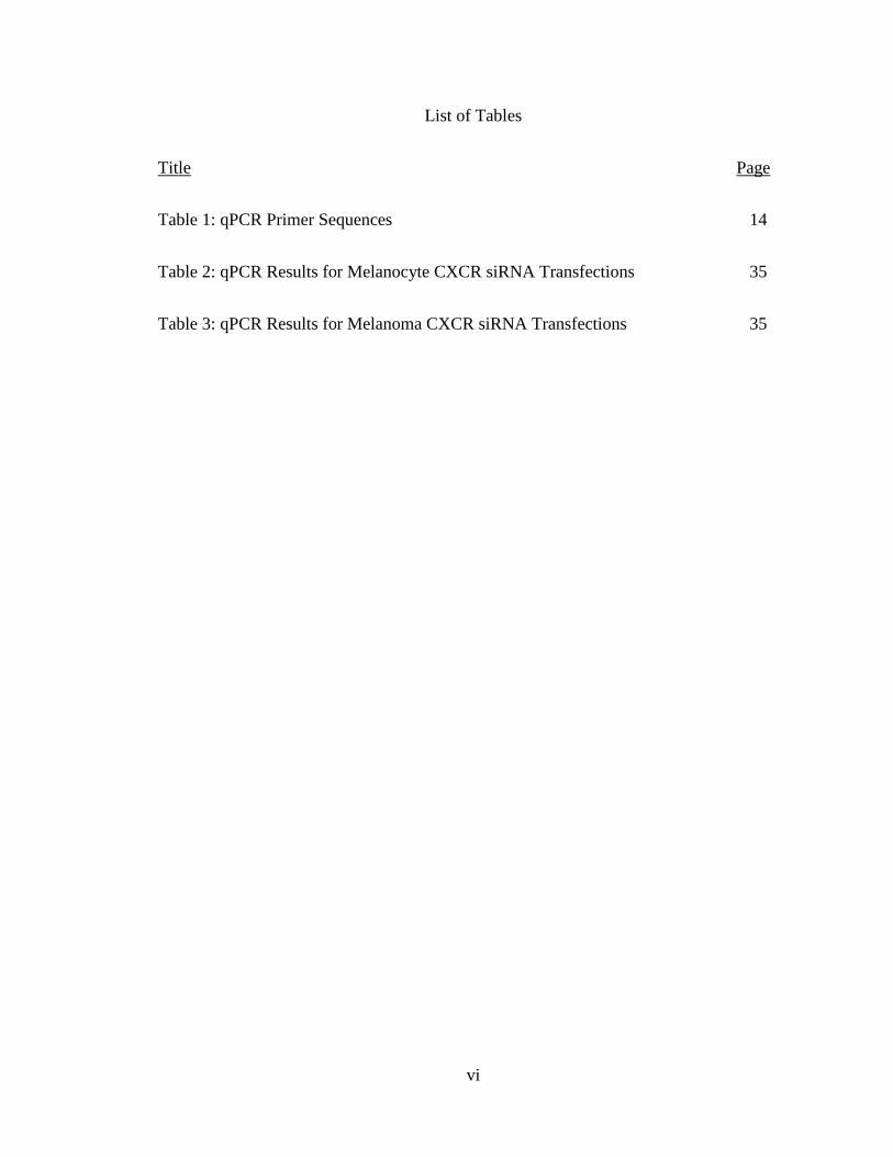
List of Tables
Title Page
Table 1: qPCR Primer Sequences 14
Table 2: qPCR Results for Melanocyte CXCR siRNA Transfections 35
Table 3: qPCR Results for Melanoma CXCR siRNA Transfections 35
vii
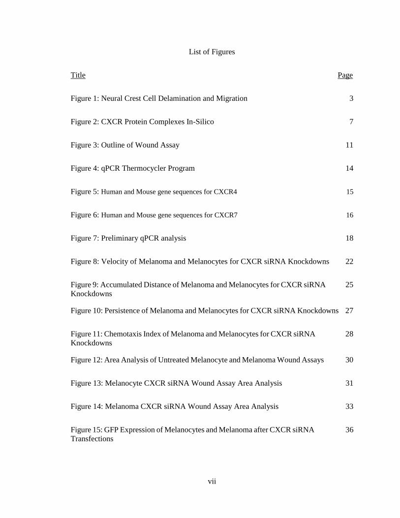
List of Figures
Title Page
Figure 1: Neural Crest Cell Delamination and Migration 3
Figure 2: CXCR Protein Complexes In-Silico 7
Figure 3: Outline of Wound Assay 11
Figure 4: qPCR Thermocycler Program 14
Figure 5: Human and Mouse gene sequences for CXCR4 15
Figure 6: Human and Mouse gene sequences for CXCR7 16
Figure 7: Preliminary qPCR analysis 18
Figure 8: Velocity of Melanoma and Melanocytes for CXCR siRNA Knockdowns 22
Figure 9: Accumulated Distance of Melanoma and Melanocytes for CXCR siRNA 25
Knockdowns
Figure 10: Persistence of Melanoma and Melanocytes for CXCR siRNA Knockdowns 27
Figure 11: Chemotaxis Index of Melanoma and Melanocytes for CXCR siRNA 28
Knockdowns
Figure 12: Area Analysis of Untreated Melanocyte and Melanoma Wound Assays 30
Figure 13: Melanocyte CXCR siRNA Wound Assay Area Analysis 31
Figure 14: Melanoma CXCR siRNA Wound Assay Area Analysis 33
Figure 15: GFP Expression of Melanocytes and Melanoma after CXCR siRNA 36
Transfections
viii
Abstract
The Roles of CXCR4 and CXCR7 in Melanocyte and Melanoma Motility
By
Samantha Hain
Master of Science in Biology
Chemokines are signaling proteins released by cells in response to chemical stimuli
in their environment. The chemokine stromal derived factor 1 (SDF1) has been regularly
studied due to its role in the growth and metastasis of multiple cancers, including melanoma.
SDF1 has two known receptors: CXCR4 and CXCR7. Previous research has mainly
focused on CXCR4 receptor signaling, which influences many cell responses, among them
the migration of neural crest cells and the amount of receptor expression is upregulated in
melanoma. CXCR7 receptor signaling is not as well studied but has been shown to
influence melanocyte migration and constrain melanoma tumor growth in vivo where as
CXCR4 could not. It is not known, however, how relevant CXCR7 may be in the capability
of melanocyte and melanoma migration. Here, I studied the potential roles of CXCR4 and
CXCR7 in melanocyte and melanoma migration in vitro by genetically silencing both
receptors. My data shows that the CXCR7 receptor is more important for migratory
capabilities of melanocytes and melanoma cells than the CXCR4 receptor. These findings
suggest that down regulating or blocking the CXCR7 receptor through targeted therapies
may show a substantial effect in melanoma treatment.

1
SECTION 1: INTRODUCTION
Chemokines are a specific type of cytokine that cause chemotactic responses in
neighboring cells that harbor reciprocal receptors; these signaling proteins are secreted by
cells to direct the movement of other cells. Chemokines are important for their role in
regulating cell migration during embryonic development, during immune responses, and
even during some cancer cell metastases. Specifically, stromal derived factor 1 (SDF1) is
a chemokine that with its two known receptors, CXCR4 and CXCR7, plays a role in tumor
growth and metastatic potential of melanoma and other cancers. My project focuses
specifically on how the migration of both melanocytes and melanoma cells are affected by
SDF1 and its receptors, CXCR4 and CXCR7.
Neural Crest Cell Migration
Melanocytes are the pigment cells of the body that are originally derived from an
embryonic pluripotent stem cell population, known as the neural crest. During embryonic
development, neural crest cells (NCC) delaminate, or split into layers, off the dorsal portion
of the neural tube and begin migrating quickly and extensively throughout the embryo,
giving rise to their derivatives. These NCC are highly migratory mesenchymal stem cells
that will contribute to the formation of many diverse structures: connective tissue, cartilage
and bone, neurons, glia, as well as the pigment cells known as melanocytes (Baggiolini et
al., 2015).
The role that SDF1 has on NCC derivatives via its CXCR4 and CXCR7 receptor
pathways, have been well studied. Most of the studies have focused on the SDF1/CXCR4
2
pathway and its importance in the regulation of NCC migration during embryonic
development (Balmadani et al., 2005). One study used Zebrafish embryos that were mutant
for the CXCR4 receptor and found that the receptor was vital for the proper migration of
NCC, specifically for the proper development of the craniofacial features. However, this
study did not find any effect after mutating CXCR7 in Zebrafish cranial NCC (Killian et
al., 2009). Experiments in chicken showed SDF1 as a chemoattractant for trunk NCCs and
that specifically CXCR4 signaling pathway was critical for guiding the specific migration
of sympathetic ganglia precursors (Kasemeier-Kulesa et al., 2005, 2006, 2010; Saito et al.,
2012). These and other studies lead scientists to conclude that SDF1/CXCR4 signaling is
required for NCC guidance and migration.
Melanocytes
Melanocytes are the cells responsible for making melanin, providing crucial
protective pigmentation against ultraviolet radiation that can have damaging effects on
DNA throughout the body (D'Orazio et al., 2013). It is well known that too much ultraviolet
radiation can damage the melanocytes, resulting in skin cancer known as melanoma (Fears
et al., 2002). Healthy skin melanocytes have a way of responding to ultraviolet radiation
through signaling mechanisms, resulting in the up regulation of pigmentation as well as the
activation of DNA repair pathways (Abdel-Malek et al., 2010).
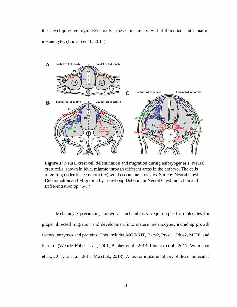
The migration of melanocyte precursors after NCC delaminate from the neural tube
is characterized by their entering what is usually referred to as the dorsolateral pathway
(Larue et al., 2003) (see Figure 1). These cells enter the space between the ectoderm and
dorsal surface of the somites, while dividing and spreading rapidly throughout the skin of
3
the developing embryo. Eventually, these precursors will differentiate into mature
melanocytes (Luciani et al., 2011).
Melanocyte precursors, known as melanoblasts, require specific molecules for
proper directed migration and development into mature melanocytes, including growth
factors, enzymes and proteins. This includes MGF/KIT, Bace2, Prex1, Cdc42, MITF, and
Fasein1 (Wehrle-Haller et al., 2001; Bebber et al., 2013; Lindsay et al., 2011; Woodham
et al., 2017; Li et al., 2011; Ma et al., 2013). A loss or mutation of any of these molecules
Figure 1: Neural crest cell delamination and migration during embryogenesis. Neural
crest cells, shown in blue, migrate through different areas in the embryo. The cells
migrating under the ectoderm (ec) will become melanocytes. Source: Neural Crest
Delamination and Migration by Jean-Loup Duband, in Neural Crest Induction and
Differentiation pp 45-77.
Integrating Regulations of Cell Interactions, Locomotion, Survival and Fate
10.1007/978-0-387-46954-6_4
4
causes the same consequence: slow and inconsistent migration, as well as
hypopigmentation and skin discoloration.
Along with its chemokine role in NCC migration, SDF1 also plays a key
chemoattractant role in melanocyte migration. SDF1 is capable of inducing melanocyte
directed migration when added at moderate and high concentrations in in vitro chemotaxis
chambers (10-200ng/ml). In these experiments, they observed that blocking CXCR7 via
antibodies showed a significant decrease in melanocyte migration, while blocking CXCR4
had no effect on migration. These findings suggest that SDF1/CXCR7 pathway plays a
vital role in melanocyte migration of melanocytes (Lee et al. 2013).
Chemokines and Melanoma
As previously mentioned, chemokines get their name because they are chemotactic
cytokines. “Chemo” comes from chemotactic, referring to movement in response to a
chemical stimulus. While “kines” from cytokines, signaling proteins secreted by cells. A
trademark of chemokine is that they can induce directed chemotaxis from nearby
responsive cells. The SDF1 chemokine that my project focuses on happens to be one of
the most evolutionarily conserved chemokine-signaling systems, using receptors CXCR4
and CXCR7 (DeVries et al., 2006). SDF1 has been regularly studied due to its role in
promoting growth and metastasis in multiple human cancers, and we desire to look further
into its mechanistic pathways. One of these SDF1-responsive cancers includes melanoma,
a deadly skin cancer.
Melanoma is the most dangerous type of skin cancer due to its low 5 year survival
rate. According to the National Cancer Institute, in 2017 there were nearly 10,000 deaths

5
due to melanoma. The majority of risk factors for melanoma are out of our control,
including exposure to ultraviolet light, fair skin, family history, older age, and sex.
Although these risk factors make some people at higher risk, melanoma can affect anyone
regardless of age, sex, or race. Individuals with fair complexions are diagnosed more
frequently than those with darker complexions. Interestingly, individuals with darker
complexions are more likely to be diagnosed at more advanced stages (Wang et al. 2016).
As melanoma progresses, it changes from a localized blemish to a metastatic tumor that
can travel throughout the body. The early stages where the melanoma is localized can be
easily treated via excision. However, as the stages progress and the cancer migrates, the
treatment is much more difficult and intense. Current therapies include radiating the entire
body via chemotherapy. Therefore, identifying the factors involved in the onset of
melanocyte transformation into melanoma is important for future clinical applications to
catch the cells before they become malignant.
Typical CXCR4 versus Atypical CXCR7 Receptors
SDF1 signals through two specific receptors, CXCR4 and CXCR7, which are very
similar in structure and are embedded in the cellular membrane (Bleul et al., 1996; Moser
et al, 2004). These receptor structures each consist of seven transmembrane domains,
resulting in three extracellular loops and three intracellular loops. The extracellular loops
bind to the chemokine SDF1 while the intracellular loop regions bind to signal molecules
known as G-proteins, in order to activate the next pathway (Wang et al., 2014). A 2014
study showed that SDF1 binding to CXCR4 initiated signaling pathways through the
recruitment of both G-proteins and another regulating protein: β-arrestin. However, in the
6
same study, CXCR7 was found to be an atypical chemokine receptor as it did not activate
G-proteins in response to SDF1, but it could signal through β-arrestin pathways (Coggins
et al., 2014). They found that after binding SDF1, the CXCR7 receptor actually had an
eight-fold higher binding affinity to β-arrestin compared to the CXCR4 receptor. This
meant that SDF1 binds to CXCR7 with a higher affinity compared to CXCR4, even up to
approximately fifty-fold greater affinity for the CXCR7 receptor compared to that for the
CXCR4 receptor (Balabanian et al., 2005; Burns et al., 2006).
The fact that CXCR7 receptor has a higher binding affinity for both SDF1 and β-
arrestin strongly suggests that it also has the ability to out-compete the CXCR4 receptor
for access to both the chemokine ligand, as well as the signaling partner. Furthermore, a
study was able to identify that CXCR4 and CXCR7 work separately in some cells, while
they work together in others (Puchert et al., 2014). This information then raises the question
whether these two receptors could form functional heterodimers and if CXCR7 can control
the CXCR4 signaling by sequestering the SDF1 ligand and/or arrestin. Therefore, we see
the development of a very promising target in the signaling axis of SDF1/CXCR4/CXCR7,
as either separate pathways or in combination, in different cancers (including melanoma)
(Hattermann et al., 2013).
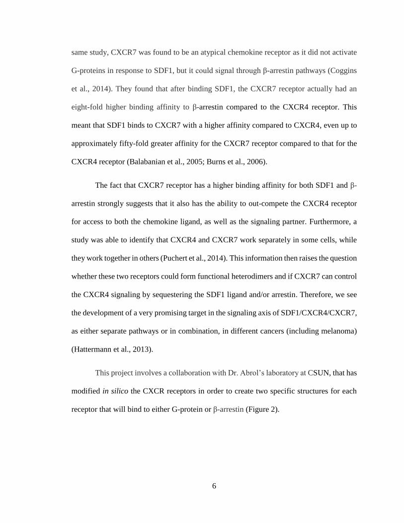
This project involves a collaboration with Dr. Abrol’s laboratory at CSUN, that has
modified in silico the CXCR receptors in order to create two specific structures for each
receptor that will bind to either G-protein or β-arrestin (Figure 2).
7
Implications of Studying the Role of CXCR4/7 in the Great Picture of Cancer Metastasis
Previous research has looked at the possible influences that SDF1/CXCR4 and
SDF1/CXCR7 pathways have on melanoma growth. In a 2014 study, Liedtke and his
colleagues studied melanoma growth and metastasis in transgenic fish lines that
overexpressed SDF1 exclusively in pigment cells. They were able to determine that the
loss of functional CXCR7 had the ability to constrain the melanoma growth in vivo.
Furthermore, experiments using CXCR4-CXCR7 linked together, showed a complete
change in the activated signaling pathways and trafficking of these receptors (Décaillot et
al., 2011). These recent studies are just some examples that show the importance of both
SDF1/CXCR4 and SDF1/CXCR7 mediated pathways in melanocyte migration and cancer.
Accordingly, a better understanding of the mechanisms behind the interactions of SDF1
with its CXCR4 and CXCR7 receptors, and its role in the onset of melanoma cancer is
necessary to take another step toward developing any type of clinical treatment.
Figure 2: Molecular Dynamics Simulation of CXCR protein complexes (red) embedded in a lipid
bilayer with bound regulating protein, either G protein or β-arrestin, in intracellular matrix (blue)
8
SECTION 2: GOAL/HYPOTHESIS
The main goal of this project was to identify if manipulation of CXCR4 and
CXCR7 chemokine receptors would contribute to a malignant transformation of
melanocytes or to the amelioration of melanoma phenotype using a melanoma cell line and
primary melanocytes. I hypothesize that: A) down regulation and inhibition of the CXCR7
receptor will show a greater change in the phenotype of the melanoma than the down
regulation and inhibition of the CXCR4 receptor in causing the cancerous melanoma
develop a less aggressive phenotype. B) Down regulation and inhibition of the CXCR7
receptor will show a greater change in the phenotype of the melanocytes than the down
regulation and inhibition of the CXCR4 receptor in causing the noncancerous melanocyte
develop a more malignant phenotype, one resembling melanoma cells.
9
SECTION 3: MATERIALS AND METHODS
Two cells lines were used for this project, a mouse melanoma B16-F0 cell line was
purchased from ATCC and a Human Epidermal Melanocyte (HEM) cell line was
purchased from Cell Applications. Media used to feed the mouse melanoma cells consisted
of DMEM, 5% FBS, 1x Pen/Strep, and 1x L-Glutamine, while the media for the human
melanocytes cell line was an all-in-one use growth media purchased from the same
company as the melanocytes. The assays performed recorded the motility of both cell lines
via live cell imaging of (i) simple movement among these cells where their motility could
be tracked, and (ii) wound assays to quantify how these cell lines reacted to an outside
influence. Then, these experiments were repeated after manipulation of the CXCR4 and
CXCR7 proteins, specifically through loss of function assays where I transfected the cells
with siRNA to silence either one or both receptors. Each experimental condition was
assessed by qPCR to examine at the gene expression of both CXCR4 and CXCR7.
Movement Assay: Cells were plated in small dishes at 80,000 cells/mL the day before the
assay. Immediately prior to imaging, 80μL of HEPES Buffer was added to the growth
media in order to maintain the desired pH conditions outside of the incubator. When SDF1
addition was required, 200ng SDF1 was added to the media and allowed to incubate for
one hour. When no SDF1 was added, imaging could begin right away. A heat lamp was
placed on top of the plated cells with a thermometer inside, in order to maintain a desired
temperature of 37oC. After finding a representative field of view under the microscope, a
video was initiated and began taking snapshots every 90 seconds for approximately four
hours (approximately 161 frames per video). The cell movement could then be tracked

10
using ImageJ software, where the manual tracking tool was used to track approximately 20
cells per video. The ImageJ software provided the Accumulated Distance values for each
cell pathway that was tracked, as well as the cell’s Displacement and Y-Axis Displacement.
However, the Velocity, Persistence, and Chemotaxic Index were all calculated in Excel.
The formulas are as follows:
• Velocity = Accumulated Distance/Number of Frames
• Persistence = Displacement/Accumulated Distance
• Chemotaxic Index = Y-Axis Displacement/Accumulated Distance
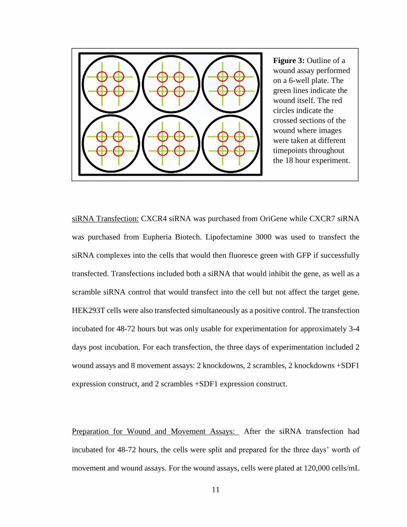
Wound Assay: Cells were plated in a multi-well plate at 120,000 cells/mL one to two days
before the experiment began. Once the cells had reach ~90-100% confluency, 200ng SDF1
was added to the proper wells and allowed to incubate for one hour prior to wounding. At
Hour 0, each well was wounded with a 10uL pipette tip, creating a “tic-tac-toe” design
(Figure 2). Images were then taken of the four crossed sections in each well at hour 0, 5, 8
and 18. At Hour 18, the cells were then collected for RNA purification, cDNA conversion,
and eventually qPCR. The images at each time point were then measured using an area
analysis – where the area left unhealed was measured using a wound healing tool in ImageJ.
11
siRNA Transfection: CXCR4 siRNA was purchased from OriGene while CXCR7 siRNA
was purchased from Eupheria Biotech. Lipofectamine 3000 was used to transfect the
siRNA complexes into the cells that would then fluoresce green with GFP if successfully
transfected. Transfections included both a siRNA that would inhibit the gene, as well as a
scramble siRNA control that would transfect into the cell but not affect the target gene.
HEK293T cells were also transfected simultaneously as a positive control. The transfection
incubated for 48-72 hours but was only usable for experimentation for approximately 3-4
days post incubation. For each transfection, the three days of experimentation included 2
wound assays and 8 movement assays: 2 knockdowns, 2 scrambles, 2 knockdowns +SDF1
expression construct, and 2 scrambles +SDF1 expression construct.
Preparation for Wound and Movement Assays: After the siRNA transfection had
incubated for 48-72 hours, the cells were split and prepared for the three days’ worth of
movement and wound assays. For the wound assays, cells were plated at 120,000 cells/mL
Figure 3: Outline of a
wound assay performed
on a 6-well plate. The
green lines indicate the
wound itself. The red
circles indicate the
crossed sections of the
wound where images
were taken at different
timepoints throughout
the 18 hour experiment.

12
and assay was performed two days later in order to obtain a single monolayer of cells to
wound. For the movement assays, for day one of experiments, the melanocytes and
melanoma cells were plated at 80,000 cells/mL in order to obtain a confluency of
approximately 40-50%. For day two and day three movements, the two cell lines were
plated at different concentrations since the melanoma cell line grew much faster than the
primary cell line. Therefore, day two movements of melanocytes were plated at 50,000
cells/mL and day three movements plated at 40,000 cells/mL. On the other hand, the
melanoma proliferated so quickly that day two movements were plated at 30,000 cells/mL
and day three movements plated 20,000 cells/mL.
RNA Purification: After each 18-hour wound assay, the cells from each well were collected
and labeled according to each condition. GeneJET RNA Purification Kits were purchased
from ThermoFisher Scientific. The protocol used was from the kit’s user guide:
Mammalian Cultured Cells Total RNA Purification Protocol for adherent cells. If
purification could not be performed immediately, the cells were frozen after step 2 in the
lysis buffer supplemented with β-mercaptoethanol until the rest of the protocol could be
completed. Upon completion of RNA purification, the concentration of each RNA sample
was calculated using a NanoDrop 2000, also a product of ThermoFisher Scientific. The
samples were then stored at -20oC until cDNA conversion could be performed.
cDNA Conversion: Purchased from ThermoFisher Scientific, SuperScript VILO cDNA
Synthesis Kit was used for cDNA conversion of the RNA samples. The protocol was
13
followed as described in the product info sheet, using a total reaction volume of 20μL.
Using the concentration given by the NanoDrop 2000, the total amount for each RNA
sample (in μL) was calculated by allowing the total concentrations in the reaction volume
to be the same (usually 500ng/μL). Since the NanoDrop 2000 cannot calculate cDNA
concentrations, this was the best way to ensure the total concentration of each individual
tube would be similar, if not the same, in order to proceed to qPCR analysis.
qPCR Analysis: All primers were purchased from Integrated DNA Technologies, Maxima
SYBR Green/ROX qPCR Master Mix and clear 96-well plates purchased from
ThermoFisher Scientific, and Bio-Rad CFX96 Real-Time PCR Thermocycler. I used
primer sequences from published papers that had previously been found to be successful
(Maishi et al., 2012; Sierro et al., 2007; Liang et al., 2001) (See Table 1). GAPDH was the
housekeeping gene used to normalize the gene expression. As for the protocol, I was able
to optimize the reaction volume to 15μL. In each well, the reaction included the following
components: 5μL of SYBR Green, 0.5μL of 10μM forward primer, 0.5μL of 10μM reverse
primer, 5μL of template cDNA, and 4μL of qPCR grade water. A master mix of all
components, minus template cDNA, was made for each set of primers, and 15μL of the
master mix was added to each well. The template cDNA was added separately as a dilution
was required prior to its addition into the well. The template cDNA for each condition was
diluted to 50ng/μL and 5μL was added to each well for a total concentration of 250 ng/μL
per reaction. The plate was then sealed, centrifuged and placed into the thermocycler. The
thermocycler was programmed based off a three-step cycling protocol and melting curve

14
analysis, both of which could be found in the user guide for the Maxima SYBR Green/ROX
qPCR Master Mix (Figure 4).
Primer Name Primer Sequence
Mouse/Human CXCR4 Forward 5’-GCWGTYCATRTCATYTACACWGTCAACCTCTA-3’
Mouse/Human CXCR4 Reverse 5’- GTSGTCTTSARGGCYTTGCGCTTCTGGTGGCC -3’
Mouse CXCR7 Forward 5’- GGTCAGTCTCGTGCAGCATA -3’
Mouse CXCR7 Reverse 5’- GTGCCGGTGAAGTAGGTGAT -3’
Human CXCR7 Forward 5’- AGCATCAAGGAGTGGCTGAT -3’
Human CXCR7 Reverse 5’- TGTGCTTCTCCTGGTCACTG -3’
Table 1: A list of primers and their sequences used for qPCR analysis of the SDF1 receptors:
CXCR4 and CXCR7. Different primers were needed for both the mouse melanoma and the
human melanocyte cell lines.
Figure 4: The thermocycler program of qPCR including the three-step cycling protocol as
well as melting curve analysis
15
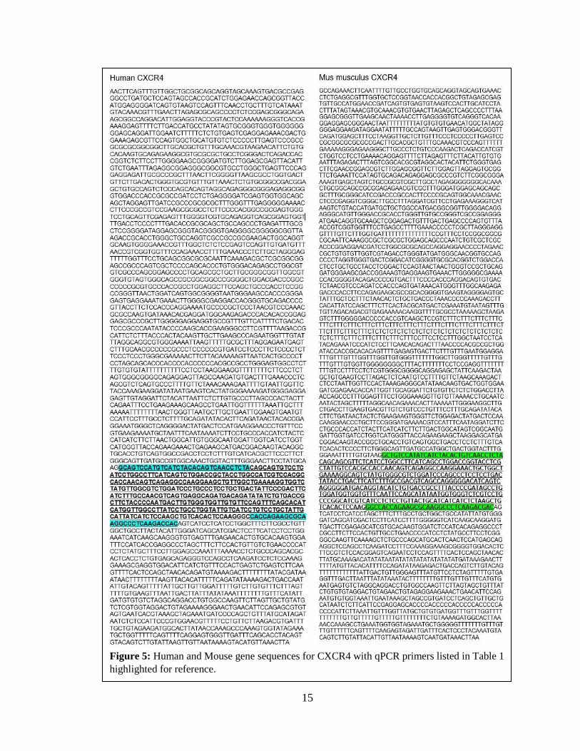
Figure 5: Human and Mouse gene sequences for CXCR4 with qPCR primers listed in Table 1
highlighted for reference.

16
Figure 6: Human and Mouse gene sequences for CXCR7 with qPCR primers listed in Table 1
highlighted for reference.
17
Statistical Analysis: For the analysis of the movement and wound assays, each experiment
was analyzed separately, and the replicates allowed for a final average to be taken. With
the average values calculated, t-tests were performed to determine if there was any
significance between two conditions – a control and an experimental. Therefore, t-tests
were able to show any significance when comparing the KD condition to either the
untreated condition (negative control) or the scramble siRNA condition (positive control).
For each of these t-tests, the two sets of data were analyzed as a two-tailed distribution,
since the results could be greater or lesser than the control, and with equal variance, since
I only compared each cell line to itself and not to each other. For the qPCR analysis, the
relative gene expressions were determined using the ΔΔCq Calculation Method as
described by horizon inspired cell solutions (Haimes & Kelley).
18
SECTION 4: RESULTS
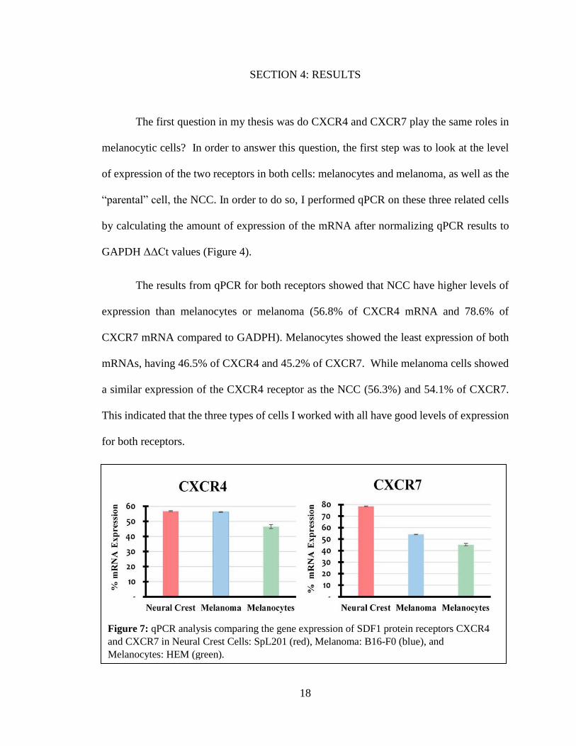
The first question in my thesis was do CXCR4 and CXCR7 play the same roles in
melanocytic cells? In order to answer this question, the first step was to look at the level
of expression of the two receptors in both cells: melanocytes and melanoma, as well as the
“parental” cell, the NCC. In order to do so, I performed qPCR on these three related cells
by calculating the amount of expression of the mRNA after normalizing qPCR results to
GAPDH ΔΔCt values (Figure 4).
The results from qPCR for both receptors showed that NCC have higher levels of
expression than melanocytes or melanoma (56.8% of CXCR4 mRNA and 78.6% of
CXCR7 mRNA compared to GADPH). Melanocytes showed the least expression of both
mRNAs, having 46.5% of CXCR4 and 45.2% of CXCR7. While melanoma cells showed
a similar expression of the CXCR4 receptor as the NCC (56.3%) and 54.1% of CXCR7.
This indicated that the three types of cells I worked with all have good levels of expression
for both receptors.
Figure 7: qPCR analysis comparing the gene expression of SDF1 protein receptors CXCR4
and CXCR7 in Neural Crest Cells: SpL201 (red), Melanoma: B16-F0 (blue), and
Melanocytes: HEM (green).
19
Effects of CXCR siRNA Knockdowns on Melanocyte and Melanoma Velocity
The next steps in looking at the role of these two receptors in melanocytic cell
behavior was to determine if CXCR receptors affected melanocyte and melanoma motility.
If CXCRs have a role in cell migratory behavior reducing or deleting them will change
their migratory capabilities, which can be quantified by measuring cell velocity. These
experiments consisted of transfecting cells with siRNA and live imaging them to gather
cells motility parameters.
Here I live cell imaged these cells after transfecting them with siRNA for CXCR4
and/or CXCR7. From now on I will refer to cells transfected with siRNA as knockdown
(KD) for each receptor. Cell movement was analyzed by manually tracking at least 15
cells at a time from each experiment using ImageJ plug-in from Ibidi website (Asano and
Horn, 2013). This plug-in generated standard cell migration parameters: velocity,
distance, persistence and chemotaxis index.
In analyzing each of these parameters, I found that within each CXCR KD
condition there was a significant change when comparing the untreated cells and the
scramble siRNA condition, as well as the untreated +SDF1 and the scramble siRNA
+SDF1 condition (Figures 6-8). This observation of a difference between control scramble
siRNA and untreated cells is likely a result of the cells being exposed to the transfection
conditions for 24-48 hours. Therefore, I focused on the CXCR siRNA KD conditions
compared strictly to the scramble siRNA condition. However, all significant change is
noted in the figures for reference. Additionally, KD of CXCR4 resulted in some off target
KD of CXCR7 in both melanocytes and melanoma, and both values will be denoted in each
figure. In the same way, KD of CXCR7 resulted in extreme off target KD of CXCR4 in
20
both melanocytes and melanoma, and thus the KD of CXCR7 in actuality is a KD of both
CXCR4 and CXCR7.
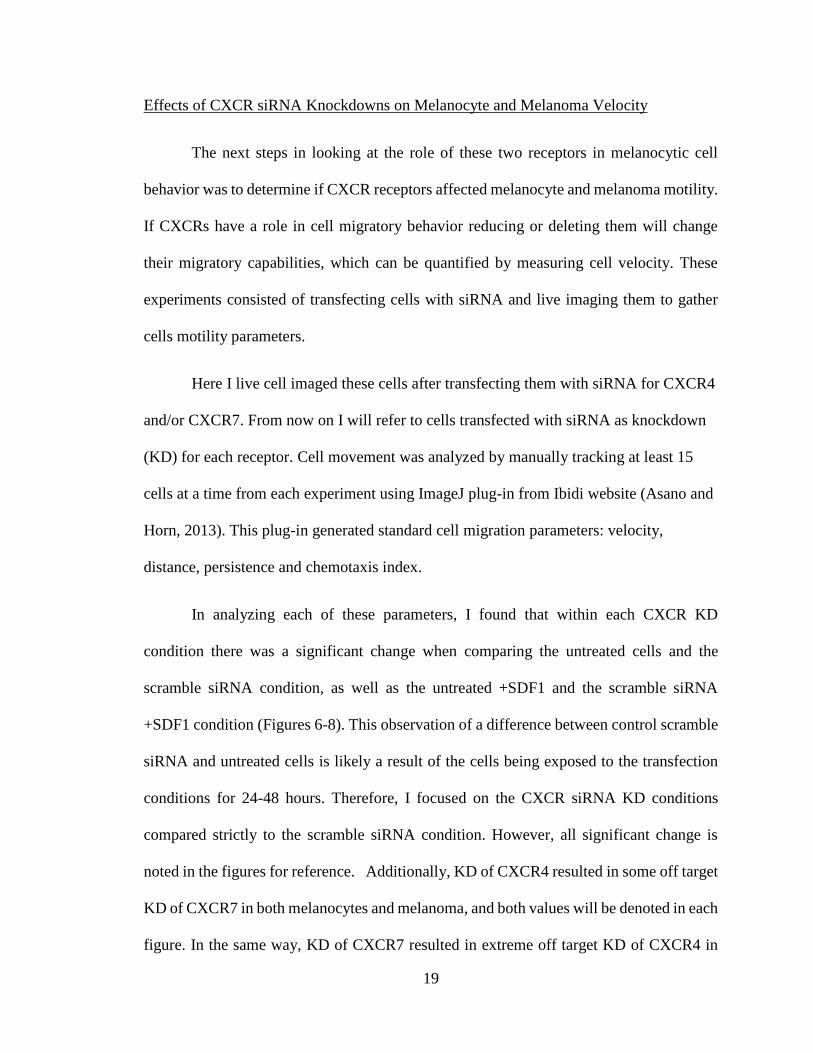
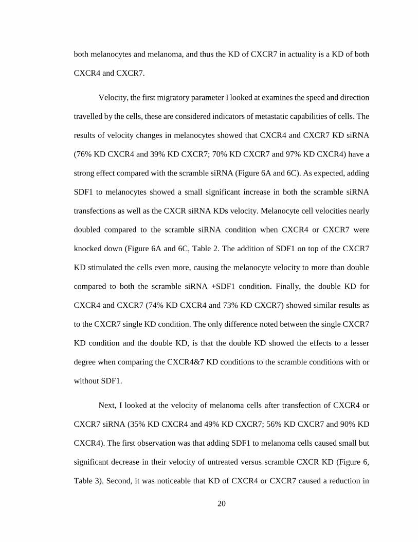
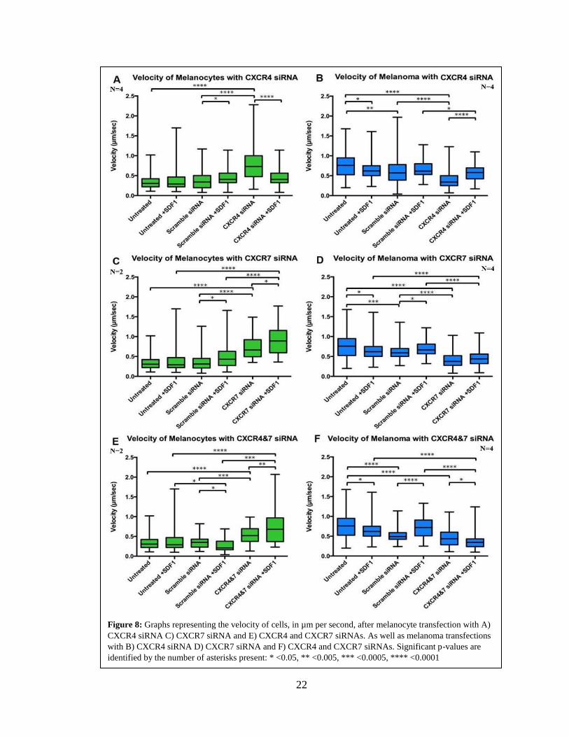
Velocity, the first migratory parameter I looked at examines the speed and direction
travelled by the cells, these are considered indicators of metastatic capabilities of cells. The
results of velocity changes in melanocytes showed that CXCR4 and CXCR7 KD siRNA
(76% KD CXCR4 and 39% KD CXCR7; 70% KD CXCR7 and 97% KD CXCR4) have a
strong effect compared with the scramble siRNA (Figure 6A and 6C). As expected, adding
SDF1 to melanocytes showed a small significant increase in both the scramble siRNA
transfections as well as the CXCR siRNA KDs velocity. Melanocyte cell velocities nearly
doubled compared to the scramble siRNA condition when CXCR4 or CXCR7 were
knocked down (Figure 6A and 6C, Table 2. The addition of SDF1 on top of the CXCR7
KD stimulated the cells even more, causing the melanocyte velocity to more than double
compared to both the scramble siRNA +SDF1 condition. Finally, the double KD for
CXCR4 and CXCR7 (74% KD CXCR4 and 73% KD CXCR7) showed similar results as
to the CXCR7 single KD condition. The only difference noted between the single CXCR7
KD condition and the double KD, is that the double KD showed the effects to a lesser
degree when comparing the CXCR4&7 KD conditions to the scramble conditions with or
without SDF1.
Next, I looked at the velocity of melanoma cells after transfection of CXCR4 or
CXCR7 siRNA (35% KD CXCR4 and 49% KD CXCR7; 56% KD CXCR7 and 90% KD
CXCR4). The first observation was that adding SDF1 to melanoma cells caused small but
significant decrease in their velocity of untreated versus scramble CXCR KD (Figure 6,
Table 3). Second, it was noticeable that KD of CXCR4 or CXCR7 caused a reduction in

21
melanoma overall velocity. In contrast with melanocytes, the double CXCR4 and CXCR7
KD in melanoma (69% KD CXCR4 and 75% KD CXCR7) caused a significant reduction
of their velocity.
22
Figure 8: Graphs representing the velocity of cells, in µm per second, after melanocyte transfection with A)
CXCR4 siRNA C) CXCR7 siRNA and E) CXCR4 and CXCR7 siRNAs. As well as melanoma transfections
with B) CXCR4 siRNA D) CXCR7 siRNA and F) CXCR4 and CXCR7 siRNAs. Significant p-values are
identified by the number of asterisks present: * <0.05, ** <0.005, *** <0.0005, **** <0.0001

23
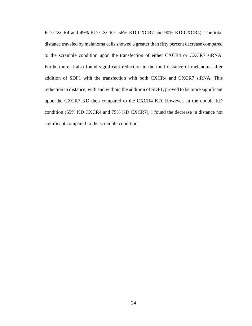
Effects of CXCR siRNA Knockdowns on Melanocyte and Melanoma Accumulated
Distance
The second migratory parameter assessed was the accumulated distance traveled
by each cell line after KD of CXCR4/7. The findings from these measurements showed
that CXCR4/7 KDs in melanocytes (76% KD CXCR4 and 39% KD CXCR7; 70% KD
CXCR7 and 97% KD CXCR4) caused, as expected from velocity results, a strong increase
in migrated distance (Figure 7A, Tables 2). The simple addition of SDF1 to the
melanocytes cells did show small but significant increases in their accumulated distance
traveled of each condition. However, what was very interesting was that transfecting
melanocytes with CXCR4 or CXCR7 siRNA caused a doubling of their total distance
traveled with CXCR4 KD, and a little less than double with the CXCR7 KD. Both CXCR
KDs were significant compared to the scramble siRNA condition as well. Furthermore, an
even larger increase was seen in the accumulated distance of the melanocytes upon the
addition of SDF1. The CXCR7 siRNA +SDF1 condition showed the distance traveled by
cells to nearly triple compared to scramble siRNA +SDF1 condition. The results for
melanocytes from double KD for CXCR4 and CXCR7 (74% KD CXCR4 and 73% KD
CXCR7) showed very similar results to the CXCR7 single KD condition. The distance
travelled by the cells nearly doubled upon CXCR4/7 KD and more than doubled when
SDF1 was added.
The results for melanoma, were also found to be similar to those of velocity change
after CXCR KD. The simple addition of SDF1 to the melanoma cells caused a small but
significant increase in their accumulated distance traveled (Figure 7, Table 3). The distance
traveled by melanomas greatly decreased upon the transfection of CXCR4/7 siRNAs (35%
24
KD CXCR4 and 49% KD CXCR7; 56% KD CXCR7 and 90% KD CXCR4). The total
distance traveled by melanoma cells showed a greater than fifty percent decrease compared
to the scramble condition upon the transfection of either CXCR4 or CXCR7 siRNA.
Furthermore, I also found significant reduction in the total distance of melanoma after
addition of SDF1 with the transfection with both CXCR4 and CXCR7 siRNA. This
reduction in distance, with and without the addition of SDF1, proved to be more significant
upon the CXCR7 KD then compared to the CXCR4 KD. However, in the double KD
condition (69% KD CXCR4 and 75% KD CXCR7), I found the decrease in distance not
significant compared to the scramble condition.
25
Figure 9: Graphs representing the accumulated distance traveled by cells in µm after melanocyte
transfection with A) CXCR4 siRNA C) CXCR7 siRNA and E) CXCR4&7 siRNAs. As well as melanoma
transfections with B) CXCR4 siRNA D) CXCR7 siRNA and F) CXCR4&7 siRNAs. Significant p-values
are identified by the number of asterisks present: * <0.05, ** <0.005, *** <0.0005, **** <0.0001
26
Effects of CXCR siRNA Knockdowns on Melanocyte and Melanoma Persistence and
Chemotaxis Index
Persistence, the third parameter of migratory capabilities, measures random versus
directional movement of cells (Petrie), simply the higher the persistence the more the cell
is moving in a single direction. Figure 8, along with Tables 2 and 3, depicts the effects
each siRNA transfection had on the persistence of each cell line. Like the other analyses,
the simple addition of SDF1 showed slight increases but only in certain transfection
conditions, but not consistently. What I also found was that there was no consistent
significant change in the KD conditions. In all three sets of CXCR KDs, no significant
change was found in the persistence of either cell line in comparison to the scramble siRNA
condition.
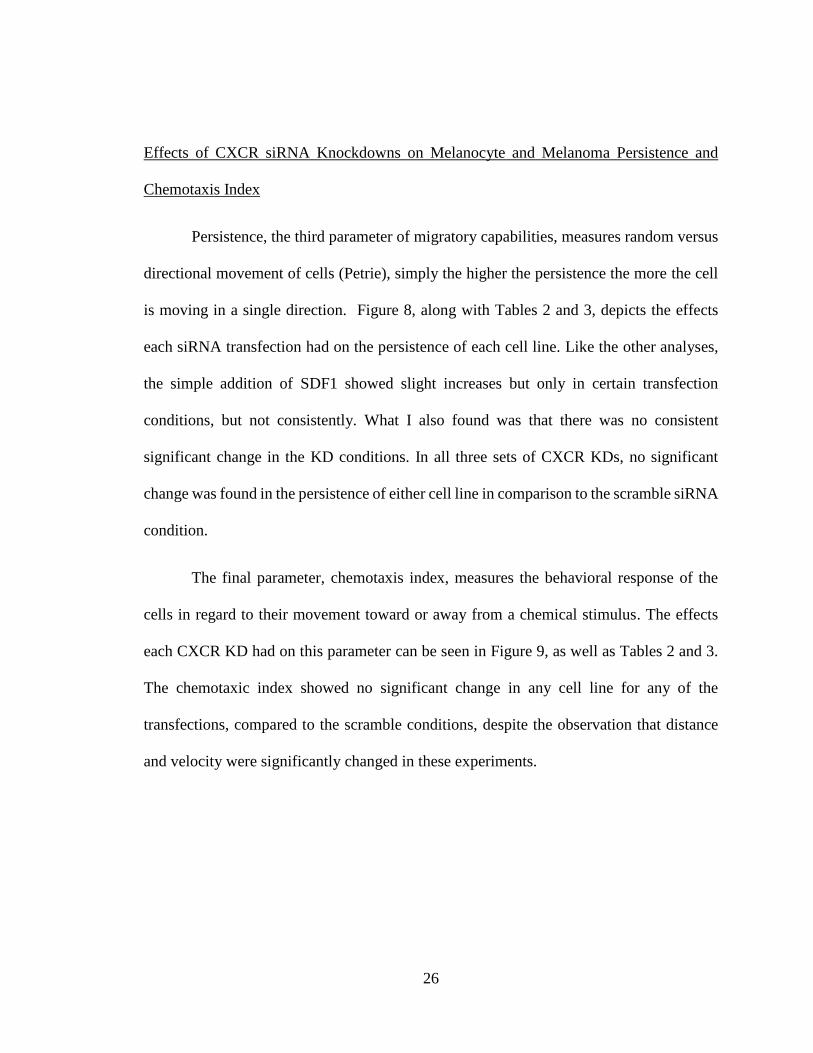
The final parameter, chemotaxis index, measures the behavioral response of the
cells in regard to their movement toward or away from a chemical stimulus. The effects
each CXCR KD had on this parameter can be seen in Figure 9, as well as Tables 2 and 3.
The chemotaxic index showed no significant change in any cell line for any of the
transfections, compared to the scramble conditions, despite the observation that distance
and velocity were significantly changed in these experiments.

27
Figure 10: Graphs representing the persistence of cells after melanocyte transfection with A) CXCR4
siRNA C) CXCR7 siRNA and E) CXCR4 and CXCR7 siRNAs. As well as melanoma transfections with B)
CXCR4 siRNA D) CXCR7 siRNA and F) CXCR4 and CXCR7 siRNAs. Significant p-values are identified
by the number of asterisks present: * <0.05, ** <0.005, *** <0.0005, **** <0.0001
28
Figure 11: Graphs representing the chemotaxis index of cells after melanocyte transfection with A) CXCR4
siRNA C) CXCR7 siRNA and E) CXCR4 and CXCR7 siRNAs. As well as melanoma transfections with B)
CXCR4 siRNA D) CXCR7 siRNA and F) CXCR4 and CXCR7 siRNAs. Significant p-values are identified
by the number of asterisks present: * <0.05, ** <0.005, *** <0.0005, **** <0.0001

29
Effects of CXCR siRNA Knockdowns on Melanocyte Wound Assays
The second set of experiments performed were 18-hour wound assays in order to
identify how the KDs can influence motility under a very different paradigm: collective
migration. Wound assays for each set of transfections were compared to the wound assay
of the untreated cell lines (Figure 10).
The first observation with melanocytes was a significant change in wound healing
capabilities in two of the three conditions: CXCR7 (-16%, p<0.02) and the double KD (-
10%, p<0.03) transfections (Figure 11, Table 2). However, the CXCR4 siRNA condition
did not show significance in the healing capabilities of melanocytes compared with the
changes observed after CXCR7 KD (76% KD CXCR4 and 39% KD CXCR7; 70% KD
CXCR7 and 97% KD CXCR4). Importantly in all these experiments, I did not observe a
significant change in the healing capabilities of any of the CXCR KDs when comparing
the scramble siRNA conditions to the untreated conditions. Similarly, there was also no
significance in any condition solely upon the addition of +SDF1.

30
Figure 12: Area analysis of images taken at each time point during an 18-hour wound assay
for each untreated cell line A) Melanocytes and B) Melanoma

31
Figure 13: Bar graphs showing progressive healing of melanocyte wounds at each time point
over an 18-hour time frame for each set of transfections A) CXCR4 siRNA B) CXCR7 siRNA
and C) CXCR4 and CXCR7 siRNAs. Significant p-values are identified by the number of
asterisks present: * <0.05, ** <0.005, *** <0.0005, **** <0.0001
32
Effects of CXCR siRNA Knockdowns on Melanoma Wound Assays
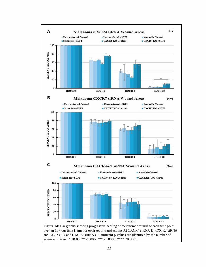
The wound assays with melanoma gave very different results from melanocytes
after CXCR4/7 KD (Figure 12, Table 3). The only experiment that showed a statistically
significant difference was in CXCR4 KD between untransfected +SDF1 and CXCR4 KD
+SDF1. There was a ten percent increase in the area left unhealed, that showed the CXCR4
KD +SDF1 condition was unable to heal the wound as efficently as the untreated +SDF1
condition. These results showed that when CXCR4 was knocked down in melanoma (35%
KD CXCR4 and 49% KD CXCR7), these cells moved less, and thus healed less efficiently.
This was the opposite of what was observed with melanocytes: they became more motile.
33
Figure 14: Bar graphs showing progressive healing of melanoma wounds at each time point
over an 18-hour time frame for each set of transfections A) CXCR4 siRNA B) CXCR7 siRNA
and C) CXCR4 and CXCR7 siRNAs. Significant p-values are identified by the number of
asterisks present: * <0.05, ** <0.005, *** <0.0005, **** <0.0001
34
qPCR and GFP Expression of Each Transfection in Melanocyte and Melanoma
Transfecting CXCR4/7 siRNA into melanocytes and melanomas does not mean
necessarily that the receptors protein expression will necessarily be knocked down or to
which extent. In order to quantify the level of KD for CXCR4 and CXCR7 after siRNA
transfections I performed qPCR analysis as well as took photos of the GFP expression after
each set of transfections, in order to confirm the successful transfection of the siRNAs and
gene KD. Table 2 summarizes the qPCR results for the melanocyte cell line and Table 3
for the melanoma cell lines, each containing all three sets siRNA transfections with and
without the addition of SDF1.
Results show a partial successful knockdown of each CXCR receptor by siRNA
transfection. The overall results showed that siRNA transfection was successful at
knocking down the corresponding receptor. However, I also noticed that CXCR7 siRNA
KD CXCR4 receptor as well (97% KD) while CXCR4 siRNA only KD ~40% of CXCR7.
The addition of SDF1 to the cells also affected the receptor KD. In melanoma instances
even doubling down the percentage KD.
When I looked at the green fluorescence levels of transfected melanocyte and
melanoma cells for knock down effects results were more consistent (Fig.13). Although
fluorescence cannot be quantified, these images did allow me to conclude that cell
transfection was successful since there was an increase identifiable difference in the GFP
expression in the cells after transfection. More recent data using specific CXCR4 or
CXCR7 antibodies shows that KD via our siRNA is truly working (data not shown from
Tyler Tran).

35

36
Figure 15: Images taken of melanocyte and melanoma cell lines
showing the cell nuclei with DAPI stain (blue) as well as the GFP
expression (green) before and after each siRNA transfection.
37
SECTION 5: DISCUSSION
The overall purpose of this thesis was to find out if CXCR7 is more relevant than
CXCR4 in melanoma metastatic capabilities. The overall data presented here may not
completely support a hypothesis where CXCR7 has a more relevant role on migratory
capabilities than CXCR4, however I can conclude that CXCR4/7 work together to regulate
the migratory capabilities of these two cell lines. Knocking-down CXCR7 caused an
unintentional knockdown of CXCR4, and vice-versa, perhaps due to the strong homology
between these two receptors. This CXCR7 KD with the accidental CXCR4 KD
significantly increased melanocyte migratory capabilities as well as significantly decreased
melanoma migratory capabilities. On the other hand, CXCR4 KD alone showed similar
but lesser significant changes in melanocyte and melanoma migratory capabilities.
A fundamental aspect in directed cell migration is the velocity of migration. Any
influences that change the normal velocity of migration does so by disrupting the basic
mechanisms of cytoskeletal cell organization (Gail and Boone, 1970). I analyzed the
velocity of melanocytes and melanoma upon KD of each CXCR receptor, in order to
determine if any effected cell motility. I observed that the most significant increase or
decrease in the velocity of melanocytes and melanoma, respectively, was after KD of the
CXCR7 receptor, which was accompanied by the unintentional KD of CXCR4 (Figure 6,
Table 2 and 3). However, CXCR4 KD alone also affected these cells’ motility to a lesser
but significant degree. This data supports the theory that the CXCR7 receptor and the
CXCR4 receptor work together to play a significant role in the migratory capabilities of
melanocytic cells.
38
Cell migration is the term used to describe the process that individual cells use to
move and migrate. The study of the mechanisms that drive cell migration can tell us about
how an organism develops to how cancer can progress. Among the cell migration assays,
the best known and widely used are the Boyden chamber (Boyden, 1962) and wound assays
(Lampugnani, 1999). Boyden chamber assays assess motile cells and their chemotactic
abilities upon the influence of soluble substances, whereas wound assays allow researchers
to study the chemokinetic transition of cells from a resting state to the migratory state.
However, there is a key distinction between stating that a chemokine is a chemokinetic or
chemotaxic molecule. Experiments looking at a cell’s chemokinetic ability require
observing changes in velocity and migrated distance. On the other hand, experiments that
affect a cell’s chemotaxis response will show changes in parameters such as persistence
and chemotaxis index, because chemoattractants will make a cell migrate toward a
chemokine source and in a specific direction. Although not necessarily true, many
researchers still consider results from Boyden assays reflect perfect chemotaxis or perfect
chemokinetic responses. I did not use the Boyden assays preferring to have a clear response
in the cells to homogeneous SDF1 presence.
Melanoblasts, the precursors of melanocytes, are known to follow the same cell
migratory mechanisms as melanocytes (Mort et al., 2015; Petit and Larue, 2016). There are
several types of molecules guiding cells, among the best known are the chemokines. These
soluble/secreted proteins exert their effect by binding to their cognate receptors, in my case
SDF1 binds to CXCR4 and CXCR7. Among the various effects that chemokines have on
melanoblasts are the stimulation of cell proliferation and migration. During development,
melanoblasts are homogenously distributed within the epidermis as they migrate to the skin,
39
eyes and hair follicles in mammals (Luciani et al., 2011; Mort et al., 2016). Research has
looked into the presence of specific receptors and ligands in order to discover the
possibility that melanoblasts may instruct each other during migration, i.e. use SDF1 which
is secreted by melanocytes themselves (Laurent-Gengoux et al., 2018). Furthermore,
research has identified that as melanocytes turn cancerous and become melanomas, their
migration patterns become aberrant and begin to proliferate extensively leading to
malignant cell invasiveness (Bonaventure et al., 2013). When studying normal versus
aberrant cell migration, most researchers examine the cell’s chemokinetic properties such
as velocity and total distance migrated (Laurent-Gengoux et al., 2018; Crawford et al.,
2017) or chemotactic properties such as persistence and chemotaxis index during migration
towards a chemokine gradient (Lee et al., 2013; Petrie et al., 2009; ).
I found that the melanocytes’ velocity doubled after what was supposed to be a KD
of CXCR7, but which included off target KD of CXCR4 as well. Furthermore, the addition
of SDF1 (CXCR7 siRNA +SDF1 condition) to melanocytes caused their velocity to nearly
triple (Figure 7). The increase after CXCR4 KD alone, though significant, did not reach
twice the velocity of the control. These findings are in opposition to those of Lee et al. who
observed that antibody blocking of CXCR7 abolished melanocyte migration, while
blocking CXCR4 had no effect on migration (Lee et al., 2013). I believe this difference in
results can be explained by looking at the specifics of the experiments both Lee et al. and
I performed. First, Lee used Boyden chambers with SDF1 in bottom wells and Ibidi
chemotaxis slides. Boyden chambers measure chemokinesis while Ibidis measure
chemotaxis. While they measured migrated distance towards SDF1 after 24 hours, they
never measured velocity. In contrast, my experiments had a symmetrical addition of SDF1,

40
and the melanocytes were recorded for several hours and individual cells had their entire
pathway tracked. Lee’s experiments focused on the chemotaxic effects of SDF1 on
melanocyte, whereas I focused on the chemokinetic response. Second, Lee’s experiments
used neutralizing antibodies to block functional receptors, while my experiments used
siRNA. Antibodies would not necessarily block receptor functions, while reducing the
amount of receptor in a cell (as siRNA does) will correlate with receptor function better.
These two factors show that the two set of experiments are not equivalent in their analysis
and therefore can show different results without necessarily contradicting each other.
On the other hand, melanoma cells showed a greater velocity decrease after CXCR7
KD (which included off target CXCR4 KD) than with CXCR4 KD only, compared to
control scramble siRNA (Figure 7, Table 3). These findings are in agreement with
published research. Li and co-workers found that knocking down CXCR7 in M14
melanoma cells significantly inhibited cell migration and invasion in the Boyden assay (Li
et al., 2017). The Boyden chamber experiment performed by Li showed similar results to
my experiments. Specifically, Li’s experiments showed that the addition of SDF1 increased
the number of M14 melanoma cells that passed through the permeable membrane, therefore
significantly increasing the invasiveness of the cells. Conversely, CXCR7 KD inhibited the
SDF1 effects and caused a significant decrease in the invasive activity of the M14
Melanoma cells. Although Li and I were analyzing different aspects of the cells in our
experiments, chemokinesis versus chemotaxic, we observed the same effects in the
melanoma cells upon the KD of CXCR7. However, another important difference between
Li’s experiments and mine, is that Li did not look at the influence of CXCR4 along with
the CXCR7 receptor. Here, we see that the CXCR7 receptor, along with the big possibility
41
of a contributing CXCR4 receptor, is influencing both the chemokinesis and chemotaxic
abilities of the melanoma cells. As research continues to determine the significance of
CXCR7 versus CXCR4 in metastasis and tumorigenesis, it has been found that it can
influence tumor growth in other types of cancers (Wang et al., 2008; Miao et al., 2007).
Furthermore, CXCR7 has more ligands than just SDF1, and knocking down this single
receptor will inhibit these other pathways as well (Richmond et al., 2018). Previous
research on the CXCR family of chemokines has found that different CXCR chemokines
can play roles in increasing melanoma tumor cell growth, depending on the location of the
tumor in the body (Richmond et al., 2018). The small influence of CXCR4 in my
experiments could be due to the increased expression of CXCR4 that is found in early
stages of melanoma metastasis in the lungs, bone marrow and liver (Bartolome et al., 2009).
An interesting finding from this thesis was the different response of melanocytes
and melanomas after CXCR4/7 KD in wound assays. While the first set of experiments
looked at the effects on cell migration of individual cells after CXCR KD, the second set
of experiments looked at cells migrating collectively after CXCR4/7 KD. The environment
in which a single cell is migrating, with little to no outside influence, such as cell to cell
contact, is quite different from the one found when cells are in a monolayer. By looking at
melanocytes and melanoma response after KD of CXCR4/7 we can study how the cell
changes from normal to aberrant behavior after CXCRs expression changes. When cells
are in a much closer proximity to one another, they will communicate and be exposed to
each other’s signals, which will create a difference in their migratory capabilities. These
wound assays are most commonly performed to look at cancerous cell lines to observe their
42
metastatic capabilities and aggressive nature, such as melanocytes and melanoma
(Crawford et al., 2017; Gallagher et al., 2013).
Since the melanocytes were able to “heal the wound” by leaving a smaller area
unoccupied after the 18-hour time point, I will propose that this was cause because the
melanocytes were developing an abnormal behavior when CXCR7 or both CXCR7 and 4
were KD. Because I could not separate CXCR4 KD from concomitant CXCR7 KD based
on my qPCRs, it is difficult to conclude about role of CXCR4 or CXCR7 in melanocytes.
As previously mentioned, knocking down CXCR7 caused an unintentional KD of CXCR4
as well. Maybe the development of the abnormal behavior in melanocytes is happening
because both receptors are being knocked down. Research investigating microarray
signatures of non-cancerous and melanocytic tumors has identified differentially expressed
genes as melanocytes develop into melanoma (Koh et al., 2009). Although CXCR4 and
CXCR7 were not included in this specific study, they may very well play a similar role as
these identified predictive genes. As my experiments show, CXCR7 and CXCR4 could,
should be included as one of the outlying genes whose increase in expression can determine
predictive melanocytic tumors. There was a ten percent increase in the area left unhealed,
that showed the CXCR4 KD +SDF1 condition was unable to heal the wound as efficently
as the untreated +SDF1 condition. These results showed that when CXCR4, and off target
CXCR7, was knocked down in melanoma, these cells moved less, and thus healed less
efficiently. This was the opposite of what was observed with melanocytes: they became
more motile.
On the other hand, the smaller response by melanomas in the wound healing assay
is likely due to their highly aggressive growth and motility, that by 18 hrs., there was almost

43
always complete healing. In other words, it is difficult to show reduced motility in highly
aggressive cancer cells.
My experiments however brought forth an interesting distinction in how these two
cell types respond to CXCRs KD. The motility assay looked at the response of individual
cells while wound assays is reflective of a collective response of cells. The motility and
wound assays showed increased cell migration after KD of CXCR4 and CXCR7 in
melanocytes. In melanoma, the motility and wound assays showed reduced cell migration
after KD of CXCR4 and CXCR7, though the wounds portrayed the reduction in a smaller
ratio. This suggests that silencing these receptors has the capability of modifying their
cytoskeletal rearrangements, causing the increased melanocyte motility and decreased
melanoma motility. These changes were not observed in the collective responses via wound
assays. Although we found a slight change, it was not as dramatic as when single cells were
live tracked.
Most researchers use Boyden chambers to assess motility, which is not as accurate
as my experiments that measured velocity of individual cells as well as collective migration
responses after CXCR KD (wound assay). The different and seemingly opposing cell
responses I observed in melanocytes and melanomas after CXCR KD in the two assays
stress the importance of doing motility assessment under different conditions because cells
behave differently when in cell-cell contact (close proximity) as in a monolayer
environment versus being dispersed, as single, isolated cells (Puliafito et al., 2012). I would
like to propose that these findings could be due to multiple factors. First, the KD conditions
do affect the cells but it could not be measurable in the wounds given that cells were in a
monolayer. Second, we have to remember that these cells also secrete SDF1 in autocrine
44
manner (Laird et al., 2008), so the response could be a result of larger amounts of SDF1
when cells were in monolayer compared with isolated conditions. Third, there’s the
possibility of an overproduction of SDF1 in response to the CXCR receptor KD by siRNA.
With the cell-cell contact provided in the wound assays, compared to the single cell
response, the cells are making up for the lack of available receptor.

45
Future Directions
For future experiments, Chick Chorioallantoic Membrane (CAM) Assays need to
be performed in order to investigate the metastatic effects of the CXCR4 and CXCR7 KDs
in-vivo. Next, the addition of Dr. Abrol’s mutated receptors (see Figure 1) need to be
incorporated into the KD experiments. The addition of the CXCR receptors that have been
mutated to recruit either G-protein or β-arrestin can be used to see which pathway has more
influence over the onset of melanoma cancer. This will allow for future assays involving
mutant receptors where the cells can be transfected with each receptor type (WT or mutant)
to establish structural mechanisms of CXCR4/7 signaling in terms of the importance of
specific receptor domains. Finally, Fluorescence Resonance Energy Transfer (FRET) can
be used in order to see interactions inside the cell using complete mutation of each CXCR
site as well as after manipulation of specific domains of each receptor.
46
CONCLUSION
In summary, I found that first, the CXCR7 and CXCR4 receptors work together in
the initiation of a possible transformation into an abnormal phenotype in melanocytes. My
observation of the drastic change in the cell velocity and accumulated distance in
melanocytes, demonstrates a direct influence of the CXCR7 and CXCR4 receptors in their
cytoskeleton. Additionally, the wound assays further support the influence of the CXCR7
and CXCR4 receptors together, as the loss of both together, although unintentional, still
caused the melanocytes to increase the speed of wound healing. The changes in
melanocytes movement capabilities may ultimately lead to malignancy. Second,
melanomas’ malignant phenotype diminished after the KD of CXCR7 (which also had off
KD target of CXCR4 receptor), as assessed by cell velocity and accumulated distance. This
suggests that CXCR7 receptor, along with CXCR4, appears to take a different role in
curbing the aggressive malignancy of the melanoma.
47
REFERENCES
1. Abdel-Malek, Z.A., Kadekaro, A.L., & Swope, V.B. (2010). Stepping up melanocytes
to the challenge of UV exposure. Pigment Cell & Melanoma Research, 23(2), 171–
186. http://doi.org/10.1111/j.1755-148X.2010.00679.x
2. Asano, Y., & Horn, E. (2013). Chemotaxis and Migration Tool 2.0. Retrieved from
https://ibidi.com/chemotaxis-analysis/171-chemotaxis-and-migration-tool.html
3. Baggiolini, A., Varum, S., Mateos, J.M., Bettosini, D., John, N., Bonalli, M., Ziegler,
U., Dimou, L., Clevers, H., Furrer, R., Sommer, L. (2015) Premigratory and Migratory
Neural Crest Cells Are Multipotent In Vivo. Cell Stem Cell. 16(3), 314-322.
4. Balabanian, K., Lagane, B., Infantino, S., Chow, K.Y., Harriague, J., Moepps, B.,
Arenzana-Seisdedos, F., Thelen M., and Bachelerie, F. (2005). The chemokine SDF-
1/CXCL12 binds to and signals through the orphan receptor RDC1 in T lymphocytes.
J. Biol. Chem. 280(42), 35760-35766.
5. Balmadani, A., Tran, P.B., Ren, D., Assimacopoulos, S., Grove, E.A., Miller, R.J.
(2005). The chemokine stromal cell-derived factor-1 regulates the migration of
sensory neuron progenitors. J. Neurosci. 25, 3995-4003
6. Bartolome, R.A., Ferreiro, S., Miquilena-Colina, M.E., Martinez-Prats, L., Soto-
Montenegro, M. L., Garcia-Bernal, D., Vaquero, J. J., Agami, R., Delgado, R., Desco,
M., Sanchez-Mateos, P., Teixido, J.. (2009) The chemokine receptor CXCR4 and the
metalloproteinase MT1-MMP are mutually required during melanoma metastasis to
lungs. Am. J. Pathol. 174, 602–612.
7. Bebber, F.V., Hruscha, A., Willem, M., Schmid, B., and Haass, C. (2013). Loss of
Bace2 in zebrafish affects melanocyte migration and is distinct from Bace1 knock out
phenotypes. J. Neurochem. 127, 471-481. Doi: 10.1111/jnc.12198
8. Bi, J., Li, P., Li, C., He, J., Wang, Y., Zhang, H., Fan, X., Jia, R., and Ge, S. (2016).
The SDF-1/CXCR4 chemokine axis in uveal melanoma cell proliferation and
migration. Tumor Biology. 3, 4175-4182. https://doi.org/10.1007/s13277-015-
4259-4
9. Bleul, C.C., Farzan, M., Choe, H., Parolin, C., Clark-Lewis, I., Sodroski, J., Springer,
T.A. (1996). The lymphocyte chemoattractant SDF-1 is a ligand for LESTR/fusin and
blocks HIV-1 entry. Nature. 382(6594), 829-33.
10. Bonaventure J., Domingues M.J., Larue L. (2013). Cellular and molecular
mechanisms controlling the migration of melanocytes and melanoma cells. Pigment
Cell Melanoma Res. 26, 316–325. Doi: 10.1111/pcmr.12080.
11. Bosgraaf, L., Waijer, A., Engel, R., Visser, A., Wessels, D., Soll, D., and Van
Haastert, P. (2005) RasGEF-containing proteins GbpC and GbpD have differential
48
effects on cell polarity and chemotaxis in Dictyostelium. J Cell Sci. 118, 1899–910.
Doi: 10.1242/jcs.02317.
12. Boyden, Stephen. (1962). The Chemotactic Effect of Mixtures of Antibody and
Antigen on Polymorphonuclear Leucocytes. J Exp Med. (115)3, 453-466. Doi:
10.1084/jem.115.3.453.
13. Burns, J.M., Summers, B.C., Wang, Y., Melikian, A., Berahovich, R., Miao, Z.,
Penfold, M.E., Sunshine, M.J., Littman, D.R., and Kuo, C.J. (2006). A novel
chemokine receptor for SDF-1 and I-TAC involved in cell survival, cell adhesion, and
tumor development. J. Exp. Med. 203, 2201-2213.
14. Coggins, N.L., Trakimas, D., Chang, S.L., Ehrlich, A., Ray, P., Luker, K.E.,
Linderman, J.J., and Luker, G.D. (2014). CXCR7 Controls Competition for
Recruitment of β-Arrestin 2 in Cells Expressing Both CXCR4 and CXCR7. PLoS
ONE 9(6), e98328. Doi: 10.1371/journal.pone.0098328
15. Crawford, M., Leclerc, V., and Dagnino, L.. (2017) A reporter mouse model for in
vivo tracing and invitro molecular studies of melanocytic lineage cells and their
diseases. The Company of Biologist 6, 1219-1228. Doi: 10.1242/bio.025833
16. D'Orazio, J., Jarrett, S., Amaro-Ortiz, A., & Scott, T. (2013). UV radiation and the
skin. International Journal of Molecular Sciences, 14(6), 12222–12248.
http://doi.org/10.3390/ijms140612222
17. Décaillot, F.M., Kazmi, M.A., Lin, Y., Ray-Saha, S., Sakmar, T.P., and Sachdev, P.
(2011). CXCR7/CXCR4 Heterodimer Constitutively Recruits β-Arrestin to Enhance
Cell Migration. J. Biol. Chem. 286, 32188-97. Doi:10.1074/jbc.M111.277038
18. DeVries, M.E., Kelvin, A.A., Xu, L., Ran, L., Robinson, J. and Kelvin, D.J. (2006)
Defining the origins and evolution of the chemokine/chemokine receptor system. J
Immunol. 176(1), 401-15.
19. Duband, Jean-Loup. (2006) Neural crest delamination and migration: Integrating
regulations of ceil interactions, locomotion, survival and fate. Advances in
Experimental Medicine and Biology. 1-33. Doi: 10.1007/978-0-387-46954-6_4
20. Fears, T.R., Bird, C.C., Guerry, D., Sagebiel, R.W., Gail, M.H., Elder, D.E., Halpern,
A., Holly, E.A., Hartge, P., and Tucker, M. A. (2002). Average Midrange Ultraviolet
Radiation Flux and Time Outdoors Predict Melanoma Risk. Cancer Research, 62(14),
3992.
21. Gail, M.H. and Boone, C.W. (1970) The locomotion of mouse fibroblasts in tissue
culture. Biophys J.10, 980–93. [PubMed: 5531614]

49
22. Gallagher, S. J., Rambow, F., Kumasaka, M., Champeval, D., Bellacosa, A., Delmas,
V., and Larue, L. (2013) Beta-catenin inhibits melanocyte migration but induces
melanoma metastasis. Oncogene, 32(17), 2230-2238. Doi: 10.1038/onc.2012.229
23. Grande-Garcia A, Echarri, A., de Rooij, J., Alderson, N.B., Waterman-Storer, C.M,
Valdivielso, J.M., and del Pozo, M.A. (2007) Caveolin-1 regulates cell polarization
and directional migration through Src kinase and Rho GTPases. J Cell Biol. 177,
683–94. Doi: 10.1083/jcb.200701006
24. Haimes, J., & Kelley, M. (n.d.). Demonstration of a ΔΔCq Calculation Method to
Compute Relative Gene Expression from qPCR Data. 4.
25. Hattermann, K. and Mentlein, R.. (2013). An infernal trio: the chemokine CXCL12 and
its receptors CXCR4 and CXCR7 in tumor biology. Annals of Anatomy =
Anatomischer Anzeiger: Official Organ of the Anatomische Gesellschaft. 195(2), 103-
110.
26. Kasemeier-Kulesa, J.C., Kulesa, P.M. and Lefcort, F. (2005). Imaging neural crest
cell dynamics during formation of dorsal root ganglia and sympathetic ganglia.
Development 132, 235-245. Doi: 10.1242/dev.01553
27. Kasemeier-Kulesa, J.C., Bradley, R., Pasquale, E.B., Lefcort, F. and Kulesa, P.M.
(2006). Eph/ephrins and N-cadherin coordinate to control the pattern of sympathetic
ganglia. Development 133, 4839-4847. Doi: 10.1242/dev.02662
28. Kasemeier-Kulesa, J.C., McLennan, R., Romine, M.H., Kulesa, P.M. and Lefcort, F.
(2010). CXCR4 controls ventral migration of sympathetic precursor cells. J Neurosci.
30, 13078-88.
29. Kasemeier-Kulesa, J.C., Morrison, J.A., Lefcort, F. and Kulesa, P.M. (2015).
TrkB/BDNF signaling patterns the sympathetic nervous system. Nat. Commun. 6,
8281.
30. Kasemeier-Kulesa J.C., Romine M.H., Morrison J.A., Bailey C.M., Welch D.R., and
Kulesa P.M. (2018). NGF reprograms metastatic melanoma to a bipotent glial-
melanocyte neural crest-like precursor. Biology Open. 7(1), bio030817.
Doi:10.1242/bio.030817
31. Killian, E. C., Birkholz, D. A., Artinger, A. B. (2009). A role for chemokine signaling
in neural crest cell migration and craniofacial development. Developmental Biology.
333, 161-172.
32. Koh, S. S., Opel, M. L., Wei, J. J., Yau, K., Shah, R., Gorre, M. E., Whitman, E.,
Shitabata, P. K., Tao, Y., Cochran, A., J., Abrishami, P., and Binder, S. W. (2009)
Molecular classification of melanoma and nevi using gene expression microarray

50
signatures and formalin-fixed and paraffin-embedded tissue. Modern Pathology. 22,
538-546.
33. Laird, D.J., von Andrian, U.H., and Wagers, A.J. (2008). Stem Cell Trafficking in
Tissue Development, Growth, and Disease. Cell, 132(4), 612–630.
https://doi.org/10.1016/j.cell.2008.01.041
34. Lampugnani, M. G. (1999). Cell Migration into a Wounded Area In Vitro. Methods in
Molecular Biology. (96), 177-182. Doi: 10.1385/1-59259-258-9:177
35. Larue, L., Kumasaka, M., and Goding, C.R. (2003). Beta-catenin in the melanocyte
lineage. Pigment Cell Res. 16, 312–317
36. Laurent-Gengoux, P., Petit, V., Aktary, Z., Gallagher, S., Tweedy, L., Machesky, L.,
and Larue, L.. (2018) Simulation of melanoblast displacements reveals new features of
developmental migration. The Company of Biologists, 145, dev 160200. Doi:
10.1242/dev.160200
37. Lee, E., Han, J., Kim, K., Choi, H., Cho, E.-G. and Lee, T. R. (2013). CXCR7
mediates SDF1-induced melanocyte migration. Pigment Cell & Melanoma Research,
26, 58–66. Doi: 10.1111/pcmr.12024
38. Li, A., Ma, Y., Yu, X., Mort, R. L., Lindsay, C. R., Stevenson, D., Strathdee, D.,
Insall, R. H., Chernoff, J., Snapper, S. B. et al. (2011). Rac1 drives melanoblasts
organization during mouse development by orchestrating pseudopod- driven motility
and cell-cycle progression. Dev. Cell 21, 722-734.
39. Li, X., Liu, P., Tian, W., Li, Z., Liu, B., and Sun, J. (2017). Mechanisms of CXCR7
induction in malignant melanoma development. Oncology Letters. 14(4), 4106-4114.
https://doi.org/10.3892/ol.2017.6720
40. Liang, T.S., Hartt, J.K., Lu, S., Martins-Green, M., Gao, J.-L., & Murphy, P.M.
(2001). Cloning, mRNA distribution, and functional expression of an avian
counterpart of the chemokine receptor/HIV coreceptor CXCR4. Journal of Leukocyte
Biology. 69, 297-305
41. Liedtke, D., Erhard, I., Abe, K., Furutani-Seiki, M., Kondoh, H., Schartl, M. (2014).
Xmrk-induced melanoma progression is affected by Sdf1 signals through CXCR7.
Pigment Cell & Melanoma Research, 2, 221-33. Doi: 10.1111/pcmr.12188
42. Lindsay, C. R., Lawn, S., Campbell, A. D., Faller, W. J., Rambow, F., Mort, R. L.,
Timpson, P., Li, A., Cammareri, P., Ridgway, R. A. et al. (2011). P-Rex1 is required
for efficient melanoblast migration and melanoma metastasis. Nat. Commun. 2, 555.
51
43. Luciani, F., Champeval, D., Herbette, A., Denat, L., Aylaj, B., Martinozzi, S.,
Ballotti, R., Kemler, R., Goding, C. R., De Vuyst, F. et al. (2011). Biological and
mathematical modeling of melanocyte development. Development 138, 3943-3954.
44. Ma, Y., Li, A., Faller, W. J., Libertini, S., Fiorito, F., Gillespie, D. A., Sansom, O. J.,
Yamashiro, S. and Machesky, L. M. (2013). Fascin 1 is transiently expressed in
mouse melanoblasts during development and promotes migration and proliferation.
Development 140, 2203-2211.
45. Maishi, N., Ohga, N., Hida, Y., Akiyama, K., Kitayama, K., Osawa, T., Onodera, Y.,
Shinohara, N., Nonomura, K., Shindoh, M., and Hida, K. (2012). CXCR7: A novel
tumor endothelial marker in renal cell carcinoma: CXCR7 in tumor endothelial cells.
Pathology International, 62(5), 309–317. https://doi.org/10.1111/j.1440-
1827.2012.02792.x
46. Miao Z., Luker K. E., Summers B. C., et al. (2007) CXCR7 (RDC1) promotes breast
and lung tumor growth in vivo and is expressed on tumor-associated
vasculature. Proc. Natl Acad. Sci. USA. 104, 15735–15740.
47. Mort, R. L., Jackson, I. J., and Patton, E. E. (2015). The melanocyte lineage in
development and disease. Development 142, 620-632.
48. Moser, B., and Willimann, K. (2004) Chemokines: role in inflammation and immune
surveillance. Ann Rheum Dis. 63(Suppl II), ii84–ii89. Doi: 10.1136/ard.2004.028316
49. Pankov, R., Endo, Y., Even-Ram, S., Araki, M., Clark, K., Cukierman, E.,
Matsumoto, K., and Yamada, K.M. (2005). A Rac switch regulates random versus
directionally persistent cell migration. J Cell Biol. 170(5), 793. Doi:
10.1083/jcb.20050352
50. Pegtel D.M., Ellenbroek, S.I.J., Mertens, A.E.E, van der Kammen, R.A, de Rooij, J.,
and Collard, J.G. (2007) The Par-Tiam1 complex controls persistent migration by
stabilizing microtubule dependent front-rear polarity. Curr Biol. 17, 1623–34. Doi:
10.1016/j.cub.2007.08.035
51. Petrie, R.J., Doyle, A.D., and Yamada, K.M. (2009). Random versus directionally
persistent cell migration. Nature reviews. Molecular cell biology, 10(8), 538–549.
Doi:10.1038/nrm2729
52. Puchert, M. and J. Engele. (2014) The peculiarities of the SDF-1/CXCL12 system: in
some cells, CXCR4 and CXCR7 sing solos, in others, they sing duets. Cell and Tissue
Research. 355(2), 239-253.
53. Puliafito, A., Hufnagel, L., Neveu, P., Streichan, S., Sigal, A., Fygenson, D. K., &
Shraiman, B. I. (2012). Collective and single cell behavior in epithelial contact
52
inhibition. Proceedings of the National Academy of Sciences. 109(3), 739-744.
https://doi.org/10.1073/pnas.1007809109
54. Richmond, A., Yang, J., & Su, Y. (2009). The good and the bad of
chemokines/chemokine receptors in melanoma. Pigment cell & melanoma
research, 22(2), 175–186. doi:10.1111/j.1755-148X.2009.00554.x
55. Saito, D., Takase, Y., Murai, H. and Takahashi, Y. (2012). The dorsal aorta initiates
a molecular cascade that instructs sympatho-adrenal specification. Science 336,
1578-1581.
56. Sierro, F., Biben, C., Martinez-Munoz, L., Mellado, M., Ransohoff, R. M., Li, M.,
Woekhl, B., Leung, H., Groom, J., Batten, M., Harvey, R.P, Martinez, C., Mackkay,
C.R. and Mackay, F. (2007). Disrupted cardiac development but normal
hematopoiesis in mice deficient in the second CXCL12/SDF-1 receptor, CXCR7.
Proceedings of the National Academy of Sciences, 104(37), 14759–14764.
https://doi.org/10.1073/pnas.0702229104
57. Wang, J. and H. Knaut. (2014). Chemokine signaling in development and disease.
Development (Cambridge, England). 141(22), 4199-4205.
58. Wang, Y., Zhao, Y., and Ma, S. (2016). Racial differences in six major subtypes of
melanoma: descriptive epidemiology. BMC Cancer. 16, 691. Doi: 10.1186/s12885-
016-2747-6
59. Wang J., Shiozawa Y., Wang Y., Jung Y., Pienta K. J., Mehra R., Loberg R.,
Taichman R. S.. (2008). The role of CXCR7/RDC1 as a chemokine receptor for
CXCL12/SDF-1 in prostate cancer. J. Biol. Chem. 283, 4283–4294.
60. Wehrle-Haller, B., Meller, M., and Westin, J. A. (2001) Analysis of Melanocyte
Precursors in Nf1 Mutants Reveals That MGF/KIT Signaling Promotes Directed Cell
Migration Independent of Its Function in Cell Survival. Developmental Biology 232,
471–483. Doi:10.1006/dbio.2001.0167
61. Woodham, E. F., Paul, N. R., Tyrrell, B., Spence, H. J., Swaminathan, K., Scribner,
M. R., Giampazolias, E., Hedley, A., Clark, W., Kage, F. et al. (2017). Coordination
by Cdc42 of actin, contractility, and adhesion for melanoblasts movement in mouse
skin. Curr. Biol. 27, 624-637.