Embed Size (px)
Citation preview
micromachines
Article
Bioprinting of Adult Dorsal Root Ganglion (DRG) NeuronsUsing Laser-Induced Side Transfer (LIST)
Katiane Roversi 1,2, Hamid Ebrahimi Orimi 2,3, Marcelo Falchetti 4, Edroaldo Lummertz da Rocha 4 ,Sebastien Talbot 1 and Christos Boutopoulos 2,5,6,*
�����������������
Citation: Roversi, K.; Ebrahimi
Orimi, H.; Falchetti, M.; Lummertz da
Rocha, E.; Talbot, S.; Boutopoulos, C.
Bioprinting of Adult Dorsal Root
Ganglion (DRG) Neurons Using
Laser-Induced Side Transfer (LIST).
Micromachines 2021, 12, 865. https://
doi.org/10.3390/mi12080865
Academic Editor: Daeha Joung
Received: 26 June 2021
Accepted: 20 July 2021
Published: 23 July 2021
Publisher’s Note: MDPI stays neutral
with regard to jurisdictional claims in
published maps and institutional affil-
iations.
Copyright: © 2021 by the authors.
Licensee MDPI, Basel, Switzerland.
This article is an open access article
distributed under the terms and
conditions of the Creative Commons
Attribution (CC BY) license (https://
creativecommons.org/licenses/by/
4.0/).
1 Département de Pharmacologie et Physiologie, Université de Montréal, Montréal, QC H3C 3J7, Canada;[email protected] (K.R.); [email protected] (S.T.)
2 Centre de Recherche Hôpital Maisonneuve-Rosemont, Montréal, QC H1T 2M4, Canada;[email protected]
3 Department of Mechanical, Industrial and Aerospace Engineering, Concordia University,Montréal, QC H3G 1M8, Canada
4 Department of Microbiology, Immunology and Parasitology, Federal University of Santa Catarina,Florianópolis 88040-900, Brazil; [email protected] (M.F.); [email protected] (E.L.d.R.)
5 Département d’Ophtalmologie, Université de Montréal, Montréal, QC H3C 3J7, Canada6 Institut de Génie Biomédical, Université de Montréal, Montréal, QC H3C 3J7, Canada* Correspondence: [email protected]; Tel.: +001-514-252-3400 (ext. 4464)
Abstract: Cell bioprinting technologies aim to fabricate tissuelike constructs by delivering biomate-rials layer-by-layer. Bioprinted constructs can reduce the use of animals in drug development andhold promise for addressing the shortage of organs for transplants. Here, we sought to validate thefeasibility of bioprinting primary adult sensory neurons using a newly developed laser-assisted cellbioprinting technology, known as Laser-Induced Side Transfer (LIST). We used dorsal root ganglionneurons (DRG; cell bodies of somatosensory neurons) to prepare our bioink. DRG-laden- dropletswere printed on fibrin-coated coverslips and their viability, calcium kinetics, neuropeptides release,and neurite outgrowth were measured. The transcriptome of the neurons was sequenced. We foundthat LIST-printed neurons maintain high viability (Printed: 86%, Control: 87% on average) andtheir capacity to release neuropeptides (Printed CGRP: 130 pg/mL, Control CGRP: 146 pg/mL). Inaddition, LIST-printed neurons do not show differences in the expressed genes compared to controlneurons. However, in printed neurons, we found compromised neurite outgrowth and lower sensi-tivity to the ligand of the TRPV1 channel, capsaicin. In conclusion, LIST-printed neurons maintainhigh viability and marginal functionality losses. Overall, this work paves the way for bioprintingfunctional 2D neuron assays.
Keywords: laser-induced side transfer; laser-assisted bioprinting; adult DRG neurons; sensoryneurons; viability; calcium kinetics; transcriptome
1. Introduction
Three-dimensional cell bioprinting technologies enable precise delivery and position-ing of cells and incorporating extracellular components for the fabrication of complex livingconstructs [1]. They find applications in the development of efficient drug screening mod-els, in vitro modelling, as well as in the generation of tissue and organs for transplantation.Recently, 3D printing technologies have been combined with biomaterials and compoundsthat change in a dynamic way (i.e., self-assembly, drug release). Printed constructs canmimic organ responses and/or interact with them. These developments are part of therapidly growing field of multidimensional printing [2,3].
Bioprinting technologies can be categorized into four main categories, namely: mate-rial jetting (e.g., ink-jet printing [4], laser-induced forward transfer (LIFT)); vat photopoly-merization (e.g., stereolithography); pneumatic or mechanical material extrusion [5]; andfree-form spatial printing [6]. Depending on the printing mechanism, these technologies
Micromachines 2021, 12, 865. https://doi.org/10.3390/mi12080865 https://www.mdpi.com/journal/micromachines
Micromachines 2021, 12, 865 2 of 10
present partial compatibility with available bioink formulations, with the bioink viscositybeing the limiting factor [7]. For instance, the bioink viscosity printability range in ink-jetprinting is 3.5–12 mPa·s, while the corresponding range for microextrusion is 30 mPa·s to> 6 × 10⁷ mPa·s. Laser-assisted bioprinting can support a wide range of bioink viscositieswith marginal effects on cell viability and function [8–10], while having the advantages ofhigh printing resolution and reproducibility [8,11]. Laser-induced forward transfer (LIFT)is the most common laser-assisted bioprinting technology. LIFT uses focused laser pulses topropel bioink drops from a donor substrate onto a collector substrate, achieving high print-ing resolution and cell density [12]. By using a nozzleless approach, LIFT is compatible withbioinks having a wide viscosity range (1–300 mPa·s). However, 3D printing capabilitiesremain limited in LIFT due to unresolved donor preparation challenges [13]. A modifiedmethodology for the laser bioprinting of cells, called Laser Induced Side Transfer (LIST),was recently developed by our group [13]. LIST uses low energy nanosecond laser pulsesto generate a transient microbubble at the distal end of a glass microcapillary supplied withbioink. Microbubble expansion results in the ejection of a cell-laden microjet perpendicularto the irradiation axis. We previously showed that LIST-printed human umbilical veinendothelial cells (HUVECs) present negligible loss of viability, and maintain their abilitiesto migrate, proliferate and form intercellular junctions [13]. This method is technicallyuncomplicated and aims to cover a technological gap in the drop-on-demand bioprintingfield: the lack of technologies that can 3D print large-scale constructs using both high andlow viscosity bioinks. Although similar technology [14] has shown compatibility witha wide bioink viscosity range (2–200 mPa·s), this is yet to be tested in cell-laden bioinksdelivered by LIST.
Three-dimensional bioprinting was recently used for neural tissue engineering as aplatform to mimic the mechanical, structural, and cellular properties of central and periph-eral nervous system tissues. The emergence of neuronal bioprinted platforms can facilitatedisease modeling and drug screening applications, as well as the fabrication of implantsfor in vivo regenerative therapies within the central and peripheral nervous systems [15].Embryonic neuronal cell types, including hippocampal, cortical and motor neurons, werepreviously printed using ink-jet printing and tested for post-printing functionality [16,17].LIFT has already shown the potential to print primary dorsal root ganglion (DRG) neu-rons [18]. However, bioprinting of the adult neuronal cells of the central nervous system(CNS) or peripheral nervous system (PNS) has been less explored—presumably, due to thelimited ability of those cells to survive thermomechanical stress and regenerate [19].
Here, we sought to validate whether LIST is compatible with primary DRG neurons.We will present a comprehensive characterization of LIST-printed DRG neurons, includingcomparative results on viability, neurite outgrowth, sensitivity to noxious stimuli, abilityto release neuropeptides, and transcriptome.
2. Materials and Methods
Neurons. The neurons were derived from dorsal root ganglions (DRG) of C57BL/6J(#000664, Jackson Laboratory, Bar Harbor, MA, USA) and VGlut2cre:td-tomatofl/wt of 6to 8 weeks of age. For the ganglion extraction, the mice were euthanized and the DRGsharvested out into DMEM medium (completed with 50 U/mL penicillin and 50 µg/mLstreptomycin (#MT-3001-Cl, Fisher, Waltham, MA, USA), 2 mM L-Glutamine (#25030-081, Life Technologies), and 10% Hi FBS (#10082-145, Life Technologies, Pasir Ris, Sin-gapore). For the cell’s dissociation, the DRGs were incubated within HEPES bufferedsaline (#51558, Sigma, Burlington, MA, USA) completed with 1 mg/mL collagenase A(#1108879300, Sigma) + 2.4 U/mL dispase II (#4942078001, Sigma) for 80 min at 37 ◦C. Theganglions were triturated with glass Pasteur pipettes in DMEM medium + DNAse (#EN052,ThermoScientific, Waltham, MA, USA), then centrifuged over a 15% BSA (#SH30574.02,HyClone/Fisher Scientific, Waltham, MA, USA) gradient in PBS, washed and then resus-pended in Neurobasal-A medium with 0.05 ng/µL NGF (#13257-019, Life Technologies),
Micromachines 2021, 12, 865 3 of 10
0.002 ng/µL GDNF (#450-51-10, Peprotech, Rocky Hill, CA, USA) and 0.01 mM AraC(#C6645, Sigma).
Bioink and printing substrate preparation. The bioink was prepared using 106 DRGneurons per ml suspended in Neurobasal-A medium with NGF, GDNF, and AraC, fib-rinogen (13.24 µM) (F8630-5G; Sigma-Aldrich, Burlington, MA, USA) and Allura red AC(458848-100G, Sigma-Aldrich), (10 mM) as a light absorber. The printing substrates werefibrin-coated 18 mm microscope round cover glasses (48380-046, VWR, Tamil Nadu, India).For the fibrin gel coating (~1 mm-thick), we used 242 µL of a Basal medium (SCME001,Millipore), containing fibrinogen (13.24 µM) (F8630-1G, Sigma) and 8 µL of a thrombinsolution (3.2 U/mL final concentration in the fibrin gel) (T7513- 100UN, Sigma-Aldrich).We used drop-casting to deposit the two solutions onto the microscope cover glasses onehour before printing.
Printing protocol. Freshly prepared bioink (~100 µL) was loaded onto a squaredcapillary (Vitrocom hollow square capillary, inner size 0.3 mm × 0.3 mm, 0.15 mm wallthickness, 50-mm long) using a syringe pump (NE-1000, New Era Pump Systems Inc.,Farmingdale, NY, USA). The laser beam was focused on the middle of the capillary, 500 µmaway from its distal end, using a 4× objective lens (plan achromat, NA = 0.1, Olympus,Tokyo, Japan). The receiving substrate was fixed on an XYZ translation stage and placed500–700 µm away from the capillary tip. Printing laser energies, measured at the sample,were 100 or 120 µJ. Printing patterns consisted of arrays of individual droplets separatedby a 500 µm gap. A detailed description of the printing setup can be found in our previouswork [13]. After printing, the samples were placed in an incubator for 20 min, then rinsedtwice with Neurobasal-A medium, completed with 2 mL of neurobasal-A medium, andput back in the incubator for 48 h.
Viability assay. Neuron viability was determined with fixable Viability Dye (VD)eFluor™ 780 (#65-0865-14, Invitrogen, Waltham, MA, USA) staining. After 48 h in culture,the coverslips containing the cells were incubated with the VD dye (1:1000 dilution) inneurobasal for 1 h in an incubator, then washed 3 times with PBS, fixed with 10% Neutralbuffered formalin for 10 min, washed 3 times with PBS, and mounted with a rectangularcoverslip using Fluoromount-G™ Mounting Medium, with DAPI (#00-4959-52, Invitrogen).Fluorescence images were captured using a Zeiss AxioImager Z2 microscope coupled to anAxioCam MRc color CCD camera. The images were analyzed using a MATLAB algorithmwhich detects the nuclei of all printed cells (blue-DAPI stained), the marker of neurons(Red-td-tomato), and the cells stained or not by the viability dye (purple).
Neurite outgrowth. For the quantification of the neurite length of each neuron, weused the NeuronJ plug-in of NIH-ImageJ software (version 2.1.0). For this, 8-bit grayscaleimages of fluorescent neurons with identifiable neurites were loaded into the softwareand calibrated according to the image magnification. The average length of the neuriteswas obtained by manually tracing the length of all neurite outgrowths from the neuron’scell body, divided by the total number of neurites per neuron. Neurite lengths and totalnumber of neurites were averaged across all neurons in each glass coverslip and plotted.
CGRP release assay. For the CGRP release assay, the neurons were cultured for48 h, then exposed to 1 uM capsaicin (#0462, Tocris, Bristol, UK) or vehicle for 10 min at37 ◦C. The supernatants were collected and the CGRP was measured using the Rat CGRPEnzyme Immunoassay Kit (#589001, Bertin Pharma/Cayman Chemical, Bristol, UK). Plateswere read at 414 nm on a Synergy H1 microplate reader (#19121628, Biotek, Orleans, VT,USA) [20].
Calcium imaging. After 48 h in culture, neurons were loaded with 5 µM Fura-2 AM(#2243-1, Biovision, Milpitas, CA, USA) at 37 ◦C for 45 min in Neurobasal-A medium,then washed with Standard Extracellular Solution (SES, 145 mM NaCl, 5 mM KCl, 2 mMCaCl2, 1 mM MgCl2, 10 mM glucose, 10 mM HEPES, pH 7.5). Their response to thenoxious ligand 100 nM capsaicin (TRPV1 agonist) and 40 mM of KCL (positive control)was analyzed at room temperature. The ligands were dispersed (30 s) onto neurons usingperfusion barrels followed by buffer washout of 210 s. For the imaging acquisition, the
Micromachines 2021, 12, 865 4 of 10
neurons were illuminated with a UV light source (Xenon lamp, 75 watts, Nikon, Melville,NY, USA), 340 nm and 380 nm excitation alternated by an LEP MAC 5000 filter wheel(Spectra services), and fluorescence emission captured by Cool SNAP ES camera (PrincetonInstruments, New Jersey, MA, USA). We processed, background corrected and analyzed340/380 ratiometric images (IPLab v2.8.0 software) and Microsoft Excel was used for posthoc analyses.
RNA sequencing. Neurons were stored in Trizol in −80 ◦C until use. The RNAwas extracted using the PureLink RNA Micro Kit (Invitrogen, #12183-016). RNA waspurified and subjected to TruSeq stranded mRNA library preparation for mouse accordingto the manufacturer’s instructions (Illumina, San Diego, CA, USA). Quality control wasperformed for RNA extraction and cDNA library preparation steps. The libraries weresequenced on an Illumina NovaSeq 6000 sequencing platform, yielding at least 25 millionreads per sample. mRNA library preparation and sequencing were performed at GenomeQuebec facilities. The reads were aligned using STAR (Spliced Transcripts Alignment toa Reference) to mouse reference genome (GRCm38, release 83), sorted the bam files bynames using SAMtools and counted reads using featureCounts.
The differential expression analysis between the groups “Printed cells” and “Controlcells” were performed in R environment and a non-specific filtering of genes with 0 countsin the six samples and of genes with the 25% lowest variance values between samples.Size factors, estimates for Negative Binomial distributed data were estimated and testedthe significance of coefficients in a Negative Binomial Generalized Linear Models (GLM)using the DESeq2 package. Were considered as differentially expressed genes those thatpresented absolute log2 Fold Change > 1 and adjusted (Benjamini and Hochberg method)p-value < 0.05. Gene symbols were obtained using the biomaRt package. The volcanoplot representation was built using the ggplot2 package. The Pearson correlation betweenthe samples regularized transcriptional profile was obtained using the cor package andvisualized it using the pheatmap package with Euclidean distance between the samples and“ward.D2” agglomeration method. These data have been deposited in the National Centerfor Biotechnology Information (NCBI)’s Gene Expression Omnibus (accession numberpending) and are accessible for download at http://www.talbotlab.com (accessed date:22 July 2021).
Statistical analysis. Results are expressed as mean ± standard error of the mean(S.E.M.) in all experiments. The statistical significance was tested by one-way ANOVAwith post hoc Tukey or two-tailed unpaired Student T-test for single comparison. Valueswere considered significantly different when p < 0.05. Statistical computations and graphswere made with GraphPad Prism software (version 9.0.2).
3. Results and Discussion3.1. Effects of the Printing Process on DRG Neurons Survival and Neurite Outgrowth
We first sought to quantify potential effects of the printing process on the cell viability. Forthis, neuron-laden droplets were printed using two laser energies (100 and 120 µJ) (Figure 1).The selection of the laser energies was based on our previous work on LIST [13]. After printing,the cells were cultured for 2 days and the cell viability was assessed (Figure 2A–L). We foundthat LIST-printed neurons maintain high viability when printed at the optimal energy of100 µJ (Printed: 86%, Control: 87% on average), while a decrease in the viability was found forprinting at the higher laser energy of 120 µJ (64%) (Figure 2M). The higher cell death observedat 120 µJ may be due to increased thermomechanical impact on the cells upon exposure tohigher energies. Orimi et al. reported marginal loss of viability in LIST-printed HUVECsusing different laser energies (90–120 µJ) [13]. However, the viability of LIST-printed DRGneurons is considerably lower at high laser energy (120 µJ) because of the inherent sensitivityof primary DRG neurons. We then tested the ability of LIST-printed neurons to grow neurites.We found compromised neurite outgrowth in printed neurons for both optimal energy (44%less than the control group) and high energy (94% less than the control group) (Figure 2N).

Micromachines 2021, 12, 865 5 of 10
Figure 1. Laser-induced side transfer (LIST) of neurons. Schematic representation of the printingsystem (A left) and indicative high-speed imaging of bioink ejection (A right). Printed droplets withDRG neurons 1 h after printing. (B,C). Scale bar = 50 µM (B,C).
Curley et al. have printed primary embryonic DRG neurons using LIFT. Although pastreports have documented minimal effects of LIFT on cell viability, their study documentedconsiderable viability loss in the printed group (84.9%) when compared to the control(95.6%) and to a cancer cell lineage printed under the same conditions (95.6%). The authorsattributed the viability compromise to the high sensibility of this population of neurons.In an inkjet printing study, reduced neurite development and loss in viability after 5 daysin culture was observed for retinal ganglion cells (RGC) [21]. The authors attributedthe impairment in the neurite growth to the greater sensitivity of the adult cells in theculture, as well as the lower density and proximity of the neurons in the printed group.Interestingly, the authors found that coculturing those adult printed RGC neurons withglial cells was able to protect them from impaired viability and neurite outgrowth [21].Therefore, in future studies, the addition of satellite glial cells could be explored as a meansto improve the neurite growth of LIST-printed adult DRG neurons. Note that we foundsuperior neuron viability in our study compared to piezoelectric inkjet printing [21]. Thismight be due to the lower liquid ejection speed in LIST (3.2 to 11.6 m/s [13]) comparedto that used in inkjet printing (10 to 13 m/s [21]). Note that the higher the ejection speed,the higher the mechanical impact on the cells upon deposition. Overall, the cell viabilitycomparison is consistent with the broader LIFT literature showing better cell viabilitycompared to inkjet printing [7].
Taken together, these results indicate that LIST with the optimal laser energy (100 µJ)does not affect the viability of the neurons but limits their ability to extend neurites.

Micromachines 2021, 12, 865 6 of 10
Figure 2. Bioprinting does not impact DRG neurons’ survival, but reduces neurite outgrowth.Representative fluorescence images of adult VGlut2cre::td-tomatofl/wt DRG neurons mice printedwith low (100 µJ, E–H) or high energy (120 µJ, I–L) or put in cell culture (control, A–D). Percentage ofviable cells 2 days post-printing (M; determined as fixable Viability Dye eFluor™ 780-) and averageneurite length (N, in µm). Results consist of the means ± S.E.M. One-way ANOVA with Tukey’smulti comparisons test. Significant differences in M and N are indicated by asterisks (p < 0.05 = *;p < 0.01 = **; p < 0.0001 = ****). Nucleus (Blue; A,E,I), VGlut2cre::td-tomatofl/wt neurons (red; B,F,J),dead cells (purple; C,G,K). Scale bar = 50 µM (A–L).
3.2. Effects of the Printing Process on Calcium Influx and Neuropeptide Release
We tested the ability of LIST-printed neurons to respond to ligands of ion channelreceptors expressed on nociceptors. TRPV1 is such a receptor, specialized in noxiousheat-sensing (~42–45 ◦C) and expressed by ~40% of nociceptors. Once activated, TRPV1triggers the uptake of sodium and calcium (Ca2+) through its ionic pore, leading to neurondepolarization and neuropeptides’ release [22]. The presence and responsiveness of thischannel can be measured by calcium microscopy. For this, DRG neurons were loadedwith the fluorescent intracellular calcium indicator Fura-2 AM, exposed to the TRPV1agonist capsaicin and potassium chloride (KCl), and calcium influxes were measured bymonitoring changes in fluorescence. We found that the LIST-printed DRG neurons have
Micromachines 2021, 12, 865 7 of 10
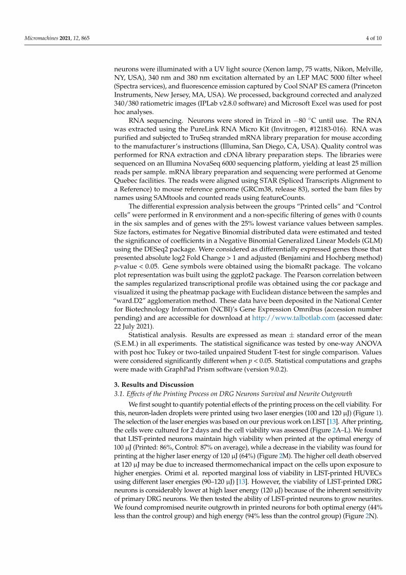
a similar response to capsaicin, while they had a decrease in the response to KCl whencompared to the control neurons (Figure 3G,H). In addition, we found a trend indicatingthat a lower percentage of neurons was activated by capsaicin, in comparison to controlneurons (Figure 3I).
Figure 3. Effects of the printing process on calcium influx triggered by capsaicin. Calcium flux (A–F,showed as heatmap of f340/f380; (G), showed as time–response curve of f340/f380) in cultured (A–C)and printed (D–F) DRG neurons after exposure to vehicle (A,D), capsaicin (100 nM; B,E) and KCl(40 mM; C,F). The maximum amplitude of response evoked by capsaicin and KCl (H). Percentageof capsaicin-responsive neurons (I). Results consist of the means ± S.E.M. Two-tailed unpairedStudent t-test. Significant differences are indicated by asterisks (p < 0.05 = *; ns: not significant asp > 0.05). Amplitude represents the point in which the change in the ratio f340/f380 is maximal.Scale bar = 100 µM (A–F).
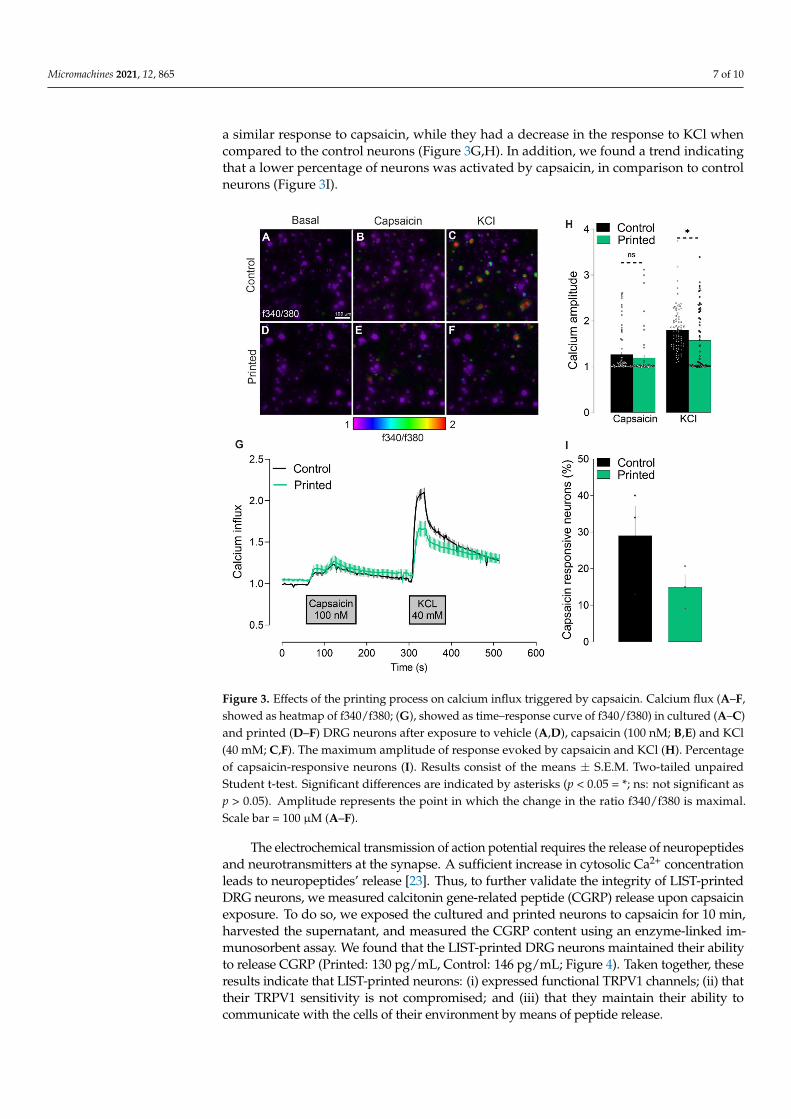
The electrochemical transmission of action potential requires the release of neuropeptidesand neurotransmitters at the synapse. A sufficient increase in cytosolic Ca2+ concentrationleads to neuropeptides’ release [23]. Thus, to further validate the integrity of LIST-printedDRG neurons, we measured calcitonin gene-related peptide (CGRP) release upon capsaicinexposure. To do so, we exposed the cultured and printed neurons to capsaicin for 10 min,harvested the supernatant, and measured the CGRP content using an enzyme-linked im-munosorbent assay. We found that the LIST-printed DRG neurons maintained their abilityto release CGRP (Printed: 130 pg/mL, Control: 146 pg/mL; Figure 4). Taken together, theseresults indicate that LIST-printed neurons: (i) expressed functional TRPV1 channels; (ii) thattheir TRPV1 sensitivity is not compromised; and (iii) that they maintain their ability tocommunicate with the cells of their environment by means of peptide release.
Micromachines 2021, 12, 865 8 of 10
Figure 4. Printed-DRG neurons release neuropeptide. DRG neurons were printed and cultured for48 h and exposed to capsaicin (1 uM). Supernatant was harvested 10 min after capsaicin exposure andCGRP release was measured by ELISA. Results consist of the means ± S.E.M. Individual values arerepresented with “◦”.One-way ANOVA with Tukey’s multi comparisons test. Significant differencesare indicated by asterisks (p < 0.05 = *; ns: not significant as u > 0.05).
To the best of our knowledge, this is the first study measuring the calcium influxand neuropeptide release in bioprinted adult DRG neurons. Using 3D bioprinted iPSC-derived spinal neurons, Joung and collaborators [24] have shown a normal calcium influxin response to KCl and glutamate, which is evidence of active and functionally matureneurons. Other studies have measured neurons activity using whole-cell patch-clamp, atechnique that records the electrophysiological properties of the neurons following theinjection of current. Xu and collaborators have found similar electrophysiological behaviorbetween control and thermally inkjet-printed embryonic primary hippocampal and corticalneurons [17]. On the other hand, Kador and collaborators found that inkjet-printed RGCneurons required higher current to develop the same response as control neurons [25].These findings show that the functional activity of printed neurons varies according to thesubtype of neurons, as well as the printing methodology used.
3.3. Effects of the Printing Process on the Expressed Genes
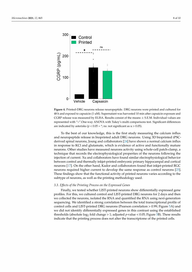
Finally, we tested whether LIST-printed neurons show differentially expressed geneprofiles. For this, we cultured control and LIST-printed DRG neurons for 2 days and thenwe collected the neurons, isolated the RNA and quantified the RNA using next-generationsequencing. We identified a strong correlation between the total transcriptional profile ofcontrol cells and LIST-printed DRG neurons (Pearson correlation > 0.99; Figure 5A) andwe did not identify differentially expressed genes in this contrast using the establishedthresholds (absolute log2 fold change > 1; adjusted p-value < 0.05; Figure 5B). These resultsindicate that the printing process does not alter the transcriptome of the printed cells.
Micromachines 2021, 12, 865 9 of 10
Figure 5. Printed nociceptor neurons transcriptome. Clustering of the samples showing the Pearsoncorrelation value between each pair of samples (A). Volcano plot representation of differentiallyexpressed genes between Control and Printed neurons (B).
4. Conclusions
In conclusion, we show that LIST-printed adult sensory neurons maintain high viabil-ity and functional integrity. Overall, this work paves the way for bioprinting functional 3Dsensory neuron assays and opens possibilities for developing bioprinted grafts for use innerve recovering medicine.
Author Contributions: Conceptualization, K.R., S.T., C.B.; methodology, K.R., H.E.O., S.T., C.B.;formal analysis, K.R., H.E.O.; transcriptome analysis, E.L.d.R. and M.F.; writing—original draftpreparation, K.R.; writing—review and editing, H.E.O.; M.F., S.T., C.B.; supervision, S.T., C.B.;funding acquisition, S.T. and C.B. All authors have read and agreed to the published version ofthe manuscript.
Funding: This research was funded by the Natural Sciences and Engineering Research Council ofCanada (Discovery grant RGPIN-2018-06767; CB), the Canadian Foundation for Innovation (ST,#37439), and the Canada Research Chair program (ST, #950-231859). CB is the recipient of a JuniorI salary award from the Fonds de la Recherche en Santé du Québec (#253123, #265459). KR holdspostdoctoral fellowships from the Fonds de Recherche du Québec Nature et technologies (FRQNT)and the Fonds de recherche en ophtalmologie de l’Université de Montréal. HEO was the recipient ofa PhD scholarship from the FRQNT.
Institutional Review Board Statement: The Institutional Animal Care and Use Committees ofUniversité de Montréal (CDEA #20046, #20047) approved all animal procedures.
Informed Consent Statement: Not applicable.
Data Availability Statement: The data that support the findings of this study are available from thecorresponding author upon reasonable request. Sequencing data are deposited on www.talbotlab.com(accessed date: 22 July 2021).
Acknowledgments: We would like to thank Mikhail Sergeev for his assistance in using the confocalmicroscopy system.
Conflicts of Interest: The authors declare no conflict of interest.
References1. Knowlton, S.; Anand, S.; Shah, T.; Tasoglu, S. Bioprinting for neural tissue engineering. Trends Neurosci. 2018, 41, 31–46. [CrossRef]2. Foresti, R.; Rossi, S.; Pinelli, S.; Alinovi, R.; Sciancalepore, C.; Delmonte, N.; Selleri, S.; Caffarra, C.; Raposio, E.; Macaluso, G.;
et al. In-vivo vascular application via ultra-fast bioprinting for future 5D personalised nanomedicine. Sci. Rep. 2020, 10, 3205.[CrossRef]
Micromachines 2021, 12, 865 10 of 10
3. Gao, B.; Yang, Q.; Zhao, X.; Jin, G.; Ma, Y.; Xu, F. 4D bioprinting for biomedical applications. Trends Biotechnol. 2016, 34, 746–756.[CrossRef]
4. Li, X.; Liu, B.; Pei, B.; Chen, J.; Zhou, D.; Peng, J.; Zhang, X.; Jia, W.; Xu, T. Inkjet bioprinting of biomaterials. Chem. Rev. 2020,120, 10793–10833. [CrossRef] [PubMed]
5. Ozbolat, I.T.; Hospodiuk, M. Current advances and future perspectives in extrusion-based bioprinting. Biomaterials 2016,76, 321–343. [CrossRef] [PubMed]
6. Lee, J.M.; Sing, S.L.; Zhou, M.; Yeong, W.Y. 3D bioprinting processes: A perspective on classification and terminology. Int. J.Bioprint. 2018, 4, 151. [CrossRef]
7. Murphy, S.V.; Atala, A. 3D bioprinting of tissues and organs. Nat. Biotechnol. 2014, 32, 773–785. [CrossRef] [PubMed]8. Hopp, B.; Smausz, T.; Kresz, N.; Barna, N.; Bor, Z.; Kolozsvari, L.; Chrisey, D.B.; Szabo, A.; Nogradi, A. Survival and proliferative
ability of various living cell types after laser-induced forward transfer. Tissue Eng. 2005, 11, 1817–1823. [CrossRef] [PubMed]9. Gruene, M.; Deiwick, A.; Koch, L.; Schlie, S.; Unger, C.; Hofmann, N.; Bernemann, I.; Glasmacher, B.; Chichkov, B. Laser printing
of stem cells for biofabrication of scaffold-free autologous grafts. Tissue Eng. Part C Methods 2011, 17, 79–87. [CrossRef] [PubMed]10. Koch, L.; Kuhn, S.; Sorg, H.; Gruene, M.; Schlie, S.; Gaebel, R.; Polchow, B.; Reimers, K.; Stoelting, S.; Ma, N.; et al. Laser printing
of skin cells and human stem cells. Tissue Eng. Part C Methods 2010, 16, 847–854. [CrossRef]11. Gruene, M.; Pflaum, M.; Hess, C.; Diamantouros, S.; Schlie, S.; Deiwick, A.; Koch, L.; Wilhelmi, M.; Jockenhoevel, S.; Haverich, A.;
et al. Laser printing of three-dimensional multicellular arrays for studies of cell-cell and cell-environment interactions. Tissue Eng.Part C Methods 2011, 17, 973–982. [CrossRef] [PubMed]
12. Guillotin, B.; Souquet, A.; Catros, S.; Duocastella, M.; Pippenger, B.; Bellance, S.; Bareille, R.; Remy, M.; Bordenave, L.; Amedee,J.; et al. Laser assisted bioprinting of engineered tissue with high cell density and microscale organization. Biomaterials 2010,31, 7250–7256. [CrossRef] [PubMed]
13. Ebrahimi Orimi, H.; Hosseini Kolkooh, S.S.; Hooker, E.; Narayanswamy, S.; Larrivee, B.; Boutopoulos, C. Drop-on-demand cellbioprinting via Laser Induced Side Transfer (LIST). Sci. Rep. 2020, 10, 9730. [CrossRef] [PubMed]
14. Delrot, P.; Modestino, M.A.; Gallaire, F.; Psaltis, D.; Moser, C. Inkjet printing of viscous monodisperse microdroplets byLaser-Induced Flow Focusing. Phys. Rev. Appl. 2016, 6, 024003. [CrossRef]
15. Cadena, M.; Ning, L.; King, A.; Hwang, B.; Jin, L.; Serpooshan, V.; Sloan, S.A. 3D bioprinting of neural tissues. Adv. Healthc. Mater.2020, e2001600. [CrossRef] [PubMed]
16. Xu, T.; Jin, J.; Gregory, C.; Hickman, J.J.; Boland, T. Inkjet printing of viable mammalian cells. Biomaterials 2005, 26, 93–99.[CrossRef]
17. Xu, T.; Gregory, C.A.; Molnar, P.; Cui, X.; Jalota, S.; Bhaduri, S.B.; Boland, T. Viability and electrophysiology of neural cellstructures generated by the inkjet printing method. Biomaterials 2006, 27, 3580–3588. [CrossRef]
18. Curley, J.L.; Sklare, S.C.; Bowser, D.A.; Saksena, J.; Moore, M.J.; Chrisey, D.B. Isolated node engineering of neuronal systemsusing laser direct write. Biofabrication 2016, 8, 015013. [CrossRef]
19. Nicholls, J.G.; Adams, W.B.; Eugenin, J.; Geiser, R.; Lepre, M.; Luque, J.M.; Wintzer, M. Why does the central nervous system notregenerate after injury? Surv. Ophthalmol. 1999, 43 (Suppl. 1), S136–S141. [CrossRef]
20. Lee, S.; Jo, S.; Talbot, S.; Zhang, H.B.; Kotoda, M.; Andrews, N.A.; Puopolo, M.; Liu, P.W.; Jacquemont, T.; Pascal, M.; et al. Novelcharged sodium and calcium channel inhibitor active against neurogenic inflammation. Elife 2019, 8, e48118. [CrossRef]
21. Lorber, B.; Hsiao, W.K.; Hutchings, I.M.; Martin, K.R. Adult rat retinal ganglion cells and glia can be printed by piezoelectricinkjet printing. Biofabrication 2014, 6, 015001. [CrossRef] [PubMed]
22. Latremoliere, A.; Woolf, C.J. Central sensitization: A generator of pain hypersensitivity by central neural plasticity. J. Pain 2009,10, 895–926. [CrossRef] [PubMed]
23. Russell, F.A.; King, R.; Smillie, S.J.; Kodji, X.; Brain, S.D. Calcitonin gene-related peptide: Physiology and pathophysiology.Physiol. Rev. 2014, 94, 1099–1142. [CrossRef]
24. Joung, D.; Truong, V.; Neitzke, C.C.; Guo, S.Z.; Walsh, P.J.; Monat, J.R.; Meng, F.; Park, S.H.; Dutton, J.R.; Parr, A.M.; et al. 3Dprinted stem-cell derived neural progenitors generate spinal cord scaffolds. Adv. Funct. Mater. 2018, 28, 1801850. [CrossRef][PubMed]
25. Kador, K.E.; Grogan, S.P.; Dorthe, E.W.; Venugopalan, P.; Malek, M.F.; Goldberg, J.L.; D’Lima D, D. Control of retinal ganglion cellpositioning and neurite growth: Combining 3D printing with radial electrospun scaffolds. Tissue Eng. Part A 2016, 22, 286–294.[CrossRef]