Embed Size (px)
Citation preview

DISEASES OF AQUATIC ORGANISMS Dis. aquat. Org.
Published December 31
Bitter crab disease: a fatal dinoflagellate infection and marketing problem for Alaskan Tanner crabs
Chionoecetes bairdi
Theodore R. ~ e y e r s ' , Timothy M. ~ o e n e m a n ~ , Catherine ~ o t e l h o ~ , Sally short'
' Division of Fisheries Rehabilitation, Enhancement and Development (FRED), Alaska Department of Fish and Game. PO Box 3-2000, Juneau, Alaska 99802-2000, USA
Division of Commercial Fisheries, Alaska Department of Fish and Game
ABSTRACT: A systemic dinoflagellate disease was discovered in Tanner crabs Chionoecetes bairdi harvested from two-thirds of the major production areas in the northern southeast panhandle of Alaska during 1985 and 1986. Processed meats from infected crabs were unmarketable as a result of a bitter flavor imparted by the causative agent. The disease is chronic but lethal, causing 100% mortality in naturally infected crabs when maintained in flowing seawater during a 5 mo period. Feral Tanner crabs in at least one harvest area had a parasite prevalence of 95 '10 regardless of sex or age class. Disease pathogenesis and parasite description, probable life history, distribution and management imphcations are discussed.
INTRODUCTION
The Tanner crab Chionoecetes balrdi, marketed as snow crab, has been an econom~cally important target species for commercial fishermen in southeastern Alaska for several years. The average annual catch of Tanner crab harvested in southeast Alaska for the last 3 yr (1984-1986) has been 1.2 million lbs (ca 545 t) with an ex-vessel value of US $2.3 million. In 1985, during February through April, 4 lots of Tanner crabs (about 66000 lbs 130 t] live wt) purchased by a local southeast- ern Alaska processor contained crab meats having a markedly bitter aftertaste. During this same period in 1986 more than 80282 lbs (36.4 t) of live crabs pur- chased by the same company were unmarketable due to the same bitter flavor. Also in 1986, a second local processor had similar problems with bitter crabs. Approximately 29567 lbs (13.4 t) of live crabs were not marketable and represented 11.4% of the Tanner crabs delivered to this second facility from 24 February through 29 March. This loss is conservative because crab deliveries prior to discovery of the problem on 24 February are not included and not all processors had
O Inter-Research/Printed in F. R. Germany
provided adequate catch information. Before the clos- ing of the 1986 Tanner crab season, commercial fisher- men were able to recognize affected live crabs by a pink carapace and milky hemolymph. Such crabs were being culled from their harvests on the fishing grounds and at the processors' docks. By grading bitter crabs from tender purchases on the fishing grounds, the latter processing company was able to reduce overall sea- sonal losses to ?.? %. A conservative estimate of dollars lost from unmarketable bitter Tanner crabs for both companies was over US $175 758 in 1986.
A laboratory examination of these crabs showed them to be infected with a non-motile single-celled protistan blood parasite resembling Hematodinium sp. reported in other decapod crustacean hosts. Chatton & Poisson (1931) originally described H. perezi in the hemolymph of Carcinus maenas and Portunus depurator from European waters. Years later the para- site was found in the blue crab Callinectes sapidus from the southeastern United States (Newman & John- son 1975), in 2 species of cancer crabs, Cancer irroratus and C. borealis, and in another portunid, Ovalipes oscellatus, from the mid-Atlantic Bight (MacLean &

196 Dis. aquat. Org. 3: 195-216, 1987
Ruddell 1978). Most recently, dinoflagellates resem- bling Hematodinium have been described as parasites of benthic amphipods (Johnson 1986).
We believe this dinoflagellate parasite is responsible for the bitter off-flavor occurring in cooked Tanner crabs and consequently is economically significant to the seafood industry. The purpose of this report is to provide new information regarding: gross and micro- scopic descriptions of bitter crab disease; disease pathogenesis in Tanner crabs; ultrastructural morphol- ogy of the causative agent; transmissibility of the agent in Tanner and king crab species and its in vitro culture characteristics; prevalence and distribution of the dis- ease in Alaskan Tanner crabs based on current catch information.
MATERIAL AND METHODS
Necropsy and histopathology. Necropsies per- formed on pink or milky crabs provided representative tissue samples from the major organ systems. These were preserved in Helley's fixative (ZnCl* substituted for HgC12) for standard histological processing and staining with hematoxylin and eosin (H&E). Hemolymph for smears was drawn in m1 syringes from the third joint of the right cheliped of each crab. Result- ing films on glass slides were fixed and stained using the Diff-Quik method (Dade Diagnostics. Inc. Aguada, Puerto Rico 00602). Entire hemolymph films were examined a t 100x magnification (approximately 264 fields) for diagnosis of infection. Wet hemolymph smears containing live parasites were examined with bright field and phase contrast microscopy. Measure- ments of organisms were made from both fresh and histological preparations at 400x using an ocular mi- crometer.
Disease pathogenesis. Host mortality and parasite development were examined in the following way. Infected and apparently uninfected crabs sorted by a seafood processor were brought into the laboratory, tagged for identification and maintained in separate 100 gal (ca 380 1) rectangular fiberglass tanks supplied with running seawater at approximately 3 gpm (ca 11 1 min-l) with salinities of 28 to 32 ppt. Both groups were re-sorted according to the presence or absence of the parasite in hemolymph smears. The accuracy of exter- nal disease signs used by fishermen and processors to identify infected crabs was evaluated by diagnosis with hemolymph smears. Both groups of crabs were fed herring a d libidum and held for a n extended period of time to allow for disease progression in infected crabs and cumulative mortality. Hemolymph films from sur- viving crabs were made a t the end of the experiment to confirm the infected and control status of both crab
groups. Saltwater temperatures were monitored 3 to 4 times a week beginning 1 mo after the start of the mortality study.
Electron microscopy. About 0.5 to 1.0 m1 of hemolymph or sterile seawater containing parasite vegetative stages from 2 crabs and dinospores from 2 others were preserved in 4 % glutaraldehyde in sea- water at 4 "C for 24 h. The samples were post-fixed in 1 % osmium tetroxide in 0.1 m cacodylate buffer (pH 7.2) and placed in 70 % EtOH for storage. Aliquots of fixed organisms were processed for standard transmis- sion electron microscopy (TEM) using Spurr's resin or Quetol 651 for embedding and uranyl acetate and lead citrate stains. Thin sections were examined in a Philips 200 or 300 TEM operated at 40 and 60 kV, respectively. The remainder of the samples in alcohol were filtered onto a 13 mm nucleopore polycarbonate membrane, critically point dried in trichlorotrifluoroethane, coated with gold palladium and examined in an AMRAY 1000-A scanning electron microscope (SEM) at 20 kV.
Disease transmission studies. Two control crabs from the pathogenesis study received 0.1 and 1.0 m1 inocula- tions, respectively, of hemolymph freshly drawn from one of the infected crabs (No. 9). Two additional unin- fected controls were inoculated with 0.2 and 2.0 ml, respectively, of a suspended subculture of the dino- flagellate organism which had been maintained in the laboratory for 70 d after isolation from another of the infected crabs (No. 5). Approximate hemocytometer counts of organisms in the preparations used for inocula were 5.01 X 10⁵ cells ml-' and 5.56 X 10' cells ml-' for freshly drawn hemolymph and cultured material, respectively. These 4 crabs were maintained in a separate tank of flowing seawater and were bled periodically to determine if a clinical infection had developed. Inoculations were made in the right cheliped of each crab but subsequent bleedings were done from the left cheliped. The remaining 5 control crabs were maintained in another saltwater tank.
Results from the 4 Tanner crabs allowed the design of a second experiment testing the susceptibility of another commercially important crab species to infec- tion with the vegetative stage of the Tanner crab dino- flagellate. Five female red king crabs Paralithodes camtschatica were each inoculated with 1 m1 of pooled hemolymph from 3 naturally infected male Tanner crabs. Six unparasitized male Tanner crabs were also inoculated in the same manner and served as positive controls. The inoculum for both crab species contained dinoflagellate vegetative stages averaging 5.34 X 10⁶ cells ml-l. Another 5 red king and 5 Tanner crabs served as sham inoculated controls each receiving 1 m1 of hemolymph pooled from 6 normal Tanner crabs. Inoculations, periodic bleedings and maintenance of crabs in flowing seawater were as explained above

Meyers et al . : Bitter crab disease 197
with sham inoculated crabs kept in a separate tank. The king crabs were collected locally from Auke Bay while Tanner crabs were purchased from commercial fishermen. Hemolymph from all inoculated crabs was examined prior to the experiment to confirm that ani- mals were not infected with the dinoflagellate.
In vitro culture. Clotted hemolymph from 3 unin- fected control crabs was centrifuged at 1500 g for 10 min. The supernatant fluid was sterilized through a 0.45 pm filter only after difficult prefiltration through a 0.5 ym filter. Approximately 1 m1 of hemolymph from a crab severely infected with the vegetative stages of the organism was collected aseptically in a sterile syringe after surface disinfection with an iodophor solution. The organism was pelleted a t 1500 g for 5 min and resuspended in 6 m1 of filtered hemolymph. Three m1 of this suspension were pipetted into an up-ended 25 cm2 plastic tissue culture flask. The remaining 3 m1 were placed in a similar flask containing a mixture of penicillin, streptomycin and amphotericin B at final concentrations of 100 IU ml-', 100 pg ml-' and 0.25 ~ l g ml-l, respectively. Resistance of the organism to this cocktail permitted subsequent subculturing using unfiltered aseptically collected hemolymph containing the antibiotic mixture. Hemolymph cultures were incu- bated at 4 to 6 OC.
Dinospore stages of the organism were maintained in a similar manner, but also in sterile seawater contain- ing the same antibiotic mixture.
Prevalence and distribution. Distribution of the dis- ease and resultant commercial losses of product were ascertained from landing information provided by 2 southeast Alaska processors. Affected crabs were re- cognized by visual inspection of cooked and uncooked carcasses and taste testing of the final product. Fisher- men provided additional distributional information, but both sources of data related specifically to male legal- sized crabs 2 140 mm in carapace width. Infection pre- va lence~ between crab sexes and among different size classes were ascertained by the Alaska Department of Fish and Game (ADF&G), Commercial Fisheries Divi- sion, using a systematic pot survey in a % nautical mile grid pattern within a harvest area reported by fisher- men to have had a high prevalence of diseased crabs. Twenty 2 X 2 m commercial lung crab pots webbed with 3.5 inch (ca 9 cm) stretch measure mesh were baited with herring and set at depths ranging from 15 to 60 fathoms (ca 27 to 110 m) with soak times of approximately 15 to 22 h. The same grid area was sampled again the following year by ADF&G using a 40 ft (12 m) beam trawl designed for capturing pink shrimp. Bottom times for the trawl ranged between 20 to 60 min at a speed of 1.5 knots. Hemolymph smears from randomly caught Tanner crabs were made and examined in the manner previously described.
RESULTS
Description of disease and agent
Gross appearance
Commercial seafood processors detected a bitter aspirin-like aftertaste, medicinal odor and chalky tex- ture that varied by degrees in certain lots of their cooked Tanner crab meats. Quality-control records indicated that fresh meats from condemned lots were abnormally opaque, odorless and soft with an oozing milky fluid. Adherent epidermis was a brilliant orange, but the pigment was unstable and easily transferred into the curd after cooking. Later in the season some commercial fishermen, alerted to this condition, were able to cull many affected crabs in the field. Such crabs were recognized by a peculiar pinkish carapace and a dirty white line appearing on the length of the ventral shell of the merus leg segments. In questionable crabs, one of the rear walking legs would be removed to observe for a milky fluid. Infected crabs also appeared more lethargic and died more readily from handling stress on the tender boats than did uninfected crabs.
Necropsies of 11 potentially infected crabs were per- formed in the fish disease laboratory at the University of Alaska, Juneau. These crabs had been sorted by processors using the criteria above. Eight of these crabs had varying degrees of epidermal hyperpigmentation and opalescent white hemolymph upon removal of the dorsal carapace. The internal organs of these crabs appeared 'frosted' with a friable white residue. Body and leg musculature was atrophied, soft and opaque with cloudy white hemolymph exuding from the cut surfaces. The 3 remaining crabs were normal on inter- nal examination.
Microscopic appearance
Fixed and stained hemolymph films from the 8 milky crabs and 1 of the apparently normal crabs showed varylng intensitles of a round to oval unicellular organ- ism present as single cells, as multinucleated aggregates and as binucleated forms in probable late cell division. Cell nuclei were deep purple, eccentric, had indistinct margins with no visible nucleoli and frequently exhibited the dinokaryon-type morphology described for Hema todinium (Chatton & Poisson 1931). The cell cytoplasm was non-staining, very vesicular, sometimes having a thin amorphous border with pro- truding or externally attached spherical droplets (Fig. 1). Circulating host hemocytes, although usually in considerable minority to the parasite, were frequent in living crabs. Impression smears of internal organs also demonstrated myriads of similar organisms.

gs
-;g
. 2 2, S 5. D
g
3
w5
'a
$"
,m,0
P
~
rn
z~
z.
s
g e
py
:y
P$ 5
Eg
g
E?
? D
I
c. L
i-
b~
g~
2
Sa
,:e
~Z
-~O
D
W
D
I 3
E. E.
DS
SS
Z
'9,
2z
~n
,
" 5;
$
-3
D
8.
E.$
V
35
G
Meyers et al.: Bitter crab disease 199
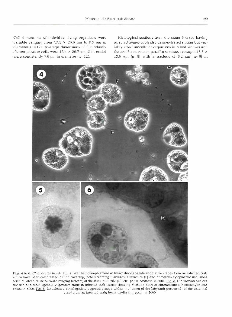
Cell dimensions of individual living organisms were Histological sections from the same 9 crabs having variable ranging from 17.1 X 26.6 pm to 9.5 pm in infected hemolymph also demonstrated similar but var- diameter (n=12). Average dimensions of 8 randomly iably sized unicellular organisms in blood sinuses and chosen parasite cells were 15.4 X 20.7 p m Cell nuclei tissues. Fixed cells in paraffin sections averaged 15.6 X
were consistently 7.6 pm in diameter (n= 12). 12.8 pm (n=8) with a nucleus of 6.2 pm (n=6) in
Figs. 4 to 6. Chionecetes bairdi. Fig. Wet hemolymph smear of living dinoflagellate vegetative stages from an infected crab which have been compressed by the coverslip; note streaming filamentous structure (F) and numerous cytoplasmic inclusions some of which cause outward bulging (arrows) of the thick refractile pellicle; phase contrast; X 2000. Flg. Dinokaryon nuclear dlvision of a dinoflagellate vegetative stage in infected crab tissues showing V-shape pairs of chromosomes; hematoxylin and eosin; X 5000 Flg. Binucleated dinoflagellate vegetative stage bmthin the lumen of the labyrinth portion (E) of the antenna1
gland from an infected crab; hernatoxyhn and eosin; X 5000

200 Dis. aquat. Org. 3: 195-216, 1987
diameter (Figs. 5 and 6). Eight of the crabs were severely infected with the organism throughout all tissues where hemolymph is normally found. The 9th crab, which had 2 to 3 organisms in a hemolymph smear, had a single small focus of organisms in the connective tissue of the pyloric stomach. The primary histopathology caused by this parasite was replace- ment of vascularized tissues throughout the body, mostly connective tissues, and severe congestion of hemal spaces. The organism was most numerous in the vesicular connective tissues of: the epidermis; the pyloric stomach directly beneath the mucosa; the hepatopancreas interstitially among the tubular ele- ments; the intestinal tract, especially outside of the circular muscular layer. Glial elements surrounding the optic ganglia of the eyestalk were also severely infil- trated by this organism. The major hemal spaces most congested with this organism included: the trabecular channels within the myocardium; the branchial sinuses within the gill filaments (Fig. 7); the sinuses of the eyestalks beneath the omatidia and surrounding the primary optic nerve fibers; the sinus in the coelomosac of the antennal gland with replacement of podocytes and some but fewer organisms visible in the lumen of the labyrinth. Actual cellular damage associated with this organism was not extensive but included: muscle cell degeneration (loss of cytoplasmic eosinophilia and striations) and necrosis in visceral organs and body musculature; epithelia1 necrosis and cell degeneration in hepatopancreatic tubules characterized by increased vacuolation, basophilic granularity of cell cytoplasm, pale nuclei (Fig. 8) and occasional organisms in the tubule lumina; necrosis and ulceration of epidermal
epithelium beneath the carapace with myriads of organisms forming an exudate on the tissue surface (Fig. 9); occasional pyknosis of urinary bladder epithelium and presence of organisms within the blad- der lumen. A host cell response to invasion by this organism was absent in most tissues. Occasional large foci of mostly agranular hemocytes and some eosinophilic granulocytes were observed in the coelomosac of the antennal gland and in the surround- ing connective tissues of the urinary bladder in infected crabs.
Disease pathogenesis
Hemolymph smears from 20 male Tanner crabs cor- related closely with previous sorting by processors into infected and uninfected groups of 10 each. However, recognition of diseased crabs by external clinical signs did produce a 10 % error (1 uninfected crab) in the infected group and a 20 % error (2 infected crabs) in the uninfected group. After re-sorting crabs by hemolymph smears infected and uninfected groups were comprised of 11 and 9 crabs, respectively. Mean live weights (g) for control and infected crabs were 1263 + 305 and 1334 -t 182, respectively. Mean carapace widths (mm) were 153 + 13 and 161 + 12, respectively. The major- ity of these crabs had 'new shells' excepting 3 and 1 old shell crabs in the control and infected groups, respec- tively. During 97 d of observation 67 % (7) of the infected crabs died while no losses occurred in the uninfected group. At this time initiation of a disease transmission study described below required use of 4
Table 1. Chionoecetes bairdi. Survival of 11 Tanner crabs naturally infected with a parasitic dinoflagellate during 158 d from 28 March to 2 September 1986=
Crab No. Approx. Survival Moribund (M) Secondary herno- Dinoflagellate water temp ( O C l b (d) or dead (D) lymph infection life stage
3 3.8 20 D Bacteria Vegetative 13 - 33 D Bacteria & ciliate Vegetative 2 5.2 58 D NEc NE 5 5.2 58 D NE NE 7 5.2 58 M Bacteria Vegetative 1 6.0 75 D Ciliate Vegetative
10 6.5 90 D Bacteria Vegetative 6 7.4 123 M None Vegetative
12 7.8 140 D Bacteria & ciliate Prespore 4 8.6 149 Dd Bacteria Dinospore 9 7.7 158 D' None Dinospore
a No mortalities in uninfected control crabs Seawater temperatures recorded every other day beginning 40 d into the experiment. Some readings are averages rather than actual observations for a given day
C NE = not examined because of severe post-mortem change Blood smear examined 2 d prior to death Blood smear examined 1 d prior to death. Prespore stages were first observed 10 d prlor to death

Meyers et al.: Bitter crab disease 201
Figs. 7 to 9. Chinoecetes bairdi. Fig. 7. Gill filament from an infected crab showing engorgement of vascular spaces with dinoflagellate vegetative stages (D); host granulocyte (arrowj; hematoxylin and eosin; X 800. Fiq. 8. Hepatopancreas from an infected crab with vacuolar degeneration and loss of nuclear staining in tubular epithelium (E) and replacement of interstitial vesicular connective tissue by myriads of &noflagellate vegetative stages (D); hematoxylin and eosin; X 315. Fig Epidermal border beneath carapace of an infected crab showing a basement membrane (arrow) devoid of an epithelia1 border except for remnant nests of cells (E) In tissue folds; myriads of dinoflagellate vegetative stages replace vesicular connective tissue of the lrisceral side (V) of the epidermis and have infiltrated to the epidermal surface (S) forming part of an exudate; hematoxylin and
eosin; x 800
control crabs leaving 5 uninfected crabs for the remain- der of the pathogenesis study. By Day 158 all naturally infected crabs were dead. No deaths had yet occurred in the control crab group (Table 1). Hemolymph from
all of the infected crabs, except one, was severely infected with the organism when initially examined. The infected crab with relatively fewer organisms in peripheral circulation was one of the last to die a t 140 d

202 Dis aquat Org 3 195-216, 1987
F ~ g s 10 to 13 Spores of dlnoflagellate Fig 10 Fixed crab hemolymph smear of dlnoflagellate multinucleated prespore stages undergoing r a p ~ d cell division, cells taken from Infected Tanner crab No 9, 9 d prior to sporulation into the smaller type of d~nospore (Figs 14 & 15). Dlff-Qulk. x 5000 FICJ l 1 Wet crab hemolymph smear of l ~ v e dinospores of the larger type 24 h after collection from Infected Tanner crab No. 4 (s l~ght lateral protrusion not shown), host granulocytes (arrows); X 2000. Fig. 12. Live dinospores in Fig 11 after 11 d in hemolymph culture showing development of distinct lateral keel (double arrow) and beaked end (slngle arrow), x 3300 F I ~ . 13. F ~ x e d hemolymph smear of 24 h-old large type dinospores in Fig. 11 showing lobed nuclei with
dist~nct outlines not apparent In vegetative stages Illustrated In Fig 1; Diff-Quick; x 5000

Meyers et al.: Bitter crab disease 203
Figs. 14 & 15. Spores of dinoflagellate. Fig. 14. Wet smear of live dinospores of the smaller type 2 d in saltwater culture after collection from infected Tanner crab No. 9. Note refrac- tile granule (arrow) at distal posterior end; X
3300; inset: same dinospore type in a fixed smear after 11 d in saltwater culture showing a bent profile, densely staining spore body and 2 flagella; Diff-Quick; X 4000. Fig. 15. Glutaral- dehyde fixed dinospores from Fig. 14 after 11 d in saltwater culture demonstrating pro- nounced corkscrew shape and 2 obvious
flagella; phase contrast; X 1320
(Table 1). The clotting characteristics of infected crab hemolymph were indistinguishable from those of con- trol crabs. Most of the crab mortalities occurred suddenly with little behavioral changes except chronic anorexia and progressive lethargy. Crabs were consi- dered moribund when movements of antennae and/or mouth parts were still discernible but the animals were unable to right themselves when turned over or failed to move when prodded.
Seawater temperatures ranged from 3.8 "C early in the mortality study to 8.9 "C near the end of the study. Respective average seawater temperatures ("C) for May, June, July, August and September were: 4.6 (n=15), 5.6 (n=18), 7.0 (n=14), 7.8 (n=12), 7.0 (n=6).
Hemolymph smears from 5 of the dead infected crabs showed assorted bacterial rods in 4 individuals and an unidentified ciliate in 3, in addition to the dinoflagellate organism (Table 1). Infected hemo- lymph from 2 of 4 additional crabs, one moribund
and the other taken 2 d prior to death, also contained obvious bacterial rods. Histological examination of one of the moribund crabs showed overwhelming numbers of the dinoflagellate cells in the tissues and tissue pathology similar to that described. Hemolymph from the control crabs remained free of any detectable infectious agents throughout the obser- vation period.
Hemolymph smears from the last 3 surviving infected crabs in the experiment exhibited dinoflagel- late cells quite different in appearance than previously noted. Multinucleated forms were present in the dead crab No. 12 (Table 1) as were single-celled stages which were much smaller than the previous larger vegetative forms. Parasite nuclei had markedly beaded chromatin and were lobed with uncharacteris- tic distinct outlines. Similar prespore stages were observed in crab No. 9 ten d prior to death (Fig. 10). One d prior to death these stages were completely

204 Dis. aquat. Org. 3: 195-216, 1987
sporulated into a single type of biflagellated dinospore with very rapid spiral motility Crab No. 4 also developed a distinct dinospore stage within the hemolymph 2 d prior to death. Sporulated hemolymph in both crabs was opaque and white wlth spore con- centrations in crab No. 4 estimated at 3.67 X 10⁷ ml-' of hemolymph. Despite identical appearing vegetative stages, the dinospores from one crab differed from those of the other in size, morphology and behavior. Both spore types underwent morphological changes during maintenance in hemolymph and saltwater cultures. Dinospores from crab No. 4 were oval, about 15.2 X 11.4 pm (n=8) , with a slight lateral protrusion when first extracted from the host (Fig. 11). After 9 d in hemolymph culture the lateral protrusion developed markedly into a keel-like structure with a beak-like
Fig. 16. Typical &noflagellate vegetative stage from the hemolyrnph of an infected Tanner crab. Transmission electron micro- graph. Nucleoplasm (N) with beaded chromatin clumps (C), cytoplasmic membrane-bound homogenous granular substance (H) associated with large vacuoles containing a lipid-like material (L) , mitochondria (M) in thin strands of compressed cytoplasm, and irregular electron dense pelli- cle (P) showing separation of lay- ers. Uranyl acetate-lead citrate: X
17 620; bar = 1 km
protrusion at one end (Fig. 12). In stained smears these spores resembled smaller vegetative stages; flagella were not visible but nuclei were longer than wide, lobed and distinct in outline (Fig. 13). Their general measurements remained the same. Dinospores from crab No. 9 were more elliptical with narrower and shorter dimensions (12.0 X 4.4 pm; n=5) with a slight lateral ridge (Fig. 14). After 24 h in saltwater culture these spores developed a bent corkscrew shape that was more pronounced 6 d later (Fig. 15). A distinct feature of this spore was a refractile round body located distally at the posterior end (Fig. 14). In stained smears the refractile body and 2 flagella were obvious as was a dense overall staining of the spore body (Fig. 14). These dinospores moved much more rapidly and errati- cally than the larger dinospores from crab No. 4.

Meyers et al.: Bitter crab disease 20,
Figs. l? & 18. Vegetative dinoflagellate cell. Fiq. l?. Ultrastructure showing cytoplasmic invagination (C) in the nucleus, and numerous granular cytoplasmic inclusions (H) some of w h ~ c h cause outward bulging of the pellicle; a break within the pellicle integrity is visible (arrow); uranyl acetate-lead citrate; X 7000; bar = 1 pm. Fig. 18. Detail of cell showng empty vacuoles (V) containing membranous debris and others filled with a lipid-like material (L); these vacuoles are within enlarged areas of cytoplasm which is usually compressed as thin strands (arrows) between voluminous homogenous granulai inclusion bodies (H);
nucleus (N); uranyl acetatelead citrate; X 15 517; bar = 1 pm
Ultrastructure the mesokaryotic detail shown for Hematodinium sp. in blue crabs (Newman & Johnson 1975), in Haplozoon axiothellae (Siebert & West 1974) and in Syndinium globiformi (Hollande 1974). Condensed chromatin sometimes caused bulging of the nuclear membrane providing a 'knobby' nuclear profile. The nucleoplasm, composed of irregular light and dark areas, was abun-
Transmission electron microscopy
A typical vegetative cell (Figs. 16 and 17) was irregular in outline with a thick electron-dense pellicle and a large nucleus having beaded and lobed chromatin similar to

206 Dis. aquat. Org. 3: 195-216, 1987
Fig. 19. Ultrastructure of the larger beaked dinospore from infected Tanner crab No. 4 after 10 d in hernolymph culture. Nucleus (N), electron-dense bodies (D), trichocysts (arrows), flagellurn (F), fibrillar polysaccharide-like material (S) associated with perinuclear space and endoplasmic reticulum and large empty vacuoles (V). Uranyl acetate-lead citrate. X 11 900, bar = 1 pm. Inset: Detail of similar dinospore showing pore-like structures (arrows) associated with pellicle. Uranyl acetate-lead citrate; X
26 000: bar = 1 pm
dantly interspersed among the chromatin while the Occas~onaUy both vacuoles and granular inclusion nuclear membrane, though visible, was not well defined. bodies would occur in outward bulges of the pellicle. A Although almost certainly present, a distinct peripheral few mitochondria and rough endoplasmic reticulum nucleolus was not observed and cytoplasmic invagina- were the only other common eukaryotic cell organelles tions were rare. The cell cytoplasm contained almost visible rarely within the thin strands of cytoplasm appres- exclusively a homogeneous membrane-bound granular sed against the cell wall or constricted between the substance associated with large vacuoles containing a homogeneous granular inclusions (Fig. 16). The cell
Somevacuoles were empty theca did not have the typical dinoflagellate structure of except for membranous debris (Fig. 18) similar to vacuo- vesicles containing plates. However, such detail may les shown for Solenodinium fallax (Hollande 1974). have been obscured by the extreme electron density of

h4eyers et al.: Bitter crab disease 207
Figs. 20 & 21. Flagellate dinospore. Fig. 20. Tangential section showing ultrastructure of smaller dinospore type from infected Tanner crab No. 9 collected immediately after sporula- tion; nuclear chromatin (C) densely inter- connected and not beaded; few trichocysts (arrows) visible; uranyl acetate-lead citrate; X
structure [W) near flagellar groove [R) and fibril- lar polysaccharide-like material [S) assoc~ated with perinuclear space as in larger spore-type; uranyl acetate-lead citrate; X 22 320; bar = 1
the pellicle in our material possibly resulting from a deposit or layer of polysaccharide-like material (Hollande 1974). Frequent breaks, separation of layers and irregular outward or inward folds were also features of the theca occasionally associated with bulging of the pellicle (Figs. 16 and l?). No cytoplasmic microtubules were apparent beneath the theca of vegetative stages, the lack of which is unusual among other parasitic dinoflagellates (Siebert & West 1974). Trichocysts were also absent.
The larger dinospore from crab No. 4 had a less dense thinner pellicle than vegetative forms, with flagella showing a 9 + 2 configuration and similar
nuclear detail as in the vegetative stage above. Nu- cleoli again were not observed. Some spores contained a fibrillar substance within dilated perinuclear spaces and associated with the endoplasmic reticulum similar to the polysaccharide inclusions reported for spores of Syndiniunl globiformi (Hollande 1974). The large granular membrane-bound cytoplasmic inclusions in vegetative stages were absent but several electron- dense bodies were abundant in most cells. The cyto- plasm was also abundant and rough endoplasmic reticulum and mitochondria were more commonly observed along with numerous peripheral trichocysts having a rhomboidal transverse profile. A series of

208 Dis, aquat. Org 3 195-216, 1987
Fig. 22. Ultrastructure of smaller &- nospore type after 23 d in saltwater culture. Longitudinal section. Cyto- plasm more electron-lucent with large vacuoles (V) and few dense bodies (D); nucleus (N) and flagellar attachment site (F) for both cir- cumferential and whiplash flagella. Uranyl acetate-lead citrate; X
11 390; bar = 1 pm.
pore-like structures probably associated with tricho- cysts were visible at or just beneath the pellicle surface. Cytoplasmic vacuoles became more abundant as this spore type aged in laboratory culture (Fig. 19).
The smaller dinospores from crab No. 9 differed from the larger type by having a more dense nucleus with chromatin in large wide whorls surrounded by very little nucleoplasm. These spores had a larger nuclear to cytoplasmic ratio with less cytoplasmic volume contain- ing much fewer electron dense bodies and trlchocysts (Fig. 20). One sectioned spore contained a concentric membrane structure directly beneath the theca near the flagellar groove and a dilated perinuclear space filled with a similar fibrillar material as in the larger spore type (Fig. 21). As these dinospores aged in vitro the cytoplasm became more electron lucent with sev- eral large vacuoles (Fig. 22).
Scanning electron microscopy
Vegetative stages of the parasite were rounded to
very irregular in shape and of varying sizes often attached to each other by intercellular bridges. The cell surface was randomly dispersed with depressions or pores of vaq ing sizes. Many of these had a homogene- ous material exuding from within appearing as smooth rounded droplets attached to each parasite cell exterior (Fig. 23). Elliptical to crescent-shaped crab hemocytes were abundant among the many vegetative parasite cells (Fig. 24).
The larger dinospores from crab No. 4 were ovate with rounded ends and had a 'warty' cell surface hav- ing a longitudinal keel and large folds (Fig. 25). The longest circumferential flagellum (15 t ~ m ) was attached very near the distal anterior end within a small laterally located elongate cleft (Fig. 26). The shorter whiplash flagellum (5 pm) was attached under- neath almost immediately behind the first but sepa- rated by a median ridge. It trailed within a deep lon- gitudinal groove alongside a ventral keel, but did not extend beyond the spore body (Fig. 27). As this spore aged in vitro the ventral keel became more pro-

Meyers et al.: Bitter crab disease 209
Figs. 23 & 24. Dinoflagellate vegeta- tive cells. Fig. 23. Scanning electron micrograph of vegetative stages (D) showng irregular thecal surface with pores and exuding droplets of un- identified material (arrows); gold palladium; X 4000; bar = 10 pm. Flg. 24. Lower magnification of represen- tative field of cells (D) showing single and multicellular forms exuding drop- lets and interspersed with some host granulocytes (arrows); gold palla-
dium; X 1000; bar = 10 pm
nounced and the anterior end of the spore became beaked (Fig. 27).
The smaller dinospore from crab No. 9 was quite different. The early spore body was elliptical, sharply pointed anteriorly and less so posteriorly with a smooth surface. A smaller ventral keel was also present (Fig. 28). The longer circumferential flagellum (18 pm) attached further back on the body in a shallow depression. As in the larger spore type, the shorter whiplash flagellum (5 gm) attached immediately behind the first but in a shallower longitudinal groove also separated by a median ridge (Fig. 29). The second flagellum extended well beyond the length of the spore. As this spore aged in vitro the anterior end
became blunted and scalloped while the body developed a distinct corkscrew twist (Fig. 30).
Disease transmission
In the first experiment vegetative forms of the para- site were detected at 55 d post-inoculation (PI) in crab No. 8 receiving 1.1 X 10⁶ of the cultured protozoan cells. At 69 d, crab No. 20 that received 2.2 X 10⁵ cells from the same preparation also developed a detectable infection. At 148 d PI, concentrations of vegetative stages in both crabs were 2.73 X 10⁶ and 1.21 X 10⁶ ml-' of hernolymph, respectively. These numbers indi-

210 Dis. aquat. Org. 3: 195-216, 1987
Figs. 25 & 26. Dinospores. Fig. 25. Scanning electron micrograph of larger dinospore taken from infected Tanner crab No. 4 soon after sporula- tion; raised protuberances on pellicle, a lateral keel and flagella visible; gold palladium; x 3000; bar = 5 pm. 26. Detail of large dinospore (similar to those in Fig. 25) showing attach- ment point of circumferential flagellum and portlon of trailing whiplash flagellum (shrunken spore body is fixation artifact); gold
palladium; X 5000; bar = 5
cated that the total concentration of organisms in each crab multiplied from inoculation doses by approxi- mately 2 to 3 log 10 when considering total body hemolymph volumes. However, unexpectedly, both crabs inoculated with vegetative stages taken directly from crab No. 9 (in which vegetative stages sporulated 60 d later) remained uninfected after 214 d PI. At 63 d PI, a hemolymph smear from the crab receiving the larger dose of crab No. 9 vegetative stages demon- strated a single corkscrew-shaped dinospore. This dis- covery suggested that the inoculated vegetative stages had sporulated in thls crab. Seawater temperatures for this duration were as indicated previously and all con- trol crabs remai.ned uninfected.
In the second experiment examining host species susceptibility vegetative stages of the parasite were detected at 83 d PI in 4 of the 5 surviving Tanner crabs receiving infected Tanner crab hemolymph. A 5th crab developed an infection at 97 d PI, while the 6th crab died early on in the experiment from undetermined causes. None of the h n g crabs inoculated with the dinoflagellate developed detectable infections 155 d PI. None of the sham inoculated crabs of either species developed infections, and only 1 control lung crab was lost from cannibalism after molting. This experiment was initiated during late February when the seawater temperature was 4.5 "C. Respective average monthly seawater temperatures ('C) during the experiment for

Meyers et al.: Bitter crab disease 211
Figs. 27 & 28. Dinospores. Fig. 27. D e t d of 2 large &nospores from Tan- ner crab No. 4 after 10 d in hemolymph culture; attachment point of whiplash flagellum is within deep ventral groove almost immediately behind depression (arrow) for cir- cumferential flagellum (missing); al- though almost obscured by particu- late debris, a pronounced beak (B) is apparent on the left spore; gold palladium; X 4000; bar = 5 pm. F* 28. Smaller type dinospores from in- - fected Tanner crab No. 9 taken soon after sporulation; a smaller ventral keel is present alongside the trailing whiplash flagellum, and the spore surface is smooth; gold palladium; x
4000; bar = 5 pm
March, April, May, and June were; 4.4 (n=19), 5.1 (n=30), 5.6 (n=27), 6.7 (n=29).
In vitro culture
Within 2 to 3 d of seeding, dinoflagellate vegetative forms attached to the surfaces of the 2 up-ended flasks and subsequently formed incomplete monolayers within 12 to 15 d (Fig. 3). The antibiotic mixture added to hemolymph did not affect parasite cell survival and growth. Consequently, it was used thereafter in all cell preparations. By 17 d, cells were detaching and loose within the supernatant fluid. The morphology of
stained cells in suspended culture was identical to those in freshly drawn hemolymph from infected crabs. On Day 19 the flask with antibiotic was gently shaken to dislodge remaining cells which were centrifuged and resuspended in 15 m1 of fresh Tanner crab hemolymph for subculturing. Subsequently, second, third and fourth generation vegetative stage cell cultures were established with monolayers becoming confluent in about 15 to 18 d. Such cell cultures were able to survive for months in undisturbed flasks. Occa- sionally, considerable cell clumping occurred rather than monolayers. These cell aggregates would periodi- cally attach by pseudopodia then detach from the flask surface. As the cell clumps aged, occasional single cells

212 Dis aquat. Org. 3: 195-216, 1987
became hypertrophied with appressed nuclei suggest- ing a degenerative or senescent change. Considerable differences in size of attached single cells also became evident and correlated with the varying sizes observed in circulating hemolymph. Small cells were able to increase in size when observed for long periods in hemolymph cultures.
Suspended vegetative stages of the parasite were able to survive for a t least 5 d in natural seawater (30 "A at 5 "C) after which the preparation was destroyed by bacterial contamination. Dinospore stages from both crabs Nos. 4 and 9 were able to survive in sterile seawater for 73 and 52 d, respectively.
Cultures of the smaller No. 9 dinospores showed
Figs. 29 & 30. Dinospores. Fig. 29. Detail of flagellar attachment in small
spore body than in large spores, and whiplash flagellurn trails beyond
I culture. One spore (arrow) has a &S- tinct corkscrew twist and blunted
I scalloped anterior end; bacterial con- tamination of culture is evident; gold
palladium; X 3800; bar = 5 gm
reduced activity 30 d after collection from the host crab and were also eventually overwhelmed by bacterial contamination that may have terminated their survival prematurely. Prespore stages from crab No. 9 collected 1 d prior to natural sporulation in the crab host were seeded into a culture flask of hemolymph. These cells readily attached in large clusters within 48 h of incuba- tion at 6 "C. After 35 d in culture, these cells began sporulating into the dinospores typical of those col- lected from the donor crab host after natural sporula- tion. Sporulation in the culture flask was not simultane- ous nor complete and proceeded slowly. Many pre- spore cell clusters did not sporulate and later died.
After 25 d in culture, some of the No. 4 large type

Meyers et al.. Bitter crab disease 213
dinospores in both hemolymph and saltwater developed into irregular shapes which no longer moved in a straight line but in a tight circular motion. In the hemolymph culture many of these cells eventually settled to the flask surface and attached by extended pseudopodia forming large clumps of non-motile cells. These attached forms were accompanied by chains of non-motile organisms loosely attached to the flask in a rosette-like pattern. In the saltwater culture only the rosette forms developed.
Disease prevalence and distribution
In 1985 bitter Tanner crabs were harvested from 3 areas in the northern end of Lynn Canal: Chilkoot, Chilkat, and Lutak Inlets (Fig. 31). During 1986, para- sitized crabs were caught from several areas including: Upper Lynn Canal (excluding Berner's Bay) from Little Island northward; the Juneau area; Port Frederick and adjacent area 114-27 in Icy Strait; area 114-21 in Cross Sound; and area 111-20 in southern Stephens Passage (Fig. 31).
Commercial fishermen reported bitter Tanner crabs approaching a prevalence of 100 % (Joe Donohue pers. comm.) in the narrow body of water between Sullivan Island and the mainland (Fig. 31). A pot survey con- ducted in this area (58'56' north latitude and 135" 21'
east longitude) by the ADF&G (Alaska Dept of Fish and Game) found an overall disease prevalence of 9 5 % among the 149 crabs examined (Table 2). Hemolymph smears from 20 randomly chosen infected crabs showed previously described prespore stages in at least 1 individual (5 %). The intensity of infection in all crabs examined ranged from very light with 4 to 5 parasite cells per smear, to moderate with 50 to 100 cells per field, to severe with hundreds or solid fields of organ- isms and no host cells visible. Numerical values of 1, 2-3, and 4-5 were assigned to hemolymph smears having light, moderate and severe infections, respec- tively. Using these very subjective values the average infection intensity from this field study was estimated as moderate (3). Since only 6 male and 2 female crabs were uninfected, there appeared to be no differences in parasite prevalence between the 2 sexes and among the size classes examined or with the depth at which gear was set. The largest male crabs were approxi- mately 6 to 7 yr old while the smallest female crabs were about 2 yr of age. Hemolymph smears from 5 red king crabs, 1 golden king crab Lithodes aequispina, and 1 blue king crab Paralithodes platypus incidentally caught in the gear did not have any detectable dino- flagellate forms.
Because of the high dinoflayellate prevalence the Sullivan Island area was closed to the commercial Tan-
Fig. 31. Distribution of dinoflagellate infection in Tanner crabs harvested from northern southeast Alaska during 1985 and 1986. 1, Lutak Inlet; 2, Chilkoot Inlet, 3, Chilkat Inlet; 4, Sullivan Island; 5, Little Island (all preceeding areas in upper Lynn Canal); 6, area 114-27 In Icy Strait; 7 , Port Frederick; 8 , area 114-21 in Cross Sound; 9, area 111-20 in southern
Stephens Passage
Table 2. Chionoecetes bairdi Prevalence of dinoflagellate parasitism in male and female Tanner crabs of different size
classes near Sullivan Island, Alaska on 1 July 1986
Carapace 'k Infected '?lo Infected width (mm) males females
71-78 (0) 67 (2/3)" 79-88 l00 (2/2) 92 (12/13) 89-98 l00 (5/5) l00 (34/34) 99-108 100 (4/4) 100 (4/4)
109-118 l00 (13/13) l00 (4/4) 119-128 82 (9/11) l00 (1/1) 129-138 83 (516) (0) 139-148 95 (19/20) (0) 149-158 92 (22124) (0) 159-163 l00 (5/5) (0)
Total % infected 93 (84/90) 97 (57/59) Total O/o new shell 97 97
I NO. infectedho. examined
214 Dis. aquat. Org. 3: 195-216, 1987
ner crab fishery in January and Februray of 1987. In March of that year the ADF&G sampling effort within the same Sullivan Island grid area recovered only 50 Tanner crabs. Three of these were legal sized males, all of which were infected with the dinoflagellate disease. The overall dlsease prevalence in this sample of crabs was 37 %.
DISCUSSION
The gross milky appearance of hemolymph and tissues in bitter tasting Tanner crabs was caused by myriads of parasitic dinoflagellate organisms resem- bling Hematodinium sp. Much of the clinical disease in the host is caused by the non-motile vegetative uni- nucleate and plasmodial stages. Most of the vegetative parasite morphology, the gross clinical signs of disease and the histopathology of infected crabs were similar to reports of Hernatodinium disease in blue crabs (New- man & Johnson 1975). However, other features of this disease are distinctly different suggesting that the bit- ter crab agent is a new entity: vegetative forms of the organism were much larger than the average cell diameter of 8.1 ym in the blue crab; no highly motile multinucleated vermiform bodies were observed; veg- etative stages did not have trichocysts in the cytoplasm when examined by TEM (Newman & Johnson 1975); diseased crabs have a bitter aftertaste when infected crab meats are cooked; sporulation does occur in the crab host producing one of two types of dinospores but not both.
In addition, this report is the first documentation of a decapod dinoflagellate disease in the Pacific Ocean and in Tanner crabs. The outcome of clinical disease in blue crabs is not documented, but mention is made that infected crabs die soon after capture and that diseased crabs, reaching a prevalence up to 3O0/0, cannot be found in late winter or early spring (Newman & John- son 1975). In the Ta.nner crab, the prevalence of .i.nfec- tion can be much higher occurring during winter and spring while the course of the disease is chronic and prolonged over several weeks or months. Lower water temperatures and a larger crab host species may pro- vide for longer development periods of this disease. The disease proved fatal to all infected crabs in the laboratory and may be capable of decimating natural Tanner crab populations. The latter possibility is supported by the ADF&G sampling of 2/3 fewer crabs from the Sullivan Island grid area in 1987 than in the previous year when 95 O/O of the crabs were infected with the disease. Also. only 6 % of the 1987 crabs were legal-sized males compared to 30 O/O in 1986. Commer- cial fishing could not have influenced the decline in population numbers because the area has been closed
to harvest. The difference in sampling gear used in both years could possibly result in differences between total numbers of crabs collected but should not cause a significant discrepancy between relative numbers of legal-sized males.
Death of host Tanner crabs having bitter crab disease may result from two possible primary mechanisms; organ and/or respiratory dysfunction. Both are prob- ably caused by the overwhelming numbers of replicat- ing parasite cells which passively infiltrate and replace hemolymph elements and all vascularized tissues. The peculiar orange discoloration of epidermis and result- ing pink carapace in infected crabs are probably caused by released carotenoid pigments from damaged epidermal cells. Crabs so affected are also not likely to survive the molting process. Overall host cell degener- ation and necrosis may be caused by pressure atrophy, anoxia and/or toxic metabolites produced by the vast numbers of the organism. Stained hemolymph smears and SEM clearly demonstrated that vegetative stages produced considerable amounts of a substance in the form of minute droplets attached to parasite cell sur- faces. Whether this material is toxic to host cells is not known. Those crabs surviving the vegetative phase of the disease die within 24 to 48 h after parasite sporula- tion that probably causes considerable mechanical dis- ruption of tissues by the newly motile organisms. Another contributing factor to crab death may be sec- ondary bacterial and ciliate protozoa1 infections in hosts already debilitated by the chronic vegetative phase of the dinoflagellate disease. The presence of these other agents in the hemolymph of 5 dead crabs could be argued as post-mortem artifact. However, the bacteremia observed in 2 crabs prior to death support the possibility of ante-mortem infection, at least by opportunistic bacteria.
Previous investigation regarding the cause of bitter flavor in Tanner crabs was done in 1985 by scientists within the National Marine Fisheries S e ~ c e in Seattle. Their unpublished report, based on organoleptic and chemical tests of processed crab meats, concluded that the bitter flavor was due to the pre-molt physiological stage of the crabs (unpubl, data, memorandum 22 March 1985). This conclusion is not likely since most of the infected crabs we examined had new shells and were not approaching a pre-molt condition. We believe the bitter flavor of cooked meat from infected crabs results from either the dinoflagellate organism ~tself or a natural metabolite produced in the hemolymph by the parasite that permeates all tissues of the crab host. This product may be the same substance represented by the visible droplets attached to parasite cell sur- faces. Whether the material within lipid-like inclusion bodies later forms the external droplets needs further study. However, chemical analysis of infected and

lvIeyers et al.. Bit ter crab d~sease 215
uninfected crab hemolymph with parallel taste tests may support this speculation on the cause of bitter flavor.
The life history of Hematodinium sp, observed in decapod hosts has not been described. However, thls present work has provided some factual information on which to propose a possible life cycle for the Tanner crab agent. Whether parasite development occurs within a yearly cycle or requires longer periods is still not resolved. Vegetative stages of the parasite divide in the host hemolymph over several months, eventually causing death in a large percentage of crabs. The vegetative stages in those few surviving crabs sporu- late into motile dinospores causing crab death and putatively serve as the infectious stage or precursors to an infectious zygote stage produced by fusion of 2 different zoospore types representing male and female sexes. The morphological differences between the 2 spore types suggest the separate sex theory supported further by other work with pandalid embryo peridinienosis (Stickney 1978, Holmes et al. 1980, Hibbits & Porter unpubl.) and syndinid parasites of Radiolarians (Hollande 1974). These investigators observed 2 different-sized spores for each parasite that were similar in some features to those in this report. However, fusion was also not evident when the 2 pan- dalid zoospore types were mixed. Nonetheless, sepa- rate-sexed dinospores might explain why 2 of our crabs receiving vegetative stages from a naturally infected crab never developed infections. The inoculated veg- etative stages may have been sufficiently advanced in development that sporulation was imminent at approx- imately the same time as in the donor host crab as long as environmental conditions remained identical. Con- sequently, vegetative stages never had time enough to multiply to detectable levels in the 2 inoculated crabs. Also, resultant dinospore numbers were still too low for detection except for 1 spore in a single smear. If such spores were haploid, they would most likely be non- infectious and eventually eliminated by the host. The artificial environmental conditions of prolonged in vitro maintenance of the vegetative stages used in the 2 successfully infected crabs must have slowed or altered their normal development towards sporulation. Thus, the vegetative stage was perpetuated with subsequent multiplication to detectable levels. In both groups of crabs, the inoculation doses were roughly equal and should not have been variables. A predictable dose response was apparent regarding the lag period of detection between the higher and lower doses in the successfully infected crabs.
A second possibility is that both spore types in bitter crab disease represent 2 closely related but different dinoflagellate parasite species indistinguishable in the vegetative stage. The pandalid agent differed in that 2
types of vegetative stages were evident, each produ- cing a different zoospore (Hibbits & Porter unpubl.). However, these investigators also found that pandalld eggs were not infected when exposed to both types of live zoospores. We have not yet conducted transmis- sion studies with the two zoospore types from bitter Tanner crab disease.
Some circumstantial evidence in both our pathogenesis and in vitl-o studies suggest that higher seawater temperatures may initiate parasite sporula- tion in the crab host if other environmental and physi- cal factors remain optimum. This evidence included sporulation in naturally and certain experimentally infected crab hosts following rising seawater tem- peratures and retardation of imminent sporulation by in vitro placement of dividing prespore cells at a lower temperature. Admittedly, other artificial factors una- voidable in in vitr-o studies could have confounded the latter result. It may be likely that vegetative forms could persist for more than one seasonal cycle in those crabs surviving this phase of the disease in areas or depths where seawater temperatures remain low. In this case yearly natural mortalities from the disease may be much lower than we observed in the laboratory or which appeared to have occurred within the Sullivan Island study area.
Further epidemiological studies must be done to establish the route of transmission which is most likely by contact with either dinospores or a zygote produced by fuslon of the 2 dlnospore types. If dinospores are directly infectious, parasitism could also be established by Ingestion of spores within infected crab tissues. Non-senescent n~orphologlcal changes in spore development outside the host crab may have some significance In the competency of spores to either infect crabs directly or fuse with the opposite spore type to produce an infectious stage. Survival of both spore types for up to 2 to 3 mo in seawater must certainly enhance the chance occurrence of host Infection. Veg- etative stages are passively Infectious and can be trans- mitted to healthy Tanner crabs experimentally by parenteral injection. Histological observations showed that vegetative stages occur in the lumina of the blad- der and digestive diverticula. Consequently, they are probably excreted with crab urine and feces. Although vegetative stages can survive for days In seawater it is unlikely they would be able to gain access to crab tissues or hemolymph by some natural process. Dino- spores may also be released in the urine and feces while the host is still alive, but most likely are released en masse during tissue decomposition following crab death. Thus, Tanner crabs In the vegetative phase of the disease probably are not infectious for other crabs. Otherwise some of our control Tanner crabs which were previously confined for several days with natur-

216 Dis. aquat. Org. 3: 195-216, 1987
ally infected crabs in commercial live holds should Acknowledqements. We thank the following people for their
ha;e developed a detectable infection during their important contributions to this report: ~itka-sbunds Seafoods
many months in captivity, The age or sex of host Tan- and Pelican Seafoods for catch and product data for the 1985 and 1986 Tanner crab seasons; the University of Alaska,
ner crabs do not seem to affect susceptibility to infec- Juneau School of Fisheries and Science and the Auke Bay tion although further observations in younger age Laboratory, NMFS for use of laboratory space and supplies; Dr classes of Tanner crabs as well as other crab swecies are Ralph Elston of Battelle Pacific Northwest Division in Sequim,
needed. ~t is encouraging to note that 7 hng crabs from Washington and Mr A1 Soeldner of Oregon State University for the quality TEM and SEM photornicrographs; Oliver Hof-
the affected Sullivan Island area did not stad and Peter philbin of the 'Steller.; George Curtiss for appear be infected and that the disease be technical assistance; Joe Donohue for catch information near transmitted to red h n g crabs in the laboratory. Sullivan Island; and Dr AI Sparks for his valuable review of
The management and economical consequences of this manuscript.
this disease for the Alaskan Tanner crab fishery are significant. Overfishing is probably the major cause for Tanner crab declines in certain areas but bitter crab LITERATURE CITED
disease be ignored as a possible contributing Chatton, E., Poisson, R. (1931). Sur l'exlstence, dans le sang factor. In addition to population reductions, the unmar- des crabs, de peridiniens parasites Hematodinium perezi ketable bitter meats can result in considerable financial n. g., n sp. (Syndinidae). C. r. Seanc. Soc. Biol. 105:
loss to processors or commercial crab fishermen. The 553-557 Hollande, A. (1974). 'Etude cornparee de la mitose Syn-
only management of this disease in feral crab popula- dinienne et de celle des peridiniens libres et des hyper- tions is in preventing parasite dissemination to mastiqines infrastructure et cycle evolutif des syndmides
unaffected Tanner crab areas. This approach could parasites de Radiolaires. ~rot is tolo~ica 3: 413:45i include the closure of severely affected areas to corn- Holmes, P. B., Mueller, G. J. , Hauck, A. K. (1980). Observa-
mercial crabbing and persistent culling of milky crabs tions and speculations on premature egg loss in Gulf of Alaska Pandalus borealis (pink shrimp). Soc. Invertebr.
on the fishing grounds before transport followed by a Pathol. XI11 Ann. Meet. Seattle (abstract) second culling at the processing fachties. Infected Johnson, P. T (1986). Parasites of benthic amphipods: dino- crabs from other areas should not be released into local flagellate (Duboscquodmnida: Syndmidae). Fish. Bull. U. S.
waters to spread the disease. It would be significant to disease management if parasite development follows a yearly cycle with sporulation and new infections occur- ring in late summer or early fall. Newly infected crabs could be harvested in October through December before the disease has progressed far enough to cause a detectable bitter flavor. No culling of infected crabs would be necessary, all crabs would be marketable and the infected crabs could be utilized before mortalities occur from the disease. This alternative would also prevent the dissemination of the disease from release of unwanted bitter crabs in other waters.
84 :- 605-6 15 MacLean, S. A., Ruddell, C. L. (1978). Three new crustacean
hosts for the parasitic dinoflagellate Hematodinium perezi (Dinoflagellata: Syndinldae). J. Parasitol. 64: 158-160
Newman, N. W., Johnson, C. A. (1975). A disease of blue crabs (Calhectes sapidus) caused by a parasitic dinoflagellate, Hematodinium sp. J. Parasitol. 61: 554-557
Siebert, A. E. , West, J. A. (1974). The fine structure of the parasitic dinoflagellate Haplozoon axiothellae. Proto- plasma 81: 17-35
Stickney, A. P. (1918). A previously unreported peridinian parasite in the eggs of the northern shrimp, Pandalus borealis. J Invertebr. Pathol. 32: 212-215
Responsible Subject Editor: Dr J . E Stewart; accepted for printing on October 8, 1987