Embed Size (px)
Citation preview
1108 BRITISH MEDICAL JOURNAL 6 NOVEMBER 1976
blind, the observers not knowing the treatment of the patient at the time offilming.The results are summarised in the table. The single patient with Hunting-
ton's chorea who was not assessed on film deteriorated both in terms ofbehaviour and involuntary movements so that the treatment had to bediscontinued. Of the remaining patients, although three showed somesubjective improvement in wellbeing, any changes in observed involuntarymovements were at best marginal and in no case did examination of thefilms show any improvement either in involuntary movements or gait whichcould be ascribed to the treatment.
Discussion
Attempts to modify brain GABA concentration hrvo not alwaysgiven consistent results. Thus Barbeau found that neither GABA itself,taken by mouth, nor a combination of isoniazid and vitamin B6,nor the GABA analogue Lioresal (f-p-chlorophenyl-y-aminobutyricacid) modified the chorea of Huntington's disease.2 On the other hand,Fisher et al noted an improvement in three out of seven patientstreated with GABA in higher dosage.3 Our finding that sodium val-proate does not appear to benefit patients with choreiform movementsis consistent with that of Shoulson et al, who found no improvementwhen sodium valproate was given either alone or in combination withGABA, although a probenecid-loading test showed there was anincrease in the central turnover of both dopamine and 5-hydroxy-tryptamine.4 Patel et al comment that the failure of sodium valproateto inhibit amphetamine-induced stereotyped behaviour raises somedoubt about the physiological role of GABA in the striatum and in thepathogenesis of chorea.5
Bird, E D, et al, Lancet, 1973, 1, 1090.2 Barbeau, A, Lancet, 1973, 2, 1499.3 Fisher, R, Norris, J W, and Gilka, L, Lancet, 1974, 1, 506.4 Shoulson, I, Kartzinel, R, and Chase, T N, Nezurology, 1976, 26, 61.5 Patel, B C, Crosset, P, and Klawans, H L, Research Communications in
Chemical Pathology and Pharmacology, 1975, 12, 635.
University Department of Medicine, Ninewells Hospital, Dundee,DD1 9SY and Dundee Royal Infirmary, DD1 9ND
J A R LENMAN, MB, FRCPED, reader in neurology and consultant neurolo-gist
I T FERGUSON, MB, MRCP, senior house officer in neurologyA M FLEMING, MB, medical assistant, clinical neurophysiologyL HERZBERG, MB, MRCP, senior registrar in neurologyJ E ROBB, BMEDSCI, MB, house physician
University Department of Pharmacology, Ninewells Hospital,Dundee, DD1 9SY
M J TURNBULL, BPHARM, PHD, lecturer
Mandril-grown graft for vascularaccess in Christmas disease
Widespread peripheral venous thrombosis resulting from intravenousinfusions of factor IX concentrate in the treatment of Christmasdisease necessitated an alternative form of vascular access. A mandril-grown graft was used after a suitable vein was found by perosseous(olecranon) venography of the arm. This vascular access has been thesole route for parenteral treatment for a severely affected patient.We report the indications, technique, and management of the case.
Case report
The patient was a 25-year-old man who had severe Christmas disease witha factor IX level of less than 1 %. In 1974 home treatment with self-administered factor IX concentrate was started, venous access being via theantecubital veins. A few months later he developed severe recurrenthaematuria that over the next year necessitated several prolonged periods inhospital and daily infusions of large doses of factor IX concentrate (EdinburghDE FIX) for weeks at a time. As a result of this he developed thrombosis ofall accessible veins, including the long saphenous, until eventually accesscould only be obtained occasionally through the smallest peripheral veins inhis hands and feet.
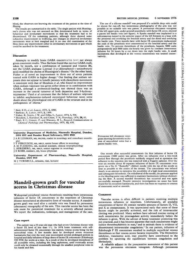
The use of a silicone mandrill was proposed if a suitable deep vein couldbe shown for run-off, but intravenous phlebography of the arm was notpossible as no suitable vein was patent. A perosseous olecranon venogramof the left upper arm, under general anaesthetic with factor IX cover, showeda patent left basilic vein (see figure). A Sparks mandril was implanted in asubcutaneous tunnel in the anterior aspect of the left forearm in a loop withthe proximal end overlying the brachial artery and the distal end overlyingthe basilic vein. Three months later with factor IX concentrate replacementthe ends of the prosthesis were anastomosed to the brachial artery and thebasilic vein. To prevent thrombosis of the prosthesis, heparin 5000 unitsperoperatively and 5000 units six-hourly was given by constant intravenousinfusion for 24 hours by a cut down into the right basilic vein. A smallhaematoma that developed at the venous anastomosis was treated conser-vatively.
Perosseous left olecranon veno-gram showing thrombotic occlu-sion of antecubital veins but apatent basilic vein.
One month after successful anastomosis the first infusion of factor IXconcentrate (1800 units) was given. After six infusions over a two-weekperiod flow through the prosthesis suddenly stopped and at operation clotadherent to the injection site was removed with a Fogarty catheter. Over thenext six months about 40 separate infusions of factor IX concentrate weregiven via a No 21 "Butterfly" (Abbot) needle with the tip of the needle asnear as possible to the centre of the stream. The concentrate was infusedslowly in an attempt to minimise the possibility of a high local concentrationand subsequent thrombosis. On withdrawal ofthe needle, the pressure appliedto the bleeding point was sufficient to maintain haemostasis without obstruct-ing the flow. A second mandril thrombosis has occurred and was againsuccessfully managed. Despite intensive investigations no cause has beenfound for his recurrent haematuria, and there has been no response to coursesof tranexamic acid or steroids.
Comment
Vascular access is often difficult in patients receiving multipleintravenous infusions or injections. Unfortunately, all availableconcentrates of factor IX contain variable amounts of factors II, VII,and X as contaminants. It was appreciated early that some of thesefactors might be partially activated, and the risk of intravascularclotting was predicted. Many authors have advised routine testing ofsuch concentrates for procoagulant activity immediately before theinfusion is given. With the advent of home treatment pretesting wasnot practical, and it has become apparent that such concentrates mightinduce thrombophlebitis in man,2 and in animals there is evidence ofdisseminated intravascular coagulation.3 In our patient, infusions ofEdinburgh F IX concentrate resulted in multiple superficial venousthromboses so that venous access became impossible. The fibrinolyticinhibitor, tranexamic acid, may possibly have contributed to theproblem by upsetting the equilibrium between coagulation andfibrinolysis.A valuable adjunct in the preoperative assessment of this patient
was the perosseous olecranon venogram. Although perosseous
on 19 March 2022 by guest. P
rotected by copyright.http://w
ww
.bmj.com
/B
r Med J: first published as 10.1136/bm
j.2.6044.1108 on 6 Novem
ber 1976. Dow
nloaded from
BRITISH MEDICAL JOURNAL 6 NOVEMBER 1976 1109
venography is used in the leg to assess the extent of deep vein throm-bosis, we believe this to be the first report of perosseous venographyof the arm in such circumstances and the first use of an arteriovenousconnection in a patient with a major clotting defect.
1 Sparks, C M, American Journal of Surgery, 1972, 124, 244.2 Kasper, C, New England Journal of Medicine, 1973, 289, 160.3Cash, J, quoted by Soulier, J P, and Steinbuch, M, in Concentrates of
Factor IX-Preparations and Clinical Use, Handbook of Haemophilia,ed K M Brinkhous and H C Hemker. New York, Elsevier PublishingHouse, 1975.
Urology Department, Glasgow Royal Infirmary, Glasgow G4 OSFANTHONY J YATES, MB, FRCSED, senior registrarUniversity Medical Department, Glasgow Royal Infirmary, GlasgowG4 OSF
ANN HARVIE, MB, CHB, senior house officerGORDON LOWE, MB, MRCP, registrarCHARLES D FORBES; MD, FRCPGLAS, senior lecturerCOLIN R M PRENTICE, MD, MRCP, senior lecturerDepartment of Surgery, Western Infirmary, Glasgow G4 OSFDAVID N H HAMILTON, PHD, FRCSGLAS, consultant
A black thyroid and minocyclinetreatmentA black thyroid has not been reported in man but has been found inmonkeys, dogs, and rats during trials with the tetracycline, mino-cycline.' We found a uniformly black thyroid in a man given mino-cycline for a year.
Case report
A 69-year-old man with respiratory difficulties due to bronchiectasis andemphysema was admitted to the Austin Hospital, Heidelberg. He hadclubbed fingers and toes and pigmentation of the alae nasi. A left pneumo-thorax with mediastinal shift was treated, but 18 days later he again becamebreathless, and despite reinsertion of an intercostal catheter he died. In
hospital he got ampicillin but no tetracycines. Minocycine 100 mg twicedaily had been taken for nearly a year up to four months before admission.At necropsy major findings were bullous emphysema, a left apical pneumo-
thorax, subcutaneous emphysema, bronchiectasis, and focal pneumonia. Thealae nasi were blue-black, the costal cartilages dark, the parietal bonesyellow-brown, and the thyroid (10 g) uniformly black.
Haematoxylin-and-eosin sections of thyroid showed slight interstitialfibrosis, pigment aggregates in colloid, and pigment granules within mostfollicular cells. In heavily pigmented areas some nuclei were pyknotic. Thepigment was iron-free, non-fluorescent, and associated with lipofuscin. Itfailed to stain with certain cationic dyes but otherwise resembled melanin.Some formalin-fixed thyroid was washed, post-fixed in glutaraldehyde
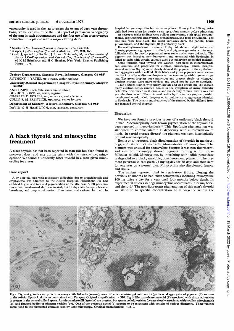
and osmium, and processed for electron microscopy. Thick, Paragon-stained sections (fig la) more clearly defined the pigment in the epitheliumand aggregates within colloid. Both black and green pigments were seen-the black usually as discrete droplets or less commonly within green drop-lets. The green droplets were numerous and present singly or clumped.Nuclear changes were more obvious and could not be due to autolysis.Thin sections stained with uranyl acetate and lead citrate (fig lb) showed
many electron-dense, rimmed bodies in the cytoplasm of many follicularcells. The rims varied in thickness, and the density of their matrix was lessgranular than colloid. These rimmed bodies lay free in the cytoplasm, withinmembrane-bound, colloid droplets or in multivesicular aggregates identicalto lipofuscin. The density and frequency of the rimmed bodies differed fromage-matched control thyroids.
Discussion
We have not found a previous report of a uniformly black thyroidin man. Macroscopically dark brown pigmentation of the thyroid hasbeen reported in mucoviscidosis.' This lipofuscin pigmentation wasattributed to chronic vitamin E deficiency with auto-oxidation oflipids. In ceroid storage disease3 the pigment was seen histologicallybut not macroscopically.
Benitz et all reported black discolouration of thyroids in monkeys,dogs, and rats but not mice after administration of minocycline. Thepigment was unusual for tetracycline because it was non-fluorescent,and electron microscopy showed pigment forming within intra-follicular colloid. Minocycline, by interfering with iodide peroxidaseis degraded to a black, insoluble, non-fluorescent pigment.' The pig-ment persisted in rats given 75 mg/kg/day for 38 days and then keptfor one year on a normal diet. Minocycline also discoloured femoraand skulls.The patient reported died in respiratory failure. During the
previous 18 months he had taken tetracyclines including minocycline100 mg twice a day for a year until four months before death. Inexperimental studies in dogs minocycline accumulates in brain, bone,and thyroid.4 The non-fluorescent pigmentation of this man's thyroidwe attribute to specific concentration of minocycline within the
_ f-'-5t; tt v...
i..
FiV .Pgetgaue r rsn nmn pteilcls(ros,sm f hc oti yntcnce p.Svrlageae fpget()aeseinte3 lod pnArliescinsand ihPrgn nia mgiiain 1.Fi .Eeto-es atra P soitdwihdsotdvsce
prsn.ntecnta.oli.pc..uoyi.irvll...rs repeet u prs oli eils c)aecoey soitdwt wolnmtcodi(madime bdesorpgmntvsile pv. n o te ynoicncli p aper t b sscitd it vscls fvaiosdimees.Ths vsiecorro,ontotepgetdgauesenblihmirsoyOrgnlmgiiainx340
on 19 March 2022 by guest. P
rotected by copyright.http://w
ww
.bmj.com
/B
r Med J: first published as 10.1136/bm
j.2.6044.1108 on 6 Novem
ber 1976. Dow
nloaded from