Embed Size (px)
Citation preview
Blood and Bone Marrow Evaluation for EosinophiliaDaniel F. Boyer, MD, PhD
� Evaluation of peripheral blood and bone marrow for anindication of persistent eosinophilia can be a challengingtask because there are many causes of eosinophilia and themorphologic differences between reactive and neoplasticcauses are often subtle or lack specificity. The purpose ofthis review is to provide an overview of the differentialdiagnosis for eosinophilia, to recommend specific steps forthe pathologist evaluating blood and bone marrow, and toemphasize 2 important causes of eosinophilia that requirespecific ancillary tests for diagnosis: myeloproliferativeneoplasm with PDGFRA rearrangement and lymphocyte-variant hypereosinophilic syndrome.
(Arch Pathol Lab Med. 2016;140:1060–1067; doi:10.5858/arpa.2016-0223-RA)
Eosinophilia is graded based on absolute eosinophil count:mild (500–1500/lL), moderate (1500–5000/lL), or severe
(.5000/lL). A patient can be described as having idiopathichypereosinophilia if there is persistent moderate or severeeosinophilia for at least 6 months without an identified cause,and the diagnosis of hypereosinophilic syndrome (HES) isapplicable when end-organ damage occurs because ofpersistent moderate or severe eosinophilia.1
The most common causes of eosinophilia are often taughtwith the pneumonic device of 3 W’s: wind, worms, andwonder drugs. Wind refers to asthma and other allergiccauses, worms refer to tissue-invasive parasites, and wonderdrugs refer to a nonspecific reaction to various medicationsor drug-induced hypersensitivity. Another common cause inthe inpatient setting is critical illness, which leads tosuppressed adrenal function, resulting in decreased steroidproduction and disinhibition of eosinophil production.2
There are several multiorgan autoimmune or idiopathicdiseases that can cause significant eosinophilia. Theautoimmune/idiopathic disorders most commonly associat-ed with eosinophilia are eosinophilic granulomatosis withpolyangiitis (EGPA, also known as Churg-Strauss syn-drome), Kimura disease, immunoglobulin (Ig)G4–relateddisease, diffuse fasciitis with eosinophilia, and eosinophilicmyositis.3 Diagnosis of these rare autoimmune disorders is
challenging and requires assessment of a variety of clinicaland laboratory findings, in addition to histopathologicevaluation of affected tissues. However, EGPA is theautoimmune condition that most frequently causes sus-tained hypereosinophilia. The main features that distinguishEGPA from other causes of HES are prominent asthmaticsymptoms, perinuclear antineutrophil cytoplasmic antibod-ies with antimyeloperoxidase specificity (present in 30%–40% of patients with EGPA), and biopsy-proven eosino-philic vasculitis.4 Single-organ eosinophilic diseases can alsocause significant peripheral blood eosinophilia. The organsmost frequently involved are the lungs, gastrointestinaltract, and skin.5–7
In addition to the nonneoplastic etiologies describedabove, eosinophilia can be caused by a variety of myeloid,lymphoid, and epithelial neoplasms. Myeloid neoplasms canresult in primary eosinophilia because of overproduction ofeosinophils from abnormal stem cells. Carcinomas andlymphoproliferative disorders can trigger cytokine produc-tion causing secondary eosinophilia. Rare congenital syn-dromes can also enter into the differential diagnosis ofeosinophilia, especially in children, and include hyper-IgEsyndrome, Omenn syndrome, and familial eosinophilia.8,9
The differential diagnosis for persistent peripheral bloodeosinophilia is summarized in Table 1.
MYELOID NEOPLASMS WITH EOSINOPHILIA
When a pathologist receives a peripheral blood or bonemarrow specimen to evaluate for eosinophilia, the mainfocus of investigation should be myeloid and lymphoidneoplasia because there is little that can be done to identifyspecific nonneoplastic causes of eosinophilia in thesespecimens. The myeloid neoplasms associated with eosin-ophilia (Table 2) include chronic myelogenous leukemia(CML); genetically defined neoplasms with rearrangementsof PDGFRA, PDGFRB, FGFR1, JAK2, and FLT3; chroniceosinophilic leukemia, not otherwise specified (CEL, NOS);and acute myeloid leukemia (AML) with core-binding factortranslocations, such as inversion 16 or t(8;21). Althoughmild eosinophilia is common with core-binding factor AML,hypereosinophilia caused by AML is rare. There are rarecases of eosinophilic myeloproliferative neoplasms associ-ated with JAK2 V617F.10–13 The morphologic findings inthese cases tend to be nonspecific, such as, hypercellularbone marrow with eosinophilia and mild fibrosis, andtesting for JAK2 V617F is necessary to make the diagnosis.The myeloid neoplasms listed so far are associated withprimary eosinophilia, meaning that the eosinophils areproduced from abnormal myeloid progenitor cells. Incontrast, Langerhans cell histiocytosis and systemic masto-cytosis (SM) are myeloid neoplasms associated with
Accepted for publication June 2, 2016.From the Department of Pathology, University of Michigan, Ann Arbor.The author has no relevant financial interest in the products or
companies described in this article.Presented in part at the New Frontiers in Pathology; October 22–
24, 2015; Ann Arbor, Michigan.Corresponding author: Daniel F. Boyer, MD, PhD, Department of
Pathology, University of Michigan, 5242 Medical Science 1, 1301Catherine St, Ann Arbor, MI 48109 (email: [email protected]).
1060 Arch Pathol Lab Med—Vol 140, October 2016 Blood and Bone Marrow Eosinophilia—Boyer
secondary eosinophilia because the eosinophils appear to bereactive, rather than arising from a neoplastic clone.
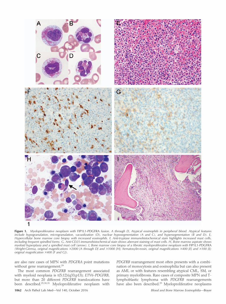
The 2008 World Health Organization classification oftumors of hematopoietic and lymphoid tissues includes thecategory of myeloid and lymphoid neoplasms withPDGFRA, PDGFRB, or FGFR1 rearrangements.14 The majordisease entities within this category are myeloproliferativeneoplasm (MPN) with PDGFRA rearrangement, MPN withPDGFRB rearrangement, and the so-called 8p11 myelopro-liferative syndrome. The MPN with PDGFRA rearrangementis a much more common cause of eosinophilia than areneoplasms with PDGFRB or FGFR1 rearrangements, andspecific testing for this entity is critically important becausePDGFRA rearrangements are usually cryptic on routinekaryotype. PDGFRA rearrangements are identified in 10% to20% of patients with HES without an identifiable reactiveetiology.15 Myeloproliferative neoplasm with PDGFRA rear-rangement shows a marked male predominance of at least30:1, based on reported case series.13,16–23 It affects a broadrange of ages with a median age of around 40 years. Initialpresentation is usually with cutaneous or pulmonarysymptoms. Splenomegaly, mucosal ulcers, and thrombo-embolic events are also common. The most seriouscomplication is cardiac dysfunction, which occurs in 20%to 30% of patients if not successfully treated. Typicallaboratory findings include markedly elevated serum vita-min B12 levels, often greater than 2000 pg/mL, and elevatedserum tryptase, approximately 30 ng/mL on average. Incontrast to reactive causes of eosinophilia, serum IgE is onlyelevated in a few patients. Eosinophils in the peripheralblood smear are often morphologically unremarkable, but,in some cases, the eosinophils show significant cytologicatypia, including hypogranulation, microgranulation, coarsebasophilic granules, vacuolization, and nuclear hyperseg-mentation or hyposegmentation (Figure 1, A through D).Bone marrow findings include hypercellularity, increasedeosinophils, increased and atypical mast cells, and reticulinfibrosis (Figure 1, E through I). The extent of reticulinfibrosis is highly variable from case to case. PDGFRArearrangement usually causes a low-grade MPN, but rarecases of AML and T-lymphoblastic lymphoma associatedwith PDGFRA rearrangement have been reported.24,25 Keyfeatures of MPN with PDGFRA rearrangement are summa-rized in Table 3.
The bone marrow and peripheral blood findings of MPNwith PDGFRA rearrangement share similarities with sys-temic mastocytosis, and accurate differentiation betweenthese diseases is important because of differences in therapyand the significant risk of cardiac disease associated withMPN with PDGFRA rearrangement.26 A comparison of the
features of MPN with PDGFRA rearrangement and systemicmastocytosis with eosinophilia (SM-EO) is presented inTable 4. Usually, MPN with PDGFRA rearrangement can bedifferentiated from SM-EO based on the presence ofPDGFRA rearrangement and the absence of urticaria, densemast cell aggregates, and KIT D816V mutation. However,microdissection experiments have shown that the atypicalmast cells in some cases of MPN with PDGFRA rearrange-ment harbor KIT D816V mutations, raising the possibilitythat the 2 diseases share more genetic similarities thanpreviously understood.27 In addition, a case has beenreported that met diagnostic criteria for both MPN withPDGFRA rearrangement and SM because of the presence ofmultiple dense mast cell aggregates in the bone marrow.28
Cases that fully meet the diagnostic criteria for both diseasesare probably best classified as SM with an associatedhematologic neoplasm, but it is still essential to documentthe presence of PDGFRA rearrangement because of theimplications for therapy (ie, PDGFRA rearrangements areassociated with excellent response to imatinib).
PDGFRA rearrangements can be detected by fluorescencein situ hybridization (FISH) or reverse transcription-polymerase chain reaction. Only a few PDGFRA rearrange-ments can be detected by routine karyotype. The mostcommon PDGFRA rearrangement is an 800-kb internaldeletion in the q12 region of chromosome 4, which causesfusion of the FIP1L1 and PDGFRA genes. Because FIP1L1and PDGFRA are relatively close together in the wild-typeconfiguration, FISH probes to evaluate FIP1L1-PDGFRAfusions usually include a probe to detect loss of interveninggenetic material, typically either the CHIC2 or LNX genes.All the PDGFRA rearrangements associated with MPNshave a breakpoint in exon 12 of PDGFRA, which disrupts aninhibitory domain.15 Regardless of the fusion partner, thePDGFRA rearrangements seem to manifest the samephenotype and show excellent response to imatinib. There
Table 1. Differential Diagnosis of Eosinophilia
Common reactions: medications, parasites, asthma/allergyAdrenal insufficiency due to critical illnessMultiorgan autoimmune/idiopathic diseases, especially
EGPA/Churg-Strauss syndromeSingle-organ eosinophilic diseases, especially lung, GI, skinMyeloid neoplasms with eosinophiliaSecondary eosinophilia due to lymphoid or epithelial
neoplasmsCongenital syndromes: hyper-IgE, Omenn syndrome,
familial eosinophilia
Abbreviations: EGPA, eosinophilic granulomatosis with polyangiitis;GI, gastrointestinal disease; IgE, immunoglobulin E.
Table 2. Myeloid Neoplasms With Eosinophilia
Primary eosinophiliaChronic myelogenous leukemiaMyeloid or lymphoid neoplasms with PDGFRA, PDGFRB,
or FGFR1 rearrangements� del(4)(q12); FIP1L1-PDGFRA� t(5;12)(q33;p13); ETV6-PDGFRB� t(8;13)(p11;q12); ZMYM2-FGFR1
Myeloid or lymphoid neoplasms with JAK2 or FLT3rearrangements� t(8;9)(p22;p24); PCM1-JAK2� t(12;13)(p13;p12); ETV6-FLT3
MPN with eosinophilia and JAK2 V617F� 1%–2% of ‘‘idiopathic’’ hypereosinophilia� Often lacks typical features of PV, ET, or PMF
Chronic eosinophilic leukemia, not otherwise specified� AEC .1500/lL, and at least 1 of the following:
* .5% blasts in BM* .2% blasts in PB* Clonal genetic abnormality
AML with inv(16)(p13.1q22) or t(8;21)(q22;q22)Secondary eosinophilia
Systemic mastocytosisLangerhans cell histiocytosis
Abbreviations: AEC, absolute eosinophil count; AML, acute myeloidleukemia; BM, bone marrow; ET, essential thrombocythemia; MPN,myeloproliferative neoplasm; PB, peripheral blood; PMF, primarymyelofibrosis; PV, polycythemia vera.
Arch Pathol Lab Med—Vol 140, October 2016 Blood and Bone Marrow Eosinophilia—Boyer 1061
are also rare cases of MPN with PDGFRA point mutationswithout gene rearrangement.29
The most common PDGFRB rearrangement associatedwith myeloid neoplasia is t(5;12)(q33;p13); ETV6-PDGFRB,but more than 20 different PDGFRB translocations havebeen described.25,30,31 Myeloproliferative neoplasm with
PDGFRB rearrangement most often presents with a combi-nation of monocytosis and eosinophilia but can also presentas AML or with features resembling atypical CML, SM, orprimary myelofibrosis. Rare cases of composite MPN and T-lymphoblastic lymphoma with PDGFRB rearrangementshave also been described.25 Myeloproliferative neoplasms
Figure 1. Myeloproliferative neoplasm with FIP1L1-PDGFRA fusion. A through D, Atypical eosinophils in peripheral blood. Atypical featuresinclude hypogranulation, microgranulation, vacuolization (D), nuclear hyposegmentation (A and C), and hypersegmentation (B and D). E,Hypercellular bone marrow core biopsy with increased eosinophils. F, Anti-tryptase immunohistochemical stain highlights increased mast cells,including frequent spindled forms. G, Anti-CD25 immunohistochemical stain shows aberrant staining of mast cells. H, Bone marrow aspirate showsmyeloid hyperplasia and a spindled mast cell (arrow). I, Bone marrow core biopsy of a fibrotic myeloproliferative neoplasm with FIP1L1-PDGFRA(Wright-Giemsa, original magnifications 32000 [A through D] and 31000 [H]; hematoxylin-eosin, original magnifications 3400 [E] and 3100 [I];original magnification 3400 [F and G]).
1062 Arch Pathol Lab Med—Vol 140, October 2016 Blood and Bone Marrow Eosinophilia—Boyer
with PDGFRB rearrangement show excellent response toimatinib.31 The 8p11 myeloproliferative syndrome is causedby FGFR1 translocations, usually t(8;13)(p11;q12); ZMYM2-FGFR1, and typically presents with a mixture of myeloid andlymphoid neoplasia.25,32 The classic presentation is the triadof nodal T-lymphoblastic lymphoma, peripheral bloodeosinophilia, and myeloid hyperplasia in the bone marrow.These patients show frequent progression to AML, but acutelymphoblastic leukemia (ALL) or mixed-lineage leukemiacan also occur. The neoplasms with FGFR1 rearrangementdo not respond to imatinib.
In addition to PDGFRA, PDGFRB, and FGFR1 generearrangements, rearrangements of the JAK2 and FLT3genes have been associated with eosinophilic myeloidneoplasms. The t(8;9)(p22;p24); PCM1-JAK2 fusion causesan MPN that can resemble atypical CML, chronic eosino-philic leukemia, or primary myelofibrosis.33–35 It shows malepredominance with a broad age range and has a high risk oftransformation to AML or Philadelphia-like B-cell ALL (BALL). Treatment and outcome data for MPN with PCM1-JAK2 fusion are limited, but some patients have respondedto treatment with JAK2 inhibitors. The t(12;13)(p13;q12);
ETV6-FLT3 fusion also causes an MPN with eosinophiliaand high-risk of AML. Only 5 cases have been reported inthe literature; of which, 3 presented with concurrent T-celllymphoma.36–39
If the genetically defined eosinophilic neoplasms havebeen ruled out, the next consideration is CEL, NOS. Thediagnostic criteria are persistent eosinophilia greater than1500/lL, no definitive features of other myeloid neoplasms,and the presence of either increased blasts (.2% in blood or.5% in bone marrow) or a nonspecific clonal geneticabnormality, such as trisomy 8 or isochromosome band17q.40 Chronic eosinophilic leukemia, NOS, shows frequenttransformation to AML and carries a poor prognosis.41
LYMPHOID NEOPLASMS WITH SECONDARYEOSINOPHILIA
In the myeloid neoplasms described above, eosinophiliaresults from overproduction of eosinophils from abnormalmyeloid progenitor cells. In contrast, lymphoid neoplasmscan cause significant eosinophilia because of cytokineproduction (Table 5). T-cell neoplasms are most commonlyassociated with eosinophilia, and significant eosinophilia ismost frequently seen in the context of cutaneous T-celllymphoma, adult T-cell leukemia/lymphoma, and angioim-munoblastic T-cell lymphoma.42 The B-cell neoplasms mostsignificantly associated with eosinophilia are classicalHodgkin lymphoma and B-lymphoblastic leukemia/lym-phoma, especially B ALL with t(5;14)(q31;q32); IGH-IL3.This uncommon translocation causes eosinophilia fromoverproduction of interleukin (IL) 3 by the neoplastic Bcells.43 The t(5;14)(q31;q32) translocation associated with BALL and eosinophilia should not be confused witht(5;14)(q35;q32); TLX3-BCL11, which is associated with T-cell ALL.
Another important lymphocytic cause of eosinophilia isproliferation of abnormal T helper 2–type T cells, which isknown as lymphocyte-variant hypereosinophilic syndrome(Table 6). The name of this disease has caused someconfusion because the term lymphocytic HES was used formany years to refer to any etiology of HES associated withincreased cytokine production by lymphocytes.44 Thecurrent usage of the term lymphocyte-variant hypereosino-philic syndrome (L-HES) refers to an eosinophilic conditioncaused by a T-cell lymphoproliferative disease characterizedby immunophenotypically aberrant and/or clonal T cells.Lymphocyte-variant HES accounts for 10% to 20% of HESwithout another definitive reactive or neoplastic cause.22,45–47
The diagnosis is often challenging because symptomsusually develop gradually and resemble an allergic reaction,and most patients do not have significant lymphadenopathyor lymphocytosis. Patients usually present with an erythem-
Table 3. Myeloproliferative Neoplasm WithPDGFRA Rearrangement
Clinical features10%–20% of patients with HESMarked male predominance (.30:1)Broad age range (average, 40 y)Usually presents with cutaneous or pulmonary symptomsSplenomegaly, mucosal ulcers, and thromboembolic
events also common20%–30% develop cardiac dysfunction if not treated.90% have elevated serum vitamin B12 at diagnosis
(often .2000 pg/mL)80% have elevated serum tryptase (median, 30 ng/mL)
Bone marrow findingsHypercellular with increased eosinophilsIncreased and atypical mast cells� Frequent spindled forms� Granulation may be decreased� Usually CD25þ but CD2�
� Dense aggregates are very rareReticulin usually increased
Abbreviation: HES, hypereosinophilic syndrome.
Table 4. Comparison of MyeloproliferativeNeoplasm (MPN) With PDGFRA Rearrangement andSystemic Mastocytosis With Eosinophilia (SM-EO)a
MPN WithPDGFRA SM-EO
Sex ratio (M:F) .30:1 1:1GI symptoms Rare CommonHeart/lung symptoms Common RareUrticaria pigmentosa, % 0 60Multifocal dense MC
aggregates, %Rare .90
.25% spindled MC, % .90 .90CD25þ MC, % .95 .95CD2þ MC Rare OccasionalKIT D816V, % Rare .90Serum AEC/tryptase,
(cells per ll/ng per mL).100 ,100
Abbreviations: AEC, absolute eosinophil count; GI, gastrointestinal;MC, mast cells.a Derived from data in Maric et al,26 2007.
Table 5. Lymphoid Neoplasms With SecondaryEosinophilia
Lymphocyte-variant hypereosinophiliaCutaneous T-cell lymphoma, especially advanced-stage MF
and SSLymphoblastic leukemia/lymphoma, especially B ALL with
t(5;14)(q31;q32)Classical Hodgkin lymphomaAdult T-cell leukemia/lymphomaAngioimmunoblastic T-cell lymphoma
Abbreviations: B ALL, B lymphoblastic leukemia; MF, mycosisfungoides; SS, Sezary syndrome.
Arch Pathol Lab Med—Vol 140, October 2016 Blood and Bone Marrow Eosinophilia—Boyer 1063
atous or papular/nodular pruritic rash. Urticaria, poikiloder-ma, and episodic angioedema can also occur. Episodicangioedema with eosinophilia is also known as Gleichsyndrome, and identification of clonal CD3�, CD4þ T cells inmost patients with Gleich syndrome suggests that it isprobably a subtype of L-HES.48 The abnormal T-cellpopulations in L-HES produce T helper 2 cytokines,including IL4, IL5, and IL13, and stimulate production ofCCL17/TARC (thymus and activation-regulated cytokine);however, testing for production of these cytokines iscurrently performed using research assays and is not readilyavailable as a clinically validated test.
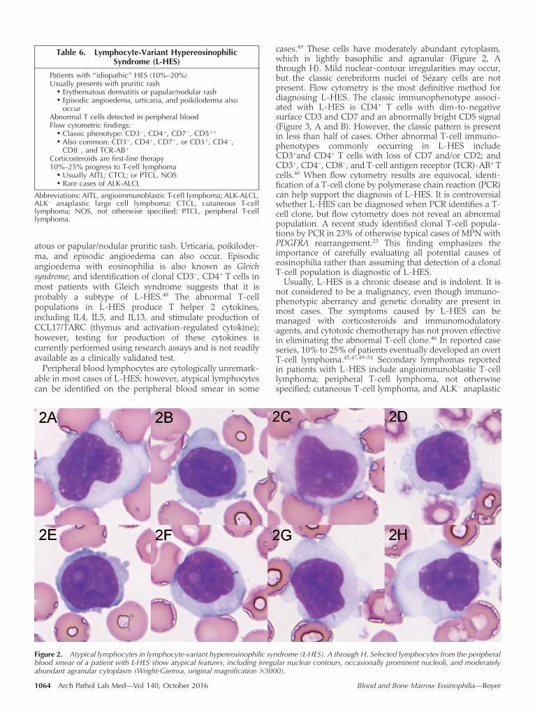
Peripheral blood lymphocytes are cytologically unremark-able in most cases of L-HES; however, atypical lymphocytescan be identified on the peripheral blood smear in some
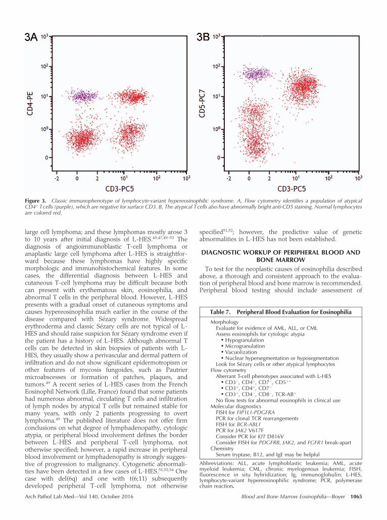
cases.49 These cells have moderately abundant cytoplasm,which is lightly basophilic and agranular (Figure 2, Athrough H). Mild nuclear-contour irregularities may occur,but the classic cerebriform nuclei of Sezary cells are notpresent. Flow cytometry is the most definitive method fordiagnosing L-HES. The classic immunophenotype associ-ated with L-HES is CD4þ T cells with dim-to-negativesurface CD3 and CD7 and an abnormally bright CD5 signal(Figure 3, A and B). However, the classic pattern is presentin less than half of cases. Other abnormal T-cell immuno-phenotypes commonly occurring in L-HES includeCD3þand CD4þ T cells with loss of CD7 and/or CD2; andCD3þ, CD4�, CD8�, and T-cell antigen receptor (TCR)-ABþ Tcells.46 When flow cytometry results are equivocal, identi-fication of a T-cell clone by polymerase chain reaction (PCR)can help support the diagnosis of L-HES. It is controversialwhether L-HES can be diagnosed when PCR identifies a T-cell clone, but flow cytometry does not reveal an abnormalpopulation. A recent study identified clonal T-cell popula-tions by PCR in 23% of otherwise typical cases of MPN withPDGFRA rearrangement.23 This finding emphasizes theimportance of carefully evaluating all potential causes ofeosinophilia rather than assuming that detection of a clonalT-cell population is diagnostic of L-HES.
Usually, L-HES is a chronic disease and is indolent. It isnot considered to be a malignancy, even though immuno-phenotypic aberrancy and genetic clonality are present inmost cases. The symptoms caused by L-HES can bemanaged with corticosteroids and immunomodulatoryagents, and cytotoxic chemotherapy has not proven effectivein eliminating the abnormal T-cell clone.46 In reported caseseries, 10% to 25% of patients eventually developed an overtT-cell lymphoma.45,47,49–51 Secondary lymphomas reportedin patients with L-HES include angioimmunoblastic T-celllymphoma; peripheral T-cell lymphoma, not otherwisespecified; cutaneous T-cell lymphoma, and ALK� anaplastic
Table 6. Lymphocyte-Variant HypereosinophilicSyndrome (L-HES)
Patients with ‘‘idiopathic’’ HES (10%–20%)Usually presents with pruritic rash� Erythematous dermatitis or papular/nodular rash� Episodic angioedema, urticaria, and poikiloderma also
occurAbnormal T cells detected in peripheral bloodFlow cytometric findings:� Classic phenotype: CD3�, CD4þ, CD7�, CD5þþ
� Also common: CD3þ, CD4þ, CD7�, or CD3þ, CD4�,CD8�, and TCR-ABþ
Corticosteroids are first-line therapy10%–25% progress to T-cell lymphoma� Usually AITL; CTCL; or PTCL, NOS� Rare cases of ALK-ALCL
Abbreviations: AITL, angioimmunoblastic T-cell lymphoma; ALK-ALCL,ALK� anaplastic large cell lymphoma; CTCL, cutaneous T-celllymphoma; NOS, not otherwise specified; PTCL, peripheral T-celllymphoma.
Figure 2. Atypical lymphocytes in lymphocyte-variant hypereosinophilic syndrome (L-HES). A through H, Selected lymphocytes from the peripheralblood smear of a patient with L-HES show atypical features, including irregular nuclear contours, occasionally prominent nucleoli, and moderatelyabundant agranular cytoplasm (Wright-Giemsa, original magnification 33000).
1064 Arch Pathol Lab Med—Vol 140, October 2016 Blood and Bone Marrow Eosinophilia—Boyer
large cell lymphoma; and these lymphomas mostly arose 3to 10 years after initial diagnosis of L-HES.45,47,49–53 Thediagnosis of angioimmunoblastic T-cell lymphoma oranaplastic large cell lymphoma after L-HES is straightfor-ward because these lymphomas have highly specificmorphologic and immunohistochemical features. In somecases, the differential diagnosis between L-HES andcutaneous T-cell lymphoma may be difficult because bothcan present with erythematous skin, eosinophilia, andabnormal T cells in the peripheral blood. However, L-HESpresents with a gradual onset of cutaneous symptoms andcauses hypereosinophilia much earlier in the course of thedisease compared with Sezary syndrome. Widespreaderythroderma and classic Sezary cells are not typical of L-HES and should raise suspicion for Sezary syndrome even ifthe patient has a history of L-HES. Although abnormal Tcells can be detected in skin biopsies of patients with L-HES, they usually show a perivascular and dermal pattern ofinfiltration and do not show significant epidermotropism orother features of mycosis fungoides, such as Pautriermicroabscesses or formation of patches, plaques, andtumors.49 A recent series of L-HES cases from the FrenchEosinophil Network (Lille, France) found that some patientshad numerous abnormal, circulating T cells and infiltrationof lymph nodes by atypical T cells but remained stable formany years, with only 2 patients progressing to overtlymphoma.49 The published literature does not offer firmconclusions on what degree of lymphadenopathy, cytologicatypia, or peripheral blood involvement defines the borderbetween L-HES and peripheral T-cell lymphoma, nototherwise specified; however, a rapid increase in peripheralblood involvement or lymphadenopathy is strongly sugges-tive of progression to malignancy. Cytogenetic abnormali-ties have been detected in a few cases of L-HES.51,52,54 Onecase with del(6q) and one with t(6;11) subsequentlydeveloped peripheral T-cell lymphoma, not otherwise
specified51,52; however, the predictive value of geneticabnormalities in L-HES has not been established.
DIAGNOSTIC WORKUP OF PERIPHERAL BLOOD ANDBONE MARROW
To test for the neoplastic causes of eosinophilia describedabove, a thorough and consistent approach to the evalua-tion of peripheral blood and bone marrow is recommended.Peripheral blood testing should include assessment of
Figure 3. Classic immunophenotype of lymphocyte-variant hypereosinophilic syndrome. A, Flow cytometry identifies a population of atypicalCD4þ Tcells (purple), which are negative for surface CD3. B, The atypical Tcells also have abnormally bright anti-CD5 staining. Normal lymphocytesare colored red.
Table 7. Peripheral Blood Evaluation for Eosinophilia
MorphologyEvaluate for evidence of AML, ALL, or CMLAssess eosinophils for cytologic atypia� Hypogranulation� Microgranulation� Vacuolization� Nuclear hypersegmentation or hyposegmentation
Look for Sezary cells or other atypical lymphocytesFlow cytometry
Aberrant T-cell phenotypes associated with L-HES� CD3�, CD4þ, CD7�, CD5þþ
� CD3þ, CD4þ, CD7�
� CD3þ, CD4�, CD8�, TCR-ABþ
No flow tests for abnormal eosinophils in clinical useMolecular diagnostics
FISH for FIP1L1-PDGFRAPCR for clonal TCR rearrangementsFISH for BCR-ABL1PCR for JAK2 V617FConsider PCR for KIT D816VConsider FISH for PDGFRB, JAK2, and FGFR1 break-apart
ChemistrySerum tryptase, B12, and IgE may be helpful
Abbreviations: ALL, acute lymphoblastic leukemia; AML, acutemyeloid leukemia; CML, chronic myelogenous leukemia; FISH,fluorescence in situ hybridization; Ig, immunoglobulin; L-HES,lymphocyte-variant hypereosinophilic syndrome; PCR, polymerasechain reaction.
Arch Pathol Lab Med—Vol 140, October 2016 Blood and Bone Marrow Eosinophilia—Boyer 1065
morphology, flow cytometry, molecular diagnostics, andserum chemistry (Table 7). The morphologic assessmentshould include looking for blasts, basophilia, and granulo-cytic left shift, which could indicate AML, ALL, or CML. Theeosinophils should be evaluated for cytologic atypia, eventhough myeloid neoplasms with primary eosinophilia oftenlack significant atypia. The peripheral blood smear shouldalso be scanned for Sezary cells or other atypical lympho-cytes because of the frequent association of secondaryeosinophilia with T-cell neoplasms. The main utility of flowcytometry for eosinophilia is detecting abnormal T-cellpopulations, which is critical for the diagnosis of L-HES.There are currently no clinically validated flow cytometricassays that differentiate reactive and neoplastic eosinophils.
The most useful molecular diagnostic tests are FISH orPCR for PDGFRA gene rearrangement (including FIP1L1-PDGFRA fusion), PCR for assessment of T-cell clonality,FISH or PCR for BCR-ABL1 fusion, and PCR for JAK2 V617F.Testing for PDGFRA gene rearrangement is especiallyimportant because it has critical diagnostic and therapeuticimplications and is usually not detected on routinekaryotype. Polymerase chain reaction for KIT D816V canbe helpful to identify SM with eosinophilia; however,consideration of patient history and clinical findings beforetesting for KIT D816V is suggested to promote efficient useof this test. Serum chemistry for levels of IgE, tryptase, andvitamin B12 can help narrow the differential diagnosis ofeosinophilia.22 Immunoglobulin E is usually elevated inpatients with secondary eosinophilia but only rarely inpatients who have myeloid neoplasms with primaryeosinophilia. Tryptase is significantly elevated in systemicmastocytosis and MPN with PDGFRA rearrangement.Vitamin B12 is usually markedly elevated in patients withMPN with PDGFRA rearrangement and is often elevated inassociation with other myeloid neoplasms.
Even if the peripheral blood findings are diagnostic of aneoplastic process, bone marrow biopsy is still recommend-ed to assess for findings that could change the diagnosis orprognosis. The key elements are blast count, aberrant mastcells, reticulin fibrosis, and karyotype (Table 8). In rare cases,bone marrow biopsy may reveal an occult metastaticmalignancy causing secondary eosinophilia. Cultured bonemarrow is usually superior to peripheral blood for cytoge-netic analysis, and the bone marrow karyotype is helpful to
identify translocations of PDGFRB, JAK2, FGFR1, and FLT3,as well as clonal abnormalities that could establish adiagnosis of CEL, NOS, in the appropriate context.
References
1. Gotlib J. World Health Organization–defined eosinophilic disorders: 2015update on diagnosis, risk stratification, and management. Am J Hematol. 2015;90(11):1077–1089.
2. Angelis M, Yu M, Takanishi D, Hasaniya NW, Brown MR. Eosinophilia as amarker of adrenal insufficiency in the surgical intensive care unit. J Am Coll Surg.1996;183(6):589–596.
3. Tamaki H, Chatterjee S, Langford CA. Eosinophilia in rheumatologic/vascular disorders. Immunol Allergy Clin North Am. 2015;35(3):453–476.
4. Greco A, Rizzo MI, De Virgilio A, et al. Churg-Strauss syndrome.Autoimmun Rev. 2015;14(4):341–348.
5. Woolnough K, Wardlaw AJ. Eosinophilia in pulmonary disorders. ImmunolAllergy Clin North Am. 2015;35(3):477–492.
6. Mehta P, Furuta GT. Eosinophils in gastrointestinal disorders: eosinophilicgastrointestinal diseases, celiac disease, inflammatory bowel diseases, andparasitic infections. Immunol Allergy Clin North Am. 2015;35(3):413–437.
7. de Graauw E, Beltraminelli H, Simon HU, Simon D. Eosinophilia indermatologic disorders. Immunol Allergy Clin North Am. 2015;35(3):545–560.
8. Valent P, Klion AD, Horny HP, et al. Contemporary consensus proposal oncriteria and classification of eosinophilic disorders and related syndromes. JAllergy Clin Immunol. 2012;130(3):607–612.
9. Curtis C, Ogbogu PU. Evaluation and differential diagnosis of persistentmarked eosinophilia. Immunol Allergy Clin North Am. 2015;35(3):387–402.
10. Jones AV, Kreil S, Zoi K, et al. Widespread occurrence of the JAK2 V617Fmutation in chronic myeloproliferative disorders. Blood. 2005;106(6):2162–2168.
11. Dahabreh IJ, Giannouli S, Zoi C, Zoi K, Loukopoulos D, Voulgarelis M.Hypereosinophilic syndrome: another face of Janus? Leuk Res. 2008;32(9):1483–1485.
12. Helbig G, Majewski M, Wieczorkiewicz A, et al. Screening for JAK2V617F point mutation in patients with hypereosinophilic syndrome-in responseto ‘‘Hypereosinophilic syndrome: another face of Janus?’’ by Dahabreh et alpublished in Leukemia Research Leuk Res. 2009;33(3):e1–e2.
13. Schwaab J, Umbach R, Metzgeroth G, et al. KIT D816V and JAK2 V617Fmutations are seen recurrently in hypereosinophilia of unknown significance. AmJ Hematol. 2015;90(9):774–777.
14. Bain BJ, Gilliland DG, Horny H-P, Vardiman JW. Myeloid and lymphoidneoplasms with eosinophilia and abnormalities of PDGFRA, PDGFRB, or FGFR1.In: Swerdlow SH, Campo E, Harris NL, et al, eds. WHO Classification of Tumoursof the Haematopoietic and Lymphoid Tissues. 4th ed. Lyon, France: IARC Press;2008:68–73. World Health Organization Classification of Tumours; vol 2.
15. Gotlib J, Cools J. Five years since the discovery of FIP1L1-PDGFRA: whatwe have learned about the fusion and other molecularly defined eosinophilias.Leukemia. 2008;22(11):1999–2010.
16. Cools J, DeAngelo DJ, Gotlib J, et al. A tyrosine kinase created by fusion ofthe PDGFRA and FIP1L1 genes as a therapeutic target of imatinib in idiopathichypereosinophilic syndrome. N Engl J Med. 2003;348(13):1201–1214.
17. Pardanani A, Brockman SR, Paternoster SF, et al. FIP1L1-PDGFRA fusion:prevalence and clinicopathologic correlates in 89 consecutive patients withmoderate to severe eosinophilia. Blood. 2004;104(10):3038–3045.
18. Vandenberghe P, Wlodarska I, Michaux L, et al. Clinical and molecularfeatures of FIP1L1-PDFGRA (þ) chronic eosinophilic leukemias. Leukemia. 2004;18(4):734–742.
19. Roche-Lestienne C, Lepers S, Soenen-Cornu V, et al. Molecular character-ization of the idiopathic hypereosinophilic syndrome (HES) in 35 French patientswith normal conventional cytogenetics. Leukemia. 2005;19(5):792–798.
20. La Starza R, Specchia G, Cuneo A, et al. The hypereosinophilic syndrome:fluorescence in situ hybridization detects the del(4)(q12)-FIP1L1/PDGFRA but notgenomic rearrangements of other tyrosine kinases. Haematologica. 2005;90(5):596–601.
21. Baccarani M, Cilloni D, Rondoni M, et al. The efficacy of imatinib mesylatein patients with FIP1L1-PDGFRa-positive hypereosinophilic syndrome: results ofa multicenter prospective study. Haematologica. 2007;92(9):1173–1179.
22. Ogbogu PU, Bochner BS, Butterfield JH, et al. Hypereosinophilicsyndrome: a multicenter, retrospective analysis of clinical characteristics andresponse to therapy. J Allergy Clin Immunol. 2009;124(6):1319–1325.e3.
23. Legrand F, Renneville A, Macintyre E, et al; for the French EosinophilNetwork. The spectrum of FIP1L1-PDGFRA-associated chronic eosinophilicleukemia: new insights based on a survey of 44 cases [published online ahead ofprint August 26, 2013]. Medicine (Baltimore). 2013;92(5):e1–e9. doi:10.1097/MD.0b013e3182a71eba
24. Metzgeroth G, Walz C, Score J, et al. Recurrent finding of the FIP1L1-PDGFRA fusion gene in eosinophilia-associated acute myeloid leukemia andlymphoblastic T-cell lymphoma. Leukemia. 2007;21(6):1183–1188.
25. Vega F, Medeiros LJ, Bueso-Ramos CE, Arboleda P, Miranda RN.Hematolymphoid neoplasms associated with rearrangements of PDGFRA,PDGFRB, and FGFR1. Am J Clin Pathol. 2015;144(3):377–392.
26. Maric I, Robyn J, Metcalfe DD, et al. KIT D816V-associated systemicmastocytosis with eosinophilia and FIP1L1/PDGFRA-associated chronic eosino-
Table 8. Bone Marrow Evaluation For Eosinophilia
Assess for increased or aberrant blasts� AML or ALL� MPN in accelerated phase� CEL, NOS
Reticulin fibrosis favors MPN over reactive eosinophiliaAssess for increased or aberrant mast cells� Morphology and IHC (CD117, tryptase, CD25)� Spindled, CD25þ mast cells are present in SM and MPN
with PDGFRA rearrangement� Dense mast cell clusters favor SM
FISH for FIP1L1-PDGFRA (if not done on blood)Karyotype� PDGFRB, JAK2, FGFR1, or FLT3 rearrangements� Other clonal abnormalities (CEL, NOS)
Assess for infiltration by an occult metastatic malignancy
Abbreviations: ALL, acute lymphoblastic leukemia; AML, acutemyeloid leukemia; CEL, chronic eosinophilic leukemia; FISH, fluores-cence in situ hybridization; IHC, immunohistochemistry; MPN,myeloproliferative neoplasm; NOS, not otherwise specified; SM,systemic mastocytosis.
1066 Arch Pathol Lab Med—Vol 140, October 2016 Blood and Bone Marrow Eosinophilia—Boyer
philic leukemia are distinct entities. J Allergy Clin Immunol. 2007;120(3):680–687.
27. Schmitt-Graeff AH, Erben P, Schwaab J, et al. The FIP1L1-PDGFRA fusiongene and the KIT D816V mutation are coexisting in a small subset of myeloid/lymphoid neoplasms with eosinophilia. Blood. 2014;123(4):595–597.
28. Florian S, Esterbauer H, Binder T, et al. Systemic mastocytosis (SM)associated with chronic eosinophilic leukemia (SM-CEL): detection of FIP1L1/PDGFRalpha, classification by WHO criteria, and response to therapy withimatinib. Leuk Res. 2006;30(9):1201–1205.
29. Elling C, Erben P, Walz C, et al. Novel imatinib-sensitive PDGFRA-activating point mutations in hypereosinophilic syndrome induce growth factorindependence and leukemia-like disease. Blood. 2011;117(10):2935–2943.
30. Arefi M, Garcia JL, Penarrubia MJ, et al. Incidence and clinicalcharacteristics of myeloproliferative neoplasms displaying a PDGFRB rearrange-ment. Eur J Haematol. 2012;89(1):37–41.
31. Cheah CY, Burbury K, Apperley JF, et al. Patients with myeloidmalignancies bearing PDGFRB fusion genes achieve durable long-termremissions with imatinib. Blood. 2014;123(23):3574–3577.
32. Jackson CC, Medeiros LJ, Miranda RN. 8p11 myeloproliferative syndrome:a review. Hum Pathol. 2010;41(4):461–476.
33. Reiter A, Walz C, Watmore A, et al. The t(8;9)(p22;p24) is a recurrentabnormality in chronic and acute leukemia that fuses PCM1 to JAK2. Cancer Res.2005;65(7):2662–2667.
34. Patterer V, Schnittger S, Kern W, Haferlach T, Haferlach C. Hematologicmalignancies with PCM1-JAK2 gene fusion share characteristics with myeloidand lymphoid neoplasms with eosinophilia and abnormalities of PDGFRA,PDGFRB, and FGFR1. Ann Hematol. 2013;92(6):759–769.
35. Bain BJ, Ahmad S. Should myeloid and lymphoid neoplasms with PCM1-JAK2 and other rearrangements of JAK2 be recognized as specific entities? Br JHaematol. 2014;166(6):809–817.
36. Vu HA, Xinh PT, Masuda M, et al. FLT3 is fused to ETV6 in amyeloproliferative disorder with hypereosinophilia and a t(12;13)(p13;q12)translocation. Leukemia. 2006;20(8):1414–1421.
37. Walz C, Erben P, Ritter M, et al. Response of ETV6-FLT3-positive myeloid/lymphoid neoplasm with eosinophilia to inhibitors of FMS-like tyrosine kinase 3.Blood. 2011;118(8):2239–2242.
38. Chonabayashi K, Hishizawa M, Matsui M, et al. Successful allogeneic stemcell transplantation with long-term remission of ETV6/FLT3-positive myeloid/lymphoid neoplasm with eosinophilia. Ann Hematol. 2014;93(3):535–537.
39. Falchi L, Mehrotra M, Newberry KJ, et al. ETV6-FLT3 fusion gene-positive,eosinophilia-associated myeloproliferative neoplasm successfully treated withsorafenib and allogeneic stem cell transplant. Leukemia. 2014;28(10):2090–2092.
40. Bain BJ, Gilliland DG, Horny H-P, Vardiman JW. Chronic eosinophilicleukaemia, not otherwise specified. In: Swerdlow SH, Campo E, Harris NL, et al,eds. WHO Classification of Tumours of the Haematopoietic and Lymphoid
Tissues. 4th ed. Lyon, France: IARC Press; 2008:51–53. World HealthOrganization Classification of Tumours; vol 2.
41. Helbig G, Soja A, Bartkowska-Chrobok A, Kyrcz-Krzemien S. Chroniceosinophilic leukemia-not otherwise specified has a poor prognosis withunresponsiveness to conventional treatment and high risk of acute transforma-tion. Am J Hematol. 2012;87(6):643–645.
42. Roufosse F, Garaud S, de Leval L. Lymphoproliferative disorders associatedwith hypereosinophilia. Semin Hematol. 2012;49(2):138–148.
43. Meeker TC, Hardy D, Willman C, Hogan T, Abrams J. Activation of theinterleukin-3 gene by chromosome translocation in acute lymphocytic leukemiawith eosinophilia. Blood. 1990;76(2):285–289.
44. Simon HU, Rothenberg ME, Bochner BS, et al. Refining the definition ofhypereosinophilic syndrome. J Allergy Clin Immunol. 2010;126(1):45–49.
45. Simon HU, Plotz SG, Dummer R, Blaser K. Abnormal clones of T cellsproducing interleukin-5 in idiopathic eosinophilia. N Engl J Med. 1999;341(15):1112–1120.
46. Roufosse F, Cogan E, Goldman M. Lymphocytic variant hypereosinophilicsyndromes. Immunol Allergy Clin North Am. 2007;27(3):389–413.
47. Vaklavas C, Tefferi A, Butterfield J, et al. ‘Idiopathic’ eosinophilia with anoccult T-cell clone: prevalence and clinical course. Leuk Res. 2007;31(5):691–694.
48. Khoury P, Herold J, Alpaugh A, et al. Episodic angioedema witheosinophilia (Gleich syndrome) is a multilineage cell cycling disorder.Haematologica. 2015;100(3):300–307.
49. Lefevre G, Copin MC, Roumier C, et al; for the French Eosinophil Network.CD3�CD4þ lymphoid variant of hypereosinophilic syndrome: nodal andextranodal histopathological and immunophenotypic features of a peripheralindolent clonal T-cell lymphoproliferative disorder. Haematologica. 2015;100(8):1086–1095.
50. Roufosse F, Schandene L, Sibille C, et al. Clonal Th2 lymphocytes inpatients with the idiopathic hypereosinophilic syndrome. Br J Haematol. 2000;109(3):540–548.
51. Helbig G, Wieczorkiewicz A, Dziaczkowska-Suszek J, Majewski M, Kyrcz-Krzemien S. T-cell abnormalities are present at high frequencies in patients withhypereosinophilic syndrome. Haematologica. 2009;94(9):1236–1241.
52. Ravoet M, Sibille C, Roufosse F, et al. 6q� is an early and persistentchromosomal aberration in CD3�CD4þ T-cell clones associated with thelymphocytic variant of hypereosinophilic syndrome. Haematologica. 2005;90(6):753–765.
53. Roufosse F, de Leval L, van Krieken H, van Deuren M. Lymphocytic varianthypereosinophilic syndrome progressing to angioimmunoblastic T-cell lympho-ma. Leuk Lymphoma. 2015;56(6):1891–1894.
54. Roumier AS, Grardel N, Laı JL, et al. Hypereosinophilia with abnormal Tcells, trisomy 7 and elevated TARC serum level. Haematologica. 2003;88(7):e104–e107.
Arch Pathol Lab Med—Vol 140, October 2016 Blood and Bone Marrow Eosinophilia—Boyer 1067