Embed Size (px)
Citation preview
nature medicine VOLUME 18 | NUMBER 6 | JUNE 2012 883
a r t i c l e s
Exosomes are small membrane vesicles (30–100 nm) derived from the luminal membranes of multivesicular bodies and constitutively released by fusion with the cell membrane1–5. In addition to diffusible factors, such as cytokines, growth factors and extracellular matrix molecules, exosomes mediate local and systemic cell communica-tion through the horizontal transfer of information, such as mRNAs, microRNAs and proteins2,5–15.
It is well recognized that bone marrow–derived cells (BMDCs) are crucial for the generation of a suitable microenvironment for the pri-mary tumor and the development of metastasis16 through a process called pre-metastatic niche formation17,18. Although secreted factors are known contributors to BMDC recruitment to both the primary tumor and to pre-metastatic niches18–21, the role of exosomes in this process has not been evaluated. Exosomes may have a role in the
crosstalk between the primary tumor and BMDCs, leading to the homing of both cell types to sites of metastasis7,22,23.
Here we identify tumor-derived exosomes as new factors that promote metastatic niche formation by educating BMDCs toward a pro-vasculogenic and pro-metastatic phenotype through upregu-lation of the MET oncoprotein24–29. Our data show that a specific expression pattern of Ras-related (Rab) proteins30,31 is associated with exosome production in melanoma. Moreover, using circulating exosomes isolated from subjects with stage 4 melanoma, we define a melanoma-specific exosome signature comprised of tyrosinase-related protein-2 (TYRP2), very late antigen 4 (VLA-4), heat shock protein 70 (HSP70), an HSP90 isoform and the MET oncoprotein. Notably, exosomal TYRP2 predicts disease progression in subjects with stage 3 melanoma. We also show that CD45−C-KITlow/+TIE2+
1Children’s Cancer and Blood Foundation Laboratories, Departments of Pediatrics, Cell and Developmental Biology, Weill Cornell Medical College, New York, New York, USA. 2Department of Molecular Biology, Princeton University, Princeton, New Jersey, USA. 3Department of Surgery, College of Physicians and Surgeons, Columbia University, New York, New York, USA. 4International Center for Research and Education, A.C. Camargo Hospital, São Paulo, Brazil. 5Departamento de Bioquímica, Universidad Autónoma de Madrid (UAM), Instituto de Investigaciones Biomédicas ‘Alberto Sols’, Consejo Superior de Investigaciones Científicas (CSIC)-UAM, IdiPAZ (Instituto de Investigación Sanitaria La Paz) & Fundación MD Anderson Cancer Center, Madrid, Spain. 6Life Sciences Division, Lawrence Berkeley National Laboratory, Berkeley, California, USA. 7Department of Neurosurgery, Weill Cornell Medical College, New York, New York, USA. 8Department of Medicine, Memorial Sloan-Kettering Cancer Center, New York, New York, USA. 9Department of Immunology, Ludwig Center for Cancer Immunotherapy, Sloan-Kettering Institute, New York, New York, USA. 10Exosome Diagnostics Inc., New York, New York, USA. 11Pediatric Oncology Branch, National Cancer Institute, US National Institutes of Health, Bethesda, Maryland, USA. 12Department of Surgery, Memorial Sloan-Kettering Cancer Center, New York, New York, USA. 13Weill Cornell Medical College, New York, New York, USA. 14Genomic Instability and Tumor Progression Program, Cancer Institute of New Jersey, New Brunswick, New Jersey, USA. 15Champalimaud Metastasis Programme, Lisbon, Portugal. 16Department of Pediatrics, Memorial Sloan-Kettering Cancer Center, New York, New York, USA. Correspondence should be addressed to J.B. ([email protected]) or D.L. ([email protected]).
Received 10 February; accepted 26 March; published online 27 May 2012; doi:10.1038/nm.2753
Melanoma exosomes educate bone marrow progenitor cells toward a pro-metastatic phenotype through METHéctor Peinado1, Maša Alecković2, Simon Lavotshkin3, Irina Matei1, Bruno Costa-Silva1,4, Gema Moreno-Bueno5, Marta Hergueta-Redondo5, Caitlin Williams1, Guillermo García-Santos1, Cyrus M Ghajar6, Ayuko Nitadori-Hoshino1, Caitlin Hoffman7, Karen Badal1, Benjamin A Garcia2, Margaret K Callahan8, Jianda Yuan9, Vilma R Martins4, Johan Skog10, Rosandra N Kaplan11, Mary S Brady12, Jedd D Wolchok8,9,13, Paul B Chapman8,13, Yibin Kang2,14,15, Jacqueline Bromberg8,13 & David Lyden1,15,16
Tumor-derived exosomes are emerging mediators of tumorigenesis. We explored the function of melanoma-derived exosomes in the formation of primary tumors and metastases in mice and human subjects. Exosomes from highly metastatic melanomas increased the metastatic behavior of primary tumors by permanently ‘educating’ bone marrow progenitors through the receptor tyrosine kinase MET. Melanoma-derived exosomes also induced vascular leakiness at pre-metastatic sites and reprogrammed bone marrow progenitors toward a pro-vasculogenic phenotype that was positive for c-Kit, the receptor tyrosine kinase Tie2 and Met. Reducing Met expression in exosomes diminished the pro-metastatic behavior of bone marrow cells. Notably, MET expression was elevated in circulating CD45−C-KITlow/+TIE2+ bone marrow progenitors from individuals with metastatic melanoma. RAB1A, RAB5B, RAB7 and RAB27A, regulators of membrane trafficking and exosome formation, were highly expressed in melanoma cells. Rab27A RNA interference decreased exosome production, preventing bone marrow education and reducing, tumor growth and metastasis. In addition, we identified an exosome-specific melanoma signature with prognostic and therapeutic potential comprised of TYRP2, VLA-4, HSP70, an HSP90 isoform and the MET oncoprotein. Our data show that exosome production, transfer and education of bone marrow cells supports tumor growth and metastasis, has prognostic value and offers promise for new therapeutic directions in the metastatic process.
npg
© 2
012
Nat
ure
Am
eric
a, In
c. A
ll rig
hts
rese
rved
.
a r t i c l e s
884 VOLUME 18 | NUMBER 6 | JUNE 2012 nature medicine
circulating bone marrow progenitors from individuals with metastatic melanoma highly express MET receptor. Our study shows that the MET oncoprotein is upregulated in bone marrow progenitor cells by tumor-derived exosomes, possibly by both direct and indirect mech-anisms, promoting the education, mobilization and pro-metastatic behavior of BMDCs.
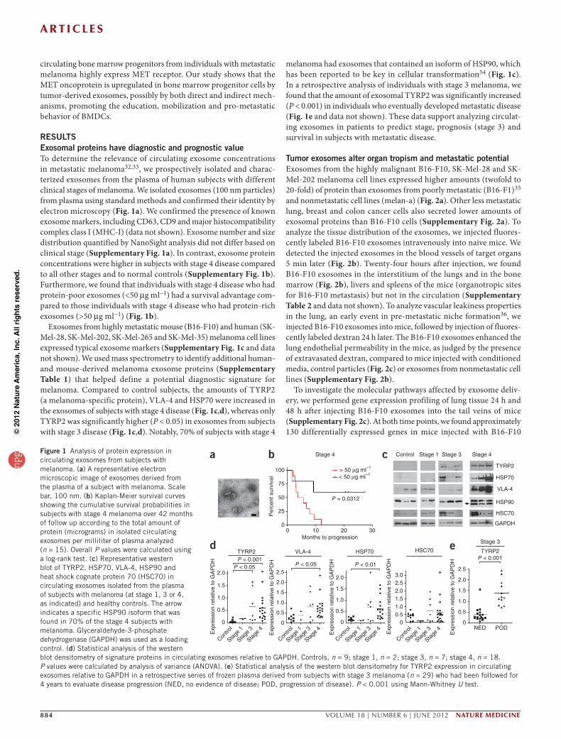
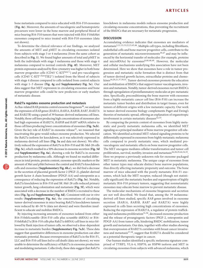
RESULTSExosomal proteins have diagnostic and prognostic valueTo determine the relevance of circulating exosome concentrations in metastatic melanoma32,33, we prospectively isolated and charac-terized exosomes from the plasma of human subjects with different clinical stages of melanoma. We isolated exosomes (100 nm particles) from plasma using standard methods and confirmed their identity by electron microscopy (Fig. 1a). We confirmed the presence of known exosome markers, including CD63, CD9 and major histocompatibility complex class I (MHC-I) (data not shown). Exosome number and size distribution quantified by NanoSight analysis did not differ based on clinical stage (Supplementary Fig. 1a). In contrast, exosome protein concentrations were higher in subjects with stage 4 disease compared to all other stages and to normal controls (Supplementary Fig. 1b). Furthermore, we found that individuals with stage 4 disease who had protein-poor exosomes (<50 µg ml−1) had a survival advantage com-pared to those individuals with stage 4 disease who had protein-rich exosomes (>50 µg ml−1) (Fig. 1b).
Exosomes from highly metastatic mouse (B16-F10) and human (SK-Mel-28, SK-Mel-202, SK-Mel-265 and SK-Mel-35) melanoma cell lines expressed typical exosome markers (Supplementary Fig. 1c and data not shown). We used mass spectrometry to identify additional human- and mouse-derived melanoma exosome proteins (Supplementary Table 1) that helped define a potential diagnostic signature for melanoma. Compared to control subjects, the amounts of TYRP2 (a melanoma-specific protein), VLA-4 and HSP70 were increased in the exosomes of subjects with stage 4 disease (Fig. 1c,d), whereas only TYRP2 was significantly higher (P < 0.05) in exosomes from subjects with stage 3 disease (Fig. 1c,d). Notably, 70% of subjects with stage 4
melanoma had exosomes that contained an isoform of HSP90, which has been reported to be key in cellular transformation34 (Fig. 1c). In a retrospective analysis of individuals with stage 3 melanoma, we found that the amount of exosomal TYRP2 was significantly increased (P < 0.001) in individuals who eventually developed metastatic disease (Fig. 1e and data not shown). These data support analyzing circulat-ing exosomes in patients to predict stage, prognosis (stage 3) and survival in subjects with metastatic disease.
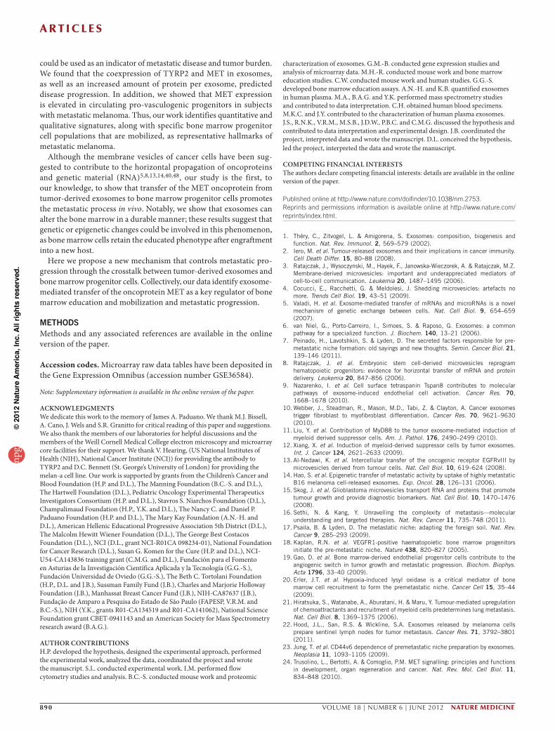
Tumor exosomes alter organ tropism and metastatic potentialExosomes from the highly malignant B16-F10, SK-Mel-28 and SK-Mel-202 melanoma cell lines expressed higher amounts (twofold to 20-fold) of protein than exosomes from poorly metastatic (B16-F1)35 and nonmetastatic cell lines (melan-a) (Fig. 2a). Other less metastatic lung, breast and colon cancer cells also secreted lower amounts of exosomal proteins than B16-F10 cells (Supplementary Fig. 2a). To analyze the tissue distribution of the exosomes, we injected fluores-cently labeled B16-F10 exosomes intravenously into naive mice. We detected the injected exosomes in the blood vessels of target organs 5 min later (Fig. 2b). Twenty-four hours after injection, we found B16-F10 exosomes in the interstitium of the lungs and in the bone marrow (Fig. 2b), livers and spleens of the mice (organotropic sites for B16-F10 metastasis) but not in the circulation (Supplementary Table 2 and data not shown). To analyze vascular leakiness properties in the lung, an early event in pre-metastatic niche formation36, we injected B16-F10 exosomes into mice, followed by injection of fluores-cently labeled dextran 24 h later. The B16-F10 exosomes enhanced the lung endothelial permeability in the mice, as judged by the presence of extravasated dextran, compared to mice injected with conditioned media, control particles (Fig. 2c) or exosomes from nonmetastatic cell lines (Supplementary Fig. 2b).
To investigate the molecular pathways affected by exosome deliv-ery, we performed gene expression profiling of lung tissue 24 h and 48 h after injecting B16-F10 exosomes into the tail veins of mice (Supplementary Fig. 2c). At both time points, we found approximately 130 differentially expressed genes in mice injected with B16-F10
a
d e
b c100
Stage 4 Control Stage 1 Stage 3
TYRP2
HSP70
VLA-4
HSP90
HSC70
GAPDH
TYRP2
2.0 2.5
Exp
ress
ion
rela
tive
to G
AP
DH
Exp
ress
ion
rela
tive
to G
AP
DH
Exp
ress
ion
rela
tive
to G
AP
DH
Exp
ress
ion
rela
tive
to G
AP
DH
Exp
ress
ion
rela
tive
to G
AP
DHP < 0.001
P < 0.05 P < 0.01P < 0.001
P < 0.05
HSP70VLA-4 HSC70
Stage 3
TYRP2
Stage 4
Contro
l
Stage
1
Stage
3
Stage
4
Contro
l
Stage
1
Stage
3
Stage
4
Contro
l
Stage
1
Stage
3
Stage
4
Contro
l
Stage
1
Stage
3
Stage
4
P = 0.0312
0
> 50 µg ml–1
< 50 µg ml–1
Per
cent
sur
viva
l
75
50
25
010 20
Months to progression30
1.5
1.0
0.5
0
2.0 3.0
1.5
1.0
0.5
0
2.0
1.5
1.0
0.5
0
2.5
2.0
1.5
1.0
0.5
0NED POD
2.52.01.51.00.5
0
Figure 1 Analysis of protein expression in circulating exosomes from subjects with melanoma. (a) A representative electron microscopic image of exosomes derived from the plasma of a subject with melanoma. Scale bar, 100 nm. (b) Kaplan-Meier survival curves showing the cumulative survival probabilities in subjects with stage 4 melanoma over 42 months of follow up according to the total amount of protein (micrograms) in isolated circulating exosomes per milliliter of plasma analyzed (n = 15). Overall P values were calculated using a log-rank test. (c) Representative western blot of TYRP2, HSP70, VLA-4, HSP90 and heat shock cognate protein 70 (HSC70) in circulating exosomes isolated from the plasma of subjects with melanoma (at stage 1, 3 or 4, as indicated) and healthy controls. The arrow indicates a specific HSP90 isoform that was found in 70% of the stage 4 subjects with melanoma. Glyceraldehyde-3-phosphate dehydrogenase (GAPDH) was used as a loading control. (d) Statistical analysis of the western blot densitometry of signature proteins in circulating exosomes relative to GAPDH. Controls, n = 9; stage 1, n = 2; stage 3, n = 7; stage 4, n = 18. P values were calculated by analysis of variance (ANOVA). (e) Statistical analysis of the western blot densitometry for TYRP2 expression in circulating exosomes relative to GAPDH in a retrospective series of frozen plasma derived from subjects with stage 3 melanoma (n = 29) who had been followed for 4 years to evaluate disease progression (NED, no evidence of disease; POD, progression of disease). P < 0.001 using Mann-Whitney U test.
npg
© 2
012
Nat
ure
Am
eric
a, In
c. A
ll rig
hts
rese
rved
.
a r t i c l e s
nature medicine VOLUME 18 | NUMBER 6 | JUNE 2012 885
exosomes as compared to control-particle–injected mice. Many of these genes were related to extracellular matrix remodeling and inflammation, such as the genes encoding heat-shock proteins, as well as S100A8 and S100A9 (Supplementary Table 3), which are known effectors of pre-metastatic niche formation37; we confirmed these findings by quantitative real time PCR (Supplementary Fig. 2d). TNFA was upregulated 24 h after exosome injection (Supplementary Fig. 2d), suggesting that it could be involved in early events during the metastatic process (for example, vascular permeability)38.
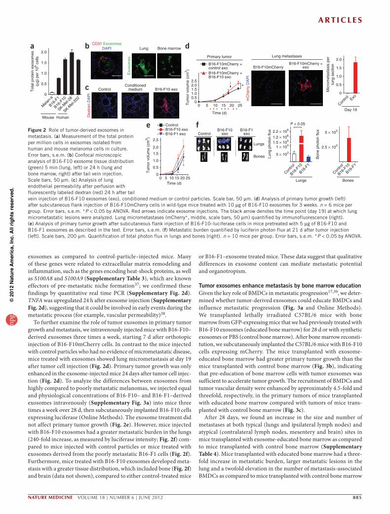
To further examine the role of tumor exosomes in primary tumor growth and metastasis, we intravenously injected mice with B16-F10–derived exosomes three times a week, starting 7 d after orthotopic injection of B16-F10mCherry cells. In contrast to the mice injected with control particles who had no evidence of micrometastatic disease, mice treated with exosomes showed lung micrometastasis at day 19 after tumor cell injection (Fig. 2d). Primary tumor growth was only enhanced in the exosome-injected mice 24 days after tumor cell injec-tion (Fig. 2d). To analyze the differences between exosomes from highly compared to poorly metastatic melanomas, we injected equal and physiological concentrations of B16-F10– and B16-F1–derived exosomes intravenously (Supplementary Fig. 3a) into mice three times a week over 28 d, then subcutaneously implanted B16-F10 cells expressing luciferase (Online Methods). The exosome treatment did not affect primary tumor growth (Fig. 2e). However, mice injected with B16-F10 exosomes had a greater metastatic burden in the lungs (240-fold increase, as measured by luciferase intensity; Fig. 2f) com-pared to mice injected with control particles or mice treated with exosomes derived from the poorly metastatic B16-F1 cells (Fig. 2f). Furthermore, mice treated with B16-F10 exosomes developed meta-stasis with a greater tissue distribution, which included bone (Fig. 2f) and brain (data not shown), compared to either control-treated mice
or B16-F1–exosome treated mice. These data suggest that qualitative differences in exosome content can mediate metastatic potential and organotropism.
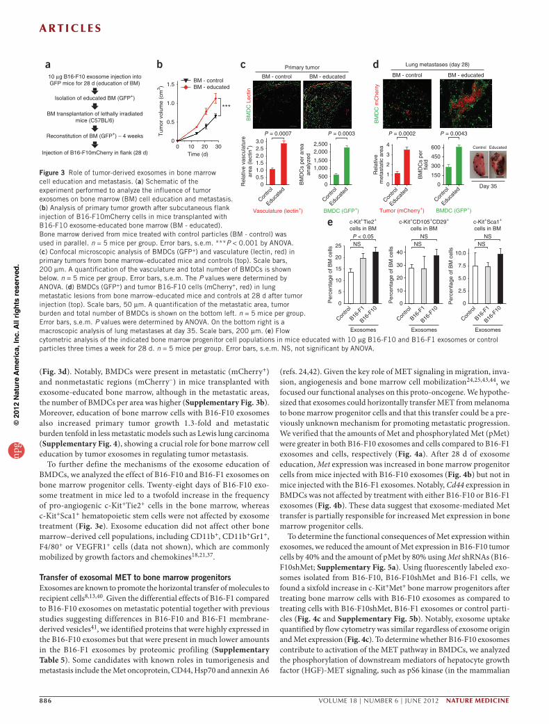
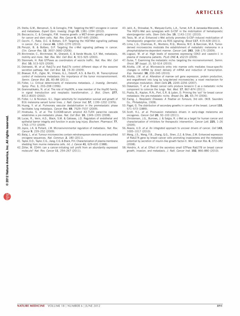
Tumor exosomes enhance metastasis by bone marrow education Given the key role of BMDCs in metastatic progression17,39, we deter-mined whether tumor-derived exosomes could educate BMDCs and influence metastatic progression (Fig. 3a and Online Methods). We transplanted lethally irradiated C57BL/6 mice with bone marrow from GFP-expressing mice that we had previously treated with B16-F10 exosomes (educated bone marrow) for 28 d or with synthetic exosomes or PBS (control bone marrow). After bone marrow reconsti-tution, we subcutaneously implanted the C57BL/6 mice with B16-F10 cells expressing mCherry. The mice transplanted with exosome- educated bone marrow had greater primary tumor growth than the mice transplanted with control bone marrow (Fig. 3b), indicating that pre-education of bone marrow cells with tumor exosomes was sufficient to accelerate tumor growth. The recruitment of BMDCs and tumor vascular density were enhanced by approximately 4.5-fold and threefold, respectively, in the primary tumors of mice transplanted with educated bone marrow compared with tumors of mice trans-planted with control bone marrow (Fig. 3c).
After 28 days, we found an increase in the size and number of metastases at both typical (lungs and ipsilateral lymph nodes) and atypical (contralateral lymph nodes, mesentery and brain) sites in mice transplanted with exosome-educated bone marrow as compared to mice transplanted with control bone marrow (Supplementary Table 4). Mice transplanted with educated bone marrow had a three-fold increase in metastatic burden, larger metastatic lesions in the lung and a twofold elevation in the number of metastasis-associated BMDCs as compared to mice transplanted with control bone marrow
2.0a b
c
dCD31 ExosomesDAPI
Dex
tran
DA
PI
mC
herr
y D
AP
IExo
som
es
Lung
ControlConditioned
medium
Lung metastases
B16-F10mCherryB16-F10mCherry +
exo
B16-F10 exo
Bone marrow
1.5
1.0
Tot
al p
rote
in e
xoso
mes
(µg)
per
106 c
ells
Tum
or v
olum
e (c
m3 )
Mic
rom
etas
tasi
s pe
rlu
ng s
ectio
n
0.52.5
2.0
1.5
1.0
0.5
0
2.01.51.00.5
00 5 10 15
Time (d)
20 25 Contro
lExo
Day 19
0
Mouse Human
Primary tumor
B16-F10mCherry +control exoB16-F10mCherry +B16-F10 exo
*
Mela
n-a
B16-F
1
B16-F
10
SK-Mel-
28
SK-Mel-
202
e fControl
Tum
or v
olum
e (c
m3 )
2.5
2.0
1.5
1.0
0.5
0
ControlB16-F10 exo B16-F10
exoB16-F1 exoB16-F1
exo
P < 0.05
Lungs
Lung
pho
ton
flux
Bon
e ph
oton
flux2.2 × 106
1.2 × 10⁶
1.5 × 10⁵
1 × 10⁴ 2.5 × 10³
5 × 10³
5 × 10³
Bones
Contro
l
Contro
l
B16-F
10
B16-F
1
B16-F
10
B16-F
1
Lungs Bones0 5 10 15
Time (d)20 25
Figure 2 Role of tumor-derived exosomes in metastasis. (a) Measurement of the total protein per million cells in exosomes isolated from human and mouse melanoma cells in culture. Error bars, s.e.m. (b) Confocal microscopic analysis of B16-F10 exosome tissue distribution (green) 5 min (lung, left) or 24 h (lung and bone marrow, right) after tail vein injection. Scale bars, 50 µm. (c) Analysis of lung endothelial permeability after perfusion with fluorescently labeled dextran (red) 24 h after tail vein injection of B16-F10 exosomes (exo), conditioned medium or control particles. Scale bar, 50 µm. (d) Analysis of primary tumor growth (left) after subcutaneous flank injection of B16-F10mCherry cells in wild-type mice treated with 10 µg of B16-F10 exosomes for 3 weeks. n = 6 mice per group. Error bars, s.e.m. *P < 0.05 by ANOVA. Red arrows indicate exosome injections. The black arrow denotes the time point (day 19) at which lung micrometastatic lesions were analyzed. Lung micrometastases (mCherry+, middle, scale bars, 50 µm) quantified by immunofluorescence (right). (e) Analysis of primary tumor growth after subcutaneous flank injection of B16-F10–luciferase cells in mice pretreated with 5 µg of B16-F10 and B16-F1 exosomes as described in the text. Error bars, s.e.m. (f) Metastatic burden quantified by luciferin photon flux at 21 d after tumor injection (left). Scale bars, 200 µm. Quantification of total photon flux in lungs and bones (right). n = 10 mice per group. Error bars, s.e.m. *P < 0.05 by ANOVA.
npg
© 2
012
Nat
ure
Am
eric
a, In
c. A
ll rig
hts
rese
rved
.
a r t i c l e s
886 VOLUME 18 | NUMBER 6 | JUNE 2012 nature medicine
(Fig. 3d). Notably, BMDCs were present in metastatic (mCherry+) and nonmetastatic regions (mCherry−) in mice transplanted with exosome-educated bone marrow, although in the metastatic areas, the number of BMDCs per area was higher (Supplementary Fig. 3b). Moreover, education of bone marrow cells with B16-F10 exosomes also increased primary tumor growth 1.3-fold and metastatic burden tenfold in less metastatic models such as Lewis lung carcinoma (Supplementary Fig. 4), showing a crucial role for bone marrow cell education by tumor exosomes in regulating tumor metastasis.
To further define the mechanisms of the exosome education of BMDCs, we analyzed the effect of B16-F10 and B16-F1 exosomes on bone marrow progenitor cells. Twenty-eight days of B16-F10 exo-some treatment in mice led to a twofold increase in the frequency of pro-angiogenic c-Kit+Tie2+ cells in the bone marrow, whereas c-Kit+Sca1+ hematopoietic stem cells were not affected by exosome treatment (Fig. 3e). Exosome education did not affect other bone marrow–derived cell populations, including CD11b+, CD11b+Gr1+, F4/80+ or VEGFR1+ cells (data not shown), which are commonly mobilized by growth factors and chemokines18,21,37.
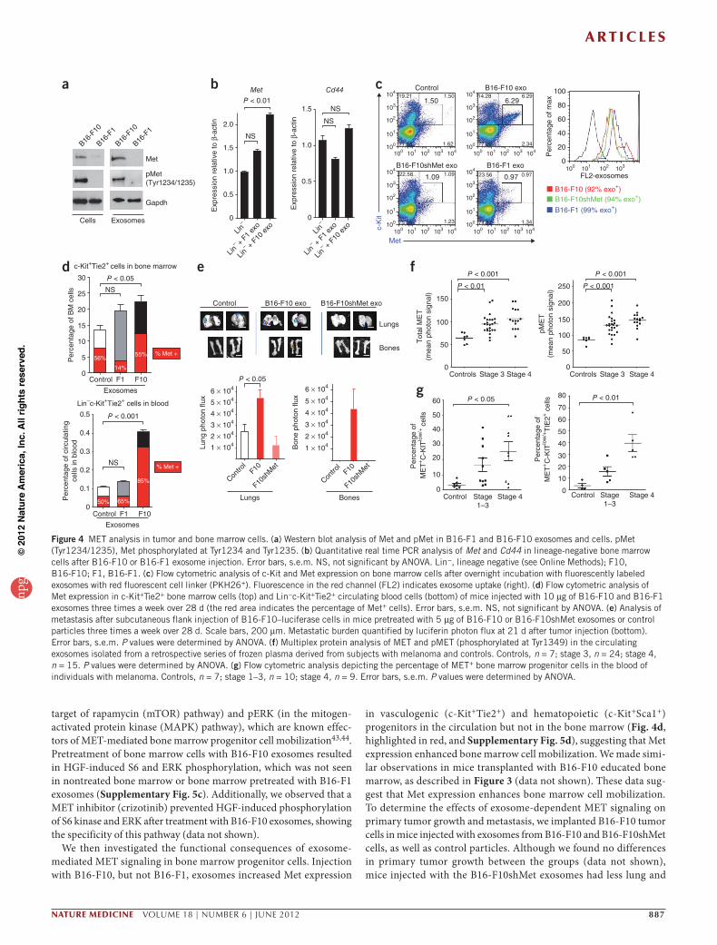
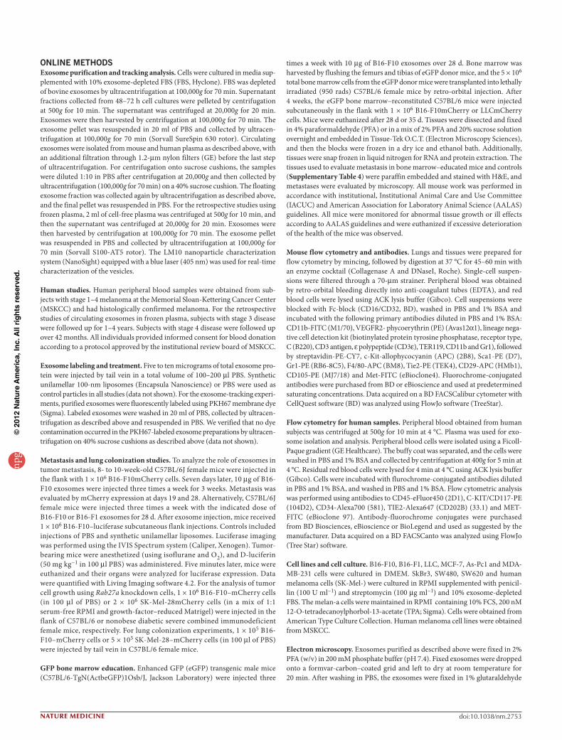
Transfer of exosomal MET to bone marrow progenitorsExosomes are known to promote the horizontal transfer of molecules to recipient cells8,13,40. Given the differential effects of B16-F1 compared to B16-F10 exosomes on metastatic potential together with previous studies suggesting differences in B16-F10 and B16-F1 membrane-derived vesicles41, we identified proteins that were highly expressed in the B16-F10 exosomes but that were present in much lower amounts in the B16-F1 exosomes by proteomic profiling (Supplementary Table 5). Some candidates with known roles in tumorigenesis and metastasis include the Met oncoprotein, CD44, Hsp70 and annexin A6
(refs. 24,42). Given the key role of MET signaling in migration, inva-sion, angiogenesis and bone marrow cell mobilization24,25,43,44, we focused our functional analyses on this proto-oncogene. We hypothe-sized that exosomes could horizontally transfer MET from melanoma to bone marrow progenitor cells and that this transfer could be a pre-viously unknown mechanism for promoting metastatic progression. We verified that the amounts of Met and phosphorylated Met (pMet) were greater in both B16-F10 exosomes and cells compared to B16-F1 exosomes and cells, respectively (Fig. 4a). After 28 d of exosome education, Met expression was increased in bone marrow progenitor cells from mice injected with B16-F10 exosomes (Fig. 4b) but not in mice injected with the B16-F1 exosomes. Notably, Cd44 expression in BMDCs was not affected by treatment with either B16-F10 or B16-F1 exosomes (Fig. 4b). These data suggest that exosome-mediated Met transfer is partially responsible for increased Met expression in bone marrow progenitor cells.
To determine the functional consequences of Met expression within exosomes, we reduced the amount of Met expression in B16-F10 tumor cells by 40% and the amount of pMet by 80% using Met shRNAs (B16-F10shMet; Supplementary Fig. 5a). Using fluorescently labeled exo-somes isolated from B16-F10, B16-F10shMet and B16-F1 cells, we found a sixfold increase in c-Kit+Met+ bone marrow progenitors after treating bone marrow cells with B16-F10 exosomes as compared to treating cells with B16-F10shMet, B16-F1 exosomes or control parti-cles (Fig. 4c and Supplementary Fig. 5b). Notably, exosome uptake quantified by flow cytometry was similar regardless of exosome origin and Met expression (Fig. 4c). To determine whether B16-F10 exosomes contribute to activation of the MET pathway in BMDCs, we analyzed the phosphorylation of downstream mediators of hepatocyte growth factor (HGF)-MET signaling, such as pS6 kinase (in the mammalian
10 µg B16-F10 exosome injection intoGFP mice for 28 d (education of BM)
a b c d
BM transplantation of lethally irradiatedmice (C57BL/6)
Reconstitution of BM (GFP+) – 4 weeks
Injection of B16-F10mCherry in flank (28 d)
Isolation of educated BM (GFP+)
1.5BM - controlBM - educated
***1.0
0.5
Tum
or v
olum
e (c
m3 )
00 10 20
Time (d)30
BM
DC
Lec
tin
Contro
l
Educa
ted
Contro
l
Educa
ted
Vasculature (lectin+) BMDC (GFP+)
BM - control
Primary tumor
BM - educated
3.0 2,5002,000
1,5001,000
5000
P = 0.0007 P = 0.0003
2.52.01.51.0
Rel
ativ
e va
scul
atur
ear
ea (
lect
in+)
BM
DC
s pe
r ar
eaan
alyz
ed
0.50
e
Contro
l
B16-F
1
B16-F
10
Contro
l
B16-F
1
B16-F
10
Contro
l
B16-F
1
B16-F
10
Exosomes Exosomes ExosomesP
erce
ntag
e of
BM
cel
ls
Per
cent
age
of B
M c
ells
Per
cent
age
of B
M c
ells
25
c-Kit+Tie2+
cells in BMc-Kit+CD105+CD29+
cells in BMc-Kit+Sca1+
cells in BM
NS NSNS
NSNSP < 0.05
40 10.0
7.5
5.0
2.5
0
30
20
10
0
20
15
10
5
0
Lung metastases (day 28)
BM - control
BM
DC
mC
herr
y
BM - educated
Control Educated
Contro
l
Contro
l
Educa
ted
Tumor (mCherry+) BMDC (GFP+)Edu
cate
dDay 35
4 600
450
300
150
0
3
2
Rel
ativ
em
etas
tatic
are
a
BM
DC
s pe
rfie
ld
1
0
P = 0.0002 P = 0.0043
Figure 3 Role of tumor-derived exosomes in bone marrow cell education and metastasis. (a) Schematic of the experiment performed to analyze the influence of tumor exosomes on bone marrow (BM) cell education and metastasis. (b) Analysis of primary tumor growth after subcutaneous flank injection of B16-F10mCherry cells in mice transplanted with B16-F10 exosome-educated bone marrow (BM - educated). Bone marrow derived from mice treated with control particles (BM - control) was used in parallel. n = 5 mice per group. Error bars, s.e.m. ***P < 0.001 by ANOVA. (c) Confocal microscopic analysis of BMDCs (GFP+) and vasculature (lectin, red) in primary tumors from bone marrow–educated mice and controls (top). Scale bars, 200 µm. A quantification of the vasculature and total number of BMDCs is shown below. n = 5 mice per group. Error bars, s.e.m. The P values were determined by ANOVA. (d) BMDCs (GFP+) and tumor B16-F10 cells (mCherry+, red) in lung metastatic lesions from bone marrow–educated mice and controls at 28 d after tumor injection (top). Scale bars, 50 µm. A quantification of the metastatic area, tumor burden and total number of BMDCs is shown on the bottom left. n = 5 mice per group. Error bars, s.e.m. P values were determined by ANOVA. On the bottom right is a macroscopic analysis of lung metastases at day 35. Scale bars, 200 µm. (e) Flow cytometric analysis of the indicated bone marrow progenitor cell populations in mice educated with 10 µg B16-F10 and B16-F1 exosomes or control particles three times a week for 28 d. n = 5 mice per group. Error bars, s.e.m. NS, not significant by ANOVA.
npg
© 2
012
Nat
ure
Am
eric
a, In
c. A
ll rig
hts
rese
rved
.
a r t i c l e s
nature medicine VOLUME 18 | NUMBER 6 | JUNE 2012 887
target of rapamycin (mTOR) pathway) and pERK (in the mitogen-activated protein kinase (MAPK) pathway), which are known effec-tors of MET-mediated bone marrow progenitor cell mobilization43,44. Pretreatment of bone marrow cells with B16-F10 exosomes resulted in HGF-induced S6 and ERK phosphorylation, which was not seen in nontreated bone marrow or bone marrow pretreated with B16-F1 exosomes (Supplementary Fig. 5c). Additionally, we observed that a MET inhibitor (crizotinib) prevented HGF-induced phosphorylation of S6 kinase and ERK after treatment with B16-F10 exosomes, showing the specificity of this pathway (data not shown).
We then investigated the functional consequences of exosome-mediated MET signaling in bone marrow progenitor cells. Injection with B16-F10, but not B16-F1, exosomes increased Met expression
in vasculogenic (c-Kit+Tie2+) and hematopoietic (c-Kit+Sca1+) progenitors in the circulation but not in the bone marrow (Fig. 4d, highlighted in red, and Supplementary Fig. 5d), suggesting that Met expression enhanced bone marrow cell mobilization. We made simi-lar observations in mice transplanted with B16-F10 educated bone marrow, as described in Figure 3 (data not shown). These data sug-gest that Met expression enhances bone marrow cell mobilization. To determine the effects of exosome-dependent MET signaling on primary tumor growth and metastasis, we implanted B16-F10 tumor cells in mice injected with exosomes from B16-F10 and B16-F10shMet cells, as well as control particles. Although we found no differences in primary tumor growth between the groups (data not shown), mice injected with the B16-F10shMet exosomes had less lung and
Lin–
Lin– +
F1 ex
o
Lin– +
F10 e
xo Lin–
Lin– +
F1 ex
o
Lin– +
F10 e
xo
bP < 0.01
Cd44Met
NS
NS
NS2.0
1.5
1.0
0.5
0
1.5
1.0
0.5
0E
xpre
ssio
n re
lativ
e to
β-a
ctin
Exp
ress
ion
rela
tive
to β
-act
in
fP < 0.001 P < 0.001
P < 0.01 P < 0.001
Controls Stage 3 Stage 4 Controls Stage 3 Stage 4
150
250
200
150
100
50
0
100
50
0
Tot
al M
ET
(mea
n ph
oton
sig
nal)
pME
T(m
ean
phot
on s
igna
l)
c Control
Met
104
103
102
101
100
104
103
102
101
100
104
103
102
101
100
104
103
102
101
100
100 101 102 103 104 100 101 102 103
100 101 102 103
104
100 101 102 103 104 100 101 102 103 104
B16-F10 exo
B16-F10 (92% exo+)B16-F10shMet (94% exo+)
B16-F1 (99% exo+)
B16-F1 exoFL2-exosomes
B16-F10shMet exo
6.29100
80
60
40
20
0
1.50
1.09 0.97
19.21 1.50 14.28 6.29
2.3477.0977.66 1.62
1.23 1.3474.12
23.56 0.971.0922.58
75.09
c-K
it
Per
cent
age
of m
ax
gP < 0.05 P < 0.01
Control ControlStage1–3
Stage1–3
Stage 4 Stage 4
80
70
60
50
40
30
20
10
0
60
50
40
30
20
10
0
Per
cent
age
of
ME
T+C
-KIT
low
/+ ce
lls
Per
cent
age
ofM
ET
+C
-KIT
low
/+T
IE2+
cells
B16-F
10
B16-F
1
B16-F
10
B16-F
1
Met
a
pMet(Tyr1234/1235)
Gapdh
Cells Exosomes
dP < 0.05
P < 0.001
Per
cent
age
of B
M c
ells
Per
cent
age
of c
ircul
atin
gce
lls in
blo
od
30
25
20
15
10
5
0
0.5
0.4
0.3
0.2
0.1
0
Control F1 F10
Control F1 F10
Exosomes
Exosomes
NS
NS
56%
14%
55%
85%
65%50%
% Met +
% Met +
c-Kit+Tie2+ cells in bone marrow
Lin–c-Kit+Tie2+ cells in blood
e
P < 0.05
Control
Lungs Bones
6 × 10⁴
5 × 10⁴
4 × 10⁴
3 × 10⁴
2 × 10⁴
1 × 10⁴
6 × 10⁴
5 × 10⁴
4 × 10⁴
3 × 10⁴
2 × 10⁴
1 × 10⁴
B16-F10 exo B16-F10shMet exo
Lungs
Bones
Lung
pho
ton
flux
Bon
e ph
oton
flux
Contro
lF10
F10sh
Met
Contro
lF10
F10sh
Met
Figure 4 MET analysis in tumor and bone marrow cells. (a) Western blot analysis of Met and pMet in B16-F1 and B16-F10 exosomes and cells. pMet (Tyr1234/1235), Met phosphorylated at Tyr1234 and Tyr1235. (b) Quantitative real time PCR analysis of Met and Cd44 in lineage-negative bone marrow cells after B16-F10 or B16-F1 exosome injection. Error bars, s.e.m. NS, not significant by ANOVA. Lin−, lineage negative (see Online Methods); F10, B16-F10; F1, B16-F1. (c) Flow cytometric analysis of c-Kit and Met expression on bone marrow cells after overnight incubation with fluorescently labeled exosomes with red fluorescent cell linker (PKH26+). Fluorescence in the red channel (FL2) indicates exosome uptake (right). (d) Flow cytometric analysis of Met expression in c-Kit+Tie2+ bone marrow cells (top) and Lin−c-Kit+Tie2+ circulating blood cells (bottom) of mice injected with 10 µg of B16-F10 and B16-F1 exosomes three times a week over 28 d (the red area indicates the percentage of Met+ cells). Error bars, s.e.m. NS, not significant by ANOVA. (e) Analysis of metastasis after subcutaneous flank injection of B16-F10–luciferase cells in mice pretreated with 5 µg of B16-F10 or B16-F10shMet exosomes or control particles three times a week over 28 d. Scale bars, 200 µm. Metastatic burden quantified by luciferin photon flux at 21 d after tumor injection (bottom). Error bars, s.e.m. P values were determined by ANOVA. (f) Multiplex protein analysis of MET and pMET (phosphorylated at Tyr1349) in the circulating exosomes isolated from a retrospective series of frozen plasma derived from subjects with melanoma and controls. Controls, n = 7; stage 3, n = 24; stage 4, n = 15. P values were determined by ANOVA. (g) Flow cytometric analysis depicting the percentage of MET+ bone marrow progenitor cells in the blood of individuals with melanoma. Controls, n = 7; stage 1–3, n = 10; stage 4, n = 9. Error bars, s.e.m. P values were determined by ANOVA.
npg
© 2
012
Nat
ure
Am
eric
a, In
c. A
ll rig
hts
rese
rved
.
a r t i c l e s
888 VOLUME 18 | NUMBER 6 | JUNE 2012 nature medicine
a Rab27aSK-Mel-161SK-Mel-28SK-Mel-202SK-Mel-265SK-Mel-23SK-Mel-264SK-Mel-35SK-Mel-202SK-Mel-197.2SK-Mel-90SK-Mel-246SK-Mel-230SK-Mel-192SK-Mel-267SK-Mel-73SK-Mel-271SK-Mel-256SK-Mel-17SK-Mel-39SK-Mel-146SK-Mel-176SK-Mel-103SK-Mel-12SK-Mel-155SK-Mel-229SK-Mel-266SK-Mel-199SK-Mel-7SK-Mel-228SK-Mel-191SK-Mel-110SK-Mel-249SK-Mel-131SK-Mel-37SK-Mel-75SK-Mel-147SK-Mel-24SK-Mel-225SK-Mel-170
AsPc1MCF7MDA231SkBr3
Rab5b Rab7 Rab1a Rab27b Rab5a b2.0
1.5
1.0
Tot
al p
rote
in e
xoso
mes
(µg
)pe
r 10
6 cel
ls
0.5
0
SK-Mel-
28
SK-Mel-
202
SK-Mel-
131
SK-Mel-
191
c1.5
1.0
0.5
Rab
27a
expr
essi
on r
elat
ive
to β
-act
in
0
shScr
amble
shRab
27a
shScr
amble
shRab
27a
B16-F10 SK-Mel-28
2.5
P = 0.0012P = 0.0001d
shScr
amble
shRab
27a
WT
shScr
amble
shRab
27a
WT
2.0
1.5
Tot
al p
rote
in e
xoso
mes
(µg)
per
106 c
ells
1.0
0.5
0
B16-F10 SK-Mel-28
e Conditioned medium
7
7
8
910
11
shRab27a
9
810
11
shScramble6,000 shScramble
shRab27a5,000
4,000
3,000
Pix
el in
tens
ity (
rela
tive
units
)
2,000
1,000
Osteop
ontin
(7)
PDGF-AA
(9) PIG
F-2
(11) PAl-1
(8) TIM
P1
(10)
0
f B16-F10
SK-Mel-28Primary tumor Lung metastases
3.0 10B16-F10shScrambleB16-F10shRab27a
***
Primary tumor Lung metastases
2.5
2.0
1.5
Tum
or v
olum
e (c
m3 )
1.0
0.5
00 10
Time (d)20
98765
Num
ber
of m
acro
met
asta
sis
per
mou
se
43210
shScr
amble
shRab
27a
SK-Mel-28shScramble
SK-Mel-28shRab27a1.5
1.0
shScr
amble
shRab
27a
1.0
***0.5
Tum
or v
olum
e (c
m3 )
00 25 50
Time (d)75
0.8
0.5
0.3
mC
herr
y ex
pres
sion
rel
ativ
eto
β-a
ctin
0
g
shScramble
shScramble
shRab27a
Primary tumor
Lung metastases
shRab27a
60P = 0.0011
P = 0.02
Tumor (mCherry+)
BM
DC
BM
DC
mC
herr
y 40
20
750R
elat
ive
met
asta
tic a
rea
0
Contro
l
shRab
27a
500
250
BM
DC
s pe
r �e
ld
0
Contro
l
BMDC (GFP+)sh
Rab27
a
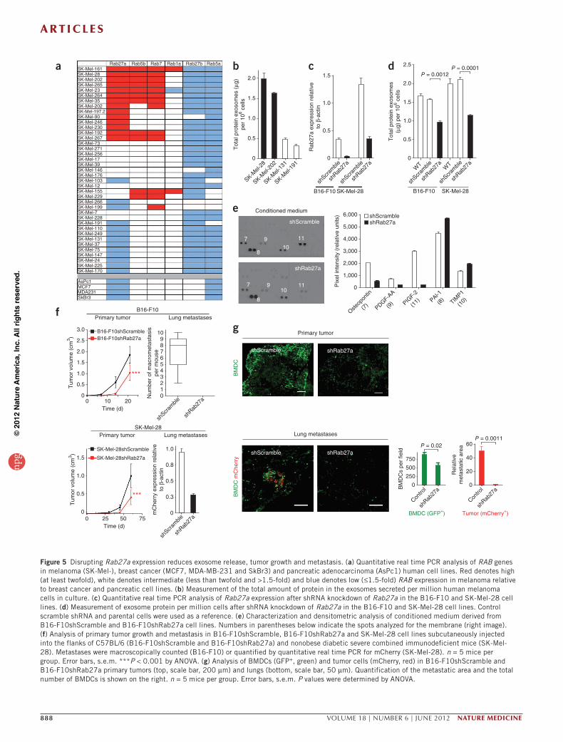
Figure 5 Disrupting Rab27a expression reduces exosome release, tumor growth and metastasis. (a) Quantitative real time PCR analysis of RAB genes in melanoma (SK-Mel-), breast cancer (MCF7, MDA-MB-231 and SkBr3) and pancreatic adenocarcinoma (AsPc1) human cell lines. Red denotes high (at least twofold), white denotes intermediate (less than twofold and >1.5-fold) and blue denotes low (≤1.5-fold) RAB expression in melanoma relative to breast cancer and pancreatic cell lines. (b) Measurement of the total amount of protein in the exosomes secreted per million human melanoma cells in culture. (c) Quantitative real time PCR analysis of Rab27a expression after shRNA knockdown of Rab27a in the B16-F10 and SK-Mel-28 cell lines. (d) Measurement of exosome protein per million cells after shRNA knockdown of Rab27a in the B16-F10 and SK-Mel-28 cell lines. Control scramble shRNA and parental cells were used as a reference. (e) Characterization and densitometric analysis of conditioned medium derived from B16-F10shScramble and B16-F10shRab27a cell lines. Numbers in parentheses below indicate the spots analyzed for the membrane (right image). (f) Analysis of primary tumor growth and metastasis in B16-F10shScramble, B16-F10shRab27a and SK-Mel-28 cell lines subcutaneously injected into the flanks of C57BL/6 (B16-F10shScramble and B16-F10shRab27a) and nonobese diabetic severe combined immunodeficient mice (SK-Mel-28). Metastases were macroscopically counted (B16-F10) or quantified by quantitative real time PCR for mCherry (SK-Mel-28). n = 5 mice per group. Error bars, s.e.m. ***P < 0.001 by ANOVA. (g) Analysis of BMDCs (GFP+, green) and tumor cells (mCherry, red) in B16-F10shScramble and B16-F10shRab27a primary tumors (top, scale bar, 200 µm) and lungs (bottom, scale bar, 50 µm). Quantification of the metastatic area and the total number of BMDCs is shown on the right. n = 5 mice per group. Error bars, s.e.m. P values were determined by ANOVA.
npg
© 2
012
Nat
ure
Am
eric
a, In
c. A
ll rig
hts
rese
rved
.
a r t i c l e s
nature medicine VOLUME 18 | NUMBER 6 | JUNE 2012 889
bone metastasis compared to mice educated with B16-F10 exosomes (Fig. 4e). Moreover, the amounts of vasculogenic and hematopoietic precursors were lower in the bone marrow and peripheral blood of mice bearing B16-F10 tumors that were injected with B16-F10shMet exosomes compared to mice treated with B16-F10 exosomes (data not shown).
To determine the clinical relevance of our findings, we analyzed the amounts of MET and pMET in circulating exosomes isolated from subjects with stage 3 or 4 melanoma (from the same cohort as described in Fig. 1). We found higher amounts of total and pMET in both the individuals with stage 3 melanoma and those with stage 4 melanoma compared to normal controls (Fig. 4f). Moreover, MET protein expression analyzed by flow cytometry was higher in both bone marrow progenitor cells (CD45−C-KITlow/+) and pro-vasculogenic cells (CD45−C-KIT+/lowTIE2+) isolated from the blood of subjects with stage 4 disease compared to cells isolated from control subjects with stage 1–3 disease (Fig. 4g and Supplementary Fig. 5e). Our data suggest that MET expression in circulating exosomes and bone marrow progenitor cells could be new predictors or early markers of metastatic disease.
Rab27a regulates exosome production and metastasisAs Ras-related RAB proteins control exosome biogenesis30, we analyzed the expression of RAB genes (RAB1A, RAB5A, RAB5B, RAB7, RAB27A and RAB27B) using a panel of 39 human-derived melanoma cell lines. Notably, those cell lines producing high concentrations of exosomes also had high expression of RAB27A, RAB5B and RAB7 mRNA relative to those cell lines producing lower concentrations of exosomes (Fig. 5a,b). Given the key role of RAB27 in exosome release31, we reasoned that inactivating this gene would reduce exosome production. We selected the Rab27a isoform for knockdown, as it was selectively expressed in melanoma-derived cell lines (Fig. 5a and data not shown). We effec-tively reduced the expression of Rab27a in B16-F10 and SK-Mel-28 cells (Fig. 5c), which resulted in a 50% decrease in exosome secretion (Fig. 5d and Supplementary Fig. 6a), showing a role for Rab27a in exosome production by melanoma cells. Although we found no marked differ-ences in total protein, protein content, exosome-specific markers or the amount of Met and pMet between the control and RAb27a knockdown cells and exosomes (Supplementary Fig. 6b,c), we did find a decrease in the secretion of placental growth factor 2 (PlGF-2), platelet-derived growth factor A chain homodimer (PDGF-AA) and osteopontin as a consequence of reducing the expression of Rab27a (Fig. 5e). Notably, Rab27a knockdown in B16-F10 and SK-Mel-28 cells reduced primary tumor growth, lung colonization and metastasis (Fig. 5f), which were associated with a decrease in the number of BMDCs recruited to these sites (Fig. 5g and Supplementary Fig. 6d). Consistent with our in vitro results (Supplementary Fig. 6a), the concentrations of circulating tumor-derived exosomes in mice bearing Rab27a knockdown tumors were reduced by 40–50 % (data not shown) and were therefore insuf-ficient to educate and mobilize BMDCs.
By injecting increasing amounts of exosomes isolated from either B16-F10shScramble (B16-F10 cells plus scramble shRNA) or B16-F10shRab27a (B16-F10 cells plus Rab27a shRNA) cell lines in mice fol-lowed by flank injection of tumor cells, we observed a dose-dependent increase in metastatic burden (Supplementary Fig. 7a,b). These data suggest that quantitative differences in exosome production can alter metastatic potential. Because overexpression of Rab27a in the B16-F1, LLC and B16-F10 cell lines led to cell death (data not shown), we were unable to determine the sufficiency of Rab27a in exosome production and modulating metastasis. Collectively, these data suggest that Rab27a
knockdown in melanoma models reduces exosome production and circulating exosome concentrations, thus preventing the recruitment of the BMDCs that are necessary for metastatic progression.
DISCUSSIONAccumulating evidence indicates that exosomes are mediators of metastasis3,11,13,15,22,23,45,46. Multiple cell types, including fibroblasts, endothelial cells and bone marrow progenitor cells, contribute to the generation of metastatic microenvironments39,47 and may be influ-enced by the horizontal transfer of molecules (for example, proteins and microRNAs) by exosomes8,40,48,49. However, the molecular and cellular mechanisms underlying this association have not been determined. Here we show that exosomes have a role in tumor pro-gression and metastatic niche formation that is distinct from that of tumor-derived growth factors, extracellular proteins and chemo-kines18,20,21,37,50,51. Tumor-derived exosomes promote the education and mobilization of BMDCs that support tumor vasculogenesis, inva-sion and metastasis. Notably, tumor-derived exosomes recruit BMDCs through upregulation of proinflammatory molecules at pre-metastatic sites. Specifically, preconditioning the bone marrow with exosomes from a highly metastatic cancer cell line (B16-F10) can increase the metastatic tumor burden and distribution in target tissues, even for tumors of different origins with a low metastatic capacity. Our work in tumor-derived exosome biology complements Paget and Ewing’s theories of metastatic spread, offering an explanation of organotropic involvement in certain metastatic diseases52,53.
By comparing the protein content of exosomes from highly meta-static and poorly metastatic melanoma cells, we identified MET signaling as a principal mediator of bone marrow progenitor cell edu-cation. We identified activated MET-related signaling proteins to be differentially expressed in exosomes from highly metastatic melanoma cells compared to poorly metastatic ones, which mediated pro- vasculogenic and metastatic effects on bone marrow progenitor cells. The MET oncogene mediates cellular transformation and tumor cell proliferation, survival, motility, invasion and metastasis26,28,29,44,54,55. Here we propose a previously unknown role for exosome-packaged MET in metastatic melanoma. The unique cargo of exosomes from other tumor types may educate distinct bone marrow populations, thus directly affecting metastatic propensity and outcome. The bone marrow of mice educated with the poorly metastatic B16-F1 exo-somes, which lack the MET receptor, reduced (though not statisti-cally significant) the metastatic burden and organotropism of highly metastatic B16-F10 primary tumors, suggesting that nonmetastatic exosomes may educate bone marrow to prevent metastatic disease.
The molecular mechanisms of exosome biogenesis and secretion are not well described. We found that, among the 39 melanoma-derived cell lines studied, specific RAB genes involved in exosome secretion (RAB1A, RAB5B, RAB7 and RAB27A) were highly expressed in cells lines secreting high concentrations of exosomes. Reducing the expression of RAB27A, a regulator of protein traffick-ing and melanoma proliferation31,56, decreased exosome production and the release of proangiogenic factors (PlGF-2, osteopontin and PDGF-AA) from tumor cells, hindering BMDC mobilization, tumor growth and metastasis. Our data, together with other studies showing that overexpression of RAB27A correlates with breast cancer invasive-ness and metastasis57,58, suggest that RAB27A should be considered as a potential therapeutic target in cancer.
Our human studies identified a specific melanoma signature com-prised of TYRP2, VLA-4, HSP70, an HSP90 isoform and MET in circulating exosomes from subjects with advanced melanoma, which
npg
© 2
012
Nat
ure
Am
eric
a, In
c. A
ll rig
hts
rese
rved
.
a r t i c l e s
890 VOLUME 18 | NUMBER 6 | JUNE 2012 nature medicine
could be used as an indicator of metastatic disease and tumor burden. We found that the coexpression of TYRP2 and MET in exosomes, as well as an increased amount of protein per exosome, predicted disease progression. In addition, we showed that MET expression is elevated in circulating pro-vasculogenic progenitors in subjects with metastatic melanoma. Thus, our work identifies quantitative and qualitative signatures, along with specific bone marrow progenitor cell populations that are mobilized, as representative hallmarks of metastatic melanoma.
Although the membrane vesicles of cancer cells have been sug-gested to contribute to the horizontal propagation of oncoproteins and genetic material (RNA)5,8,13,14,40,48, our study is the first, to our knowledge, to show that transfer of the MET oncoprotein from tumor-derived exosomes to bone marrow progenitor cells promotes the metastatic process in vivo. Notably, we show that exosomes can alter the bone marrow in a durable manner; these results suggest that genetic or epigenetic changes could be involved in this phenomenon, as bone marrow cells retain the educated phenotype after engraftment into a new host.
Here we propose a new mechanism that controls metastatic pro-gression through the crosstalk between tumor-derived exosomes and bone marrow progenitor cells. Collectively, our data identify exosome-mediated transfer of the oncoprotein MET as a key regulator of bone marrow education and mobilization and metastatic progression.
METHODSMethods and any associated references are available in the online version of the paper.
Accession codes. Microarray raw data tables have been deposited in the Gene Expression Omnibus (accession number GSE36584).
Note: Supplementary information is available in the online version of the paper.
ACKNoWLeDGMeNtSWe dedicate this work to the memory of James A. Paduano. We thank M.J. Bissell, A. Cano, J. Wels and S.R. Granitto for critical reading of this paper and suggestions. We also thank the members of our laboratories for helpful discussions and the members of the Weill Cornell Medical College electron microscopy and microarray core facilities for their support. We thank V. Hearing, (US National Institutes of Health (NIH), National Cancer Institute (NCI)) for providing the antibody to TYRP2 and D.C. Bennett (St. George’s University of London) for providing the melan-a cell line. Our work is supported by grants from the Children’s Cancer and Blood Foundation (H.P. and D.L.), The Manning Foundation (B.C.-S. and D.L.), The Hartwell Foundation (D.L.), Pediatric Oncology Experimental Therapeutics Investigators Consortium (H.P. and D.L.), Stavros S. Niarchos Foundation (D.L.), Champalimaud Foundation (H.P., Y.K. and D.L.), The Nancy C. and Daniel P. Paduano Foundation (H.P. and D.L.), The Mary Kay Foundation (A.N.-H. and D.L.), American Hellenic Educational Progressive Association 5th District (D.L.), The Malcolm Hewitt Wiener Foundation (D.L.), The George Best Costacos Foundation (D.L.), NCI (D.L., grant NCI-R01CA 098234-01), National Foundation for Cancer Research (D.L.), Susan G. Komen for the Cure (H.P. and D.L.), NCI-U54-CA143836 training grant (C.M.G. and D.L.), Fundación para el Fomento en Asturias de la Investigación Científica Aplicada y la Tecnología (G.G.-S.), Fundación Universidad de Oviedo (G.G.-S.), The Beth C. Tortolani Foundation (H.P., D.L. and J.B.), Sussman Family Fund (J.B.), Charles and Marjorie Holloway Foundation (J.B.), Manhassat Breast Cancer Fund (J.B.), NIH-CA87637 (J.B.), Fundação de Amparo a Pesquisa do Estado de São Paulo (FAPESP, V.R.M. and B.C.-S.), NIH (Y.K., grants R01-CA134519 and R01-CA141062), National Science Foundation grant CBET-0941143 and an American Society for Mass Spectrometry research award (B.A.G.).
AUtHoR CoNtRIBUtIoNSH.P. developed the hypothesis, designed the experimental approach, performed the experimental work, analyzed the data, coordinated the project and wrote the manuscript. S.L. conducted experimental work. I.M. performed flow cytometry studies and analysis. B.C.-S. conducted mouse work and proteomic
characterization of exosomes. G.M.-B. conducted gene expression studies and analysis of microarray data. M.H.-R. conducted mouse work and bone marrow education studies. C.W. conducted mouse work and human studies. G.G.-S. developed bone marrow education assays. A.N.-H. and K.B. quantified exosomes in human plasma. M.A., B.A.G. and Y.K. performed mass spectrometry studies and contributed to data interpretation. C.H. obtained human blood specimens. M.K.C. and J.Y. contributed to the characterization of human plasma exosomes. J.S., R.N.K., V.R.M., M.S.B., J.D.W., P.B.C. and C.M.G. discussed the hypothesis and contributed to data interpretation and experimental design. J.B. coordinated the project, interpreted data and wrote the manuscript. D.L. conceived the hypothesis, led the project, interpreted the data and wrote the manuscript.
CoMPetING FINANCIAL INteReStSThe authors declare competing financial interests: details are available in the online version of the paper.
Published online at http://www.nature.com/doifinder/10.1038/nm.2753. Reprints and permissions information is available online at http://www.nature.com/reprints/index.html.
1. Théry, C., Zitvogel, L. & Amigorena, S. Exosomes: composition, biogenesis and function. Nat. Rev. Immunol. 2, 569–579 (2002).
2. Iero, M. et al. Tumour-released exosomes and their implications in cancer immunity. Cell Death Differ. 15, 80–88 (2008).
3. Ratajczak, J., Wysoczynski, M., Hayek, F., Janowska-Wieczorek, A. & Ratajczak, M.Z. Membrane-derived microvesicles: important and underappreciated mediators of cell-to-cell communication. Leukemia 20, 1487–1495 (2006).
4. Cocucci, E., Racchetti, G. & Meldolesi, J. Shedding microvesicles: artefacts no more. Trends Cell Biol. 19, 43–51 (2009).
5. Valadi, H. et al. Exosome-mediated transfer of mRNAs and microRNAs is a novel mechanism of genetic exchange between cells. Nat. Cell Biol. 9, 654–659 (2007).
6. van Niel, G., Porto-Carreiro, I., Simoes, S. & Raposo, G. Exosomes: a common pathway for a specialized function. J. Biochem. 140, 13–21 (2006).
7. Peinado, H., Lavotshkin, S. & Lyden, D. The secreted factors responsible for pre-metastatic niche formation: old sayings and new thoughts. Semin. Cancer Biol. 21, 139–146 (2011).
8. Ratajczak, J. et al. Embryonic stem cell-derived microvesicles reprogram hematopoietic progenitors: evidence for horizontal transfer of mRNA and protein delivery. Leukemia 20, 847–856 (2006).
9. Nazarenko, I. et al. Cell surface tetraspanin Tspan8 contributes to molecular pathways of exosome-induced endothelial cell activation. Cancer Res. 70, 1668–1678 (2010).
10. Webber, J., Steadman, R., Mason, M.D., Tabi, Z. & Clayton, A. Cancer exosomes trigger fibroblast to myofibroblast differentiation. Cancer Res. 70, 9621–9630 (2010).
11. Liu, Y. et al. Contribution of MyD88 to the tumor exosome-mediated induction of myeloid derived suppressor cells. Am. J. Pathol. 176, 2490–2499 (2010).
12. Xiang, X. et al. Induction of myeloid-derived suppressor cells by tumor exosomes. Int. J. Cancer 124, 2621–2633 (2009).
13. Al-Nedawi, K. et al. Intercellular transfer of the oncogenic receptor EGFRvIII by microvesicles derived from tumour cells. Nat. Cell Biol. 10, 619–624 (2008).
14. Hao, S. et al. Epigenetic transfer of metastatic activity by uptake of highly metastatic B16 melanoma cell-released exosomes. Exp. Oncol. 28, 126–131 (2006).
15. Skog, J. et al. Glioblastoma microvesicles transport RNA and proteins that promote tumour growth and provide diagnostic biomarkers. Nat. Cell Biol. 10, 1470–1476 (2008).
16. Sethi, N. & Kang, Y. Unravelling the complexity of metastasis—molecular understanding and targeted therapies. Nat. Rev. Cancer 11, 735–748 (2011).
17. Psaila, B. & Lyden, D. The metastatic niche: adapting the foreign soil. Nat. Rev. Cancer 9, 285–293 (2009).
18. Kaplan, R.N. et al. VEGFR1-positive haematopoietic bone marrow progenitors initiate the pre-metastatic niche. Nature 438, 820–827 (2005).
19. Gao, D. et al. Bone marrow-derived endothelial progenitor cells contribute to the angiogenic switch in tumor growth and metastatic progression. Biochim. Biophys. Acta 1796, 33–40 (2009).
20. Erler, J.T. et al. Hypoxia-induced lysyl oxidase is a critical mediator of bone marrow cell recruitment to form the premetastatic niche. Cancer Cell 15, 35–44 (2009).
21. Hiratsuka, S., Watanabe, A., Aburatani, H. & Maru, Y. Tumour-mediated upregulation of chemoattractants and recruitment of myeloid cells predetermines lung metastasis. Nat. Cell Biol. 8, 1369–1375 (2006).
22. Hood, J.L., San, R.S. & Wickline, S.A. Exosomes released by melanoma cells prepare sentinel lymph nodes for tumor metastasis. Cancer Res. 71, 3792–3801 (2011).
23. Jung, T. et al. CD44v6 dependence of premetastatic niche preparation by exosomes. Neoplasia 11, 1093–1105 (2009).
24. Trusolino, L., Bertotti, A. & Comoglio, P.M. MET signalling: principles and functions in development, organ regeneration and cancer. Nat. Rev. Mol. Cell Biol. 11, 834–848 (2010).
npg
© 2
012
Nat
ure
Am
eric
a, In
c. A
ll rig
hts
rese
rved
.
a r t i c l e s
nature medicine VOLUME 18 | NUMBER 6 | JUNE 2012 891
25. Stella, G.M., Benvenuti, S. & Comoglio, P.M. Targeting the MET oncogene in cancer and metastases. Expert Opin. Investig. Drugs 19, 1381–1394 (2010).
26. Boccaccio, C. & Comoglio, P.M. Invasive growth: a MET-driven genetic programme for cancer and stem cells. Nat. Rev. Cancer 6, 637–645 (2006).
27. Cecchi, F., Rabe, D.C. & Bottaro, D.P. Targeting the HGF/Met signalling pathway in cancer. Eur. J. Cancer 46, 1260–1270 (2010).
28. Peruzzi, B. & Bottaro, D.P. Targeting the c-Met signaling pathway in cancer. Clin. Cancer Res. 12, 3657–3660 (2006).
29. Birchmeier, C., Birchmeier, W., Gherardi, E. & Vande Woude, G.F. Met, metastasis, motility and more. Nat. Rev. Mol. Cell Biol. 4, 915–925 (2003).
30. Stenmark, H. Rab GTPases as coordinators of vesicle traffic. Nat. Rev. Mol. Cell Biol. 10, 513–525 (2009).
31. Ostrowski, M. et al. Rab27a and Rab27b control different steps of the exosome secretion pathway. Nat. Cell Biol. 12, 19–30 (2009).
32. Braeuer, R.R., Zigler, M., Villares, G.J., Dobroff, A.S. & Bar-Eli, M. Transcriptional control of melanoma metastasis: the importance of the tumor microenvironment. Semin. Cancer Biol. 21, 83–88 (2011).
33. Fidler, I.J. Critical determinants of melanoma metastasis. J. Investig. Dermatol. Symp. Proc. 1, 203–208 (1996).
34. Grammatikakis, N. et al. The role of Hsp90N, a new member of the Hsp90 family, in signal transduction and neoplastic transformation. J. Biol. Chem. 277, 8312–8320 (2002).
35. Fidler, I.J. & Nicolson, G.L. Organ selectivity for implantation survival and growth of B16 melanoma variant tumor lines. J. Natl. Cancer Inst. 57, 1199–1202 (1976).
36. Huang, Y. et al. Pulmonary vascular destabilization in the premetastatic phase facilitates lung metastasis. Cancer Res. 69, 7529–7537 (2009).
37. Hiratsuka, S. et al. The S100A8-serum amyloid A3–TLR4 paracrine cascade establishes a pre-metastatic phase. Nat. Cell Biol. 10, 1349–1355 (2008).
38. Lucas, R., Verin, A.D., Black, S.M. & Catravas, J.D. Regulators of endothelial and epithelial barrier integrity and function in acute lung injury. Biochem. Pharmacol. 77, 1763–1772 (2009).
39. Joyce, J.A. & Pollard, J.W. Microenvironmental regulation of metastasis. Nat. Rev. Cancer 9, 239–252 (2009).
40. Balaj, L. et al. Tumour microvesicles contain retrotransposon elements and amplified oncogene sequences. Nat. Commun. 2, 180 (2011).
41. Taylor, D.D., Taylor, C.G., Jiang, C.G. & Black, P.H. Characterization of plasma membrane shedding from murine melanoma cells. Int. J. Cancer 41, 629–635 (1988).
42. Zöller, M. CD44: can a cancer-initiating cell profit from an abundantly expressed molecule? Nat. Rev. Cancer 11, 254–267 (2011).
43. Jalili, A., Shirvaikar, N., Marquez-Curtis, L.A., Turner, A.R. & Janowska-Wieczorek, A. The HGF/c-Met axis synergizes with G-CSF in the mobilization of hematopoietic stem/progenitor cells. Stem Cells Dev. 19, 1143–1151 (2010).
44. Tesio, M. et al. Enhanced c-Met activity promotes G-CSF–induced mobilization of hematopoietic progenitor cells via ROS signaling. Blood 117, 419–428 (2011).
45. Lima, L.G., Chammas, R., Monteiro, R.Q., Moreira, M.E. & Barcinski, M.A. Tumor-derived microvesicles modulate the establishment of metastatic melanoma in a phosphatidylserine-dependent manner. Cancer Lett. 283, 168–175 (2009).
46. Logozzi, M. et al. High levels of exosomes expressing CD63 and caveolin-1 in plasma of melanoma patients. PLoS ONE 4, e5219 (2009).
47. Guise, T. Examining the metastatic niche: targeting the microenvironment. Semin. Oncol. 37 (suppl. 2), S2–S14 (2010).
48. Aliotta, J.M. et al. Microvesicle entry into marrow cells mediates tissue-specific changes in mRNA by direct delivery of mRNA and induction of transcription. Exp. Hematol. 38, 233–245 (2010).
49. Aliotta, J.M. et al. Alteration of marrow cell gene expression, protein production, and engraftment into lung by lung-derived microvesicles: a novel mechanism for phenotype modulation. Stem Cells 25, 2245–2256 (2007).
50. Oskarsson, T. et al. Breast cancer cells produce tenascin C as a metastatic niche component to colonize the lungs. Nat. Med. 17, 867–874 (2011).
51. Psaila, B., Kaplan, R.N., Port, E.R. & Lyden, D. Priming the ‘soil’ for breast cancer metastasis: the pre-metastatic niche. Breast Dis. 26, 65–74 (2006).
52. Ewing, J. Neoplastic Diseases: A Treatise on Tumours, 3rd edn. (W.B. Saunders Co., Philadelphia, 1928).
53. Paget, G. The distribution of secondary growths in cancer of the breast. Lancet 133, 571–573 (1889).
54. Scott, K.L. et al. Proinvasion metastasis drivers in early-stage melanoma are oncogenes. Cancer Cell 20, 92–103 (2011).
55. Christensen, J.G., Burrows, J. & Salgia, R. c-Met as a target for human cancer and characterization of inhibitors for therapeutic intervention. Cancer Lett. 225, 1–26 (2005).
56. Akavia, U.D. et al. An integrated approach to uncover drivers of cancer. Cell 143, 1005–1017 (2010).
57. Wang, J.S., Wang, F.B., Zhang, Q.G., Shen, Z.Z. & Shao, Z.M. Enhanced expression of Rab27A gene by breast cancer cells promoting invasiveness and the metastasis potential by secretion of insulin-like growth factor-II. Mol. Cancer Res. 6, 372–382 (2008).
58. Hendrix, A. et al. Effect of the secretory small GTPase Rab27B on breast cancer growth, invasion, and metastasis. J. Natl. Cancer Inst. 102, 866–880 (2010).
npg
© 2
012
Nat
ure
Am
eric
a, In
c. A
ll rig
hts
rese
rved
.
nature medicine doi:10.1038/nm.2753
ONLINE METHODSExosome purification and tracking analysis. Cells were cultured in media sup-plemented with 10% exosome-depleted FBS (FBS, Hyclone). FBS was depleted of bovine exosomes by ultracentrifugation at 100,000g for 70 min. Supernatant fractions collected from 48–72 h cell cultures were pelleted by centrifugation at 500g for 10 min. The supernatant was centrifuged at 20,000g for 20 min. Exosomes were then harvested by centrifugation at 100,000g for 70 min. The exosome pellet was resuspended in 20 ml of PBS and collected by ultracen-trifugation at 100,000g for 70 min (Sorvall SureSpin 630 rotor). Circulating exosomes were isolated from mouse and human plasma as described above, with an additional filtration through 1.2-µm nylon filters (GE) before the last step of ultracentrifugation. For centrifugation onto sucrose cushions, the samples were diluted 1:10 in PBS after centrifugation at 20,000g and then collected by ultracentrifugation (100,000g for 70 min) on a 40% sucrose cushion. The floating exosome fraction was collected again by ultracentrifugation as described above, and the final pellet was resuspended in PBS. For the retrospective studies using frozen plasma, 2 ml of cell-free plasma was centrifuged at 500g for 10 min, and then the supernatant was centrifuged at 20,000g for 20 min. Exosomes were then harvested by centrifugation at 100,000g for 70 min. The exosome pellet was resuspended in PBS and collected by ultracentrifugation at 100,000g for 70 min (Sorvall S100-AT5 rotor). The LM10 nanoparticle characterization system (NanoSight) equipped with a blue laser (405 nm) was used for real-time characterization of the vesicles.
Human studies. Human peripheral blood samples were obtained from sub-jects with stage 1–4 melanoma at the Memorial Sloan-Kettering Cancer Center (MSKCC) and had histologically confirmed melanoma. For the retrospective studies of circulating exosomes in frozen plasma, subjects with stage 3 disease were followed up for 1–4 years. Subjects with stage 4 disease were followed up over 42 months. All individuals provided informed consent for blood donation according to a protocol approved by the institutional review board of MSKCC.
Exosome labeling and treatment. Five to ten micrograms of total exosome pro-tein were injected by tail vein in a total volume of 100–200 µl PBS. Synthetic unilamellar 100-nm liposomes (Encapsula Nanoscience) or PBS were used as control particles in all studies (data not shown). For the exosome-tracking experi-ments, purified exosomes were fluorescently labeled using PKH67 membrane dye (Sigma). Labeled exosomes were washed in 20 ml of PBS, collected by ultracen-trifugation as described above and resuspended in PBS. We verified that no dye contamination occurred in the PKH67-labeled exosome preparations by ultracen-trifugation on 40% sucrose cushions as described above (data not shown).
Metastasis and lung colonization studies. To analyze the role of exosomes in tumor metastasis, 8- to 10-week-old C57BL/6J female mice were injected in the flank with 1 × 10⁶ B16-F10mCherry cells. Seven days later, 10 µg of B16-F10 exosomes were injected three times a week for 3 weeks. Metastasis was evaluated by mCherry expression at days 19 and 28. Alternatively, C57BL/6J female mice were injected three times a week with the indicated dose of B16-F10 or B16-F1 exosomes for 28 d. After exosome injection, mice received 1 × 10⁶ B16-F10–luciferase subcutaneous flank injections. Controls included injections of PBS and synthetic unilamellar liposomes. Luciferase imaging was performed using the IVIS Spectrum system (Caliper, Xenogen). Tumor-bearing mice were anesthetized (using isoflurane and O2), and D-luciferin (50 mg kg−1 in 100 µl PBS) was administered. Five minutes later, mice were euthanized and their organs were analyzed for luciferase expression. Data were quantified with Living Imaging software 4.2. For the analysis of tumor cell growth using Rab27a knockdown cells, 1 × 10⁶ B16-F10–mCherry cells (in 100 µl of PBS) or 2 × 10⁶ SK-Mel-28mCherry cells (in a mix of 1:1 serum-free RPMI and growth-factor–reduced Matrigel) were injected in the flank of C57BL/6 or nonobese diabetic severe combined immunodeficient female mice, respectively. For lung colonization experiments, 1 × 10⁵ B16-F10–mCherry cells or 5 × 10⁵ SK-Mel-28–mCherry cells (in 100 µl of PBS) were injected by tail vein in C57BL/6 female mice.
GFP bone marrow education. Enhanced GFP (eGFP) transgenic male mice (C57BL/6-TgN(ActbeGFP)1Osb/J, Jackson Laboratory) were injected three
times a week with 10 µg of B16-F10 exosomes over 28 d. Bone marrow was harvested by flushing the femurs and tibias of eGFP donor mice, and the 5 × 10⁶ total bone marrow cells from the eGFP donor mice were transplanted into lethally irradiated (950 rads) C57BL/6 female mice by retro-orbital injection. After 4 weeks, the eGFP bone marrow–reconstituted C57BL/6 mice were injected subcutaneously in the flank with 1 × 10⁶ B16-F10mCherry or LLCmCherry cells. Mice were euthanized after 28 d or 35 d. Tissues were dissected and fixed in 4% paraformaldehyde (PFA) or in a mix of 2% PFA and 20% sucrose solution overnight and embedded in Tissue-Tek O.C.T. (Electron Microscopy Sciences), and then the blocks were frozen in a dry ice and ethanol bath. Additionally, tissues were snap frozen in liquid nitrogen for RNA and protein extraction. The tissues used to evaluate metastasis in bone marrow–educated mice and controls (Supplementary Table 4) were paraffin embedded and stained with H&E, and metastases were evaluated by microscopy. All mouse work was performed in accordance with institutional, Institutional Animal Care and Use Committee (IACUC) and American Association for Laboratory Animal Science (AALAS) guidelines. All mice were monitored for abnormal tissue growth or ill effects according to AALAS guidelines and were euthanized if excessive deterioration of the health of the mice was observed.
Mouse flow cytometry and antibodies. Lungs and tissues were prepared for flow cytometry by mincing, followed by digestion at 37 °C for 45–60 min with an enzyme cocktail (Collagenase A and DNaseI, Roche). Single-cell suspen-sions were filtered through a 70-µm strainer. Peripheral blood was obtained by retro-orbital bleeding directly into anti-coagulant tubes (EDTA), and red blood cells were lysed using ACK lysis buffer (Gibco). Cell suspensions were blocked with Fc-block (CD16/CD32, BD), washed in PBS and 1% BSA and incubated with the following primary antibodies diluted in PBS and 1% BSA: CD11b-FITC (M1/70), VEGFR2- phycoerythrin (PE) (Avas12α1), lineage nega-tive cell detection kit (biotinylated protein tyrosine phosphatase, receptor type, C (B220), CD3 antigen, ε polypeptide (CD3ε), TER119, CD11b and Gr1), followed by streptavidin-PE-CY7, c-Kit-allophycocyanin (APC) (2B8), Sca1-PE (D7), Gr1-PE (RB6-8C5), F4/80-APC (BM8), Tie2-PE (TEK4), CD29-APC (HMb1), CD105-PE (MJ7/18) and Met-FITC (eBioclone4). Fluorochrome-conjugated antibodies were purchased from BD or eBioscience and used at predetermined saturating concentrations. Data acquired on a BD FACSCalibur cytometer with CellQuest software (BD) was analyzed using FlowJo software (TreeStar).
Flow cytometry for human samples. Peripheral blood obtained from human subjects was centrifuged at 500g for 10 min at 4 °C. Plasma was used for exo-some isolation and analysis. Peripheral blood cells were isolated using a Ficoll-Paque gradient (GE Healthcare). The buffy coat was separated, and the cells were washed in PBS and 1% BSA and collected by centrifugation at 400g for 5 min at 4 °C. Residual red blood cells were lysed for 4 min at 4 °C using ACK lysis buffer (Gibco). Cells were incubated with flurochrome-conjugated antibodies diluted in PBS and 1% BSA, and washed in PBS and 1% BSA. Flow cytometric analysis was performed using antibodies to CD45-eFluor450 (2D1), C-KIT/CD117-PE (104D2), CD34-Alexa700 (581), TIE2-Alexa647 (CD202B) (33.1) and MET-FITC (eBioclone 97). Antibody-fluorochrome conjugates were purchased from BD Biosciences, eBioscience or BioLegend and used as suggested by the manufacturer. Data acquired on a BD FACSCanto was analyzed using FlowJo (Tree Star) software.
Cell lines and cell culture. B16-F10, B16-F1, LLC, MCF-7, As-Pc1 and MDA-MB-231 cells were cultured in DMEM. SkBr3, SW480, SW620 and human melanoma cells (SK-Mel-) were cultured in RPMI supplemented with penicil-lin (100 U ml−1) and streptomycin (100 µg ml−1) and 10% exosome-depleted FBS. The melan-a cells were maintained in RPMI containing 10% FCS, 200 nM 12-O-tetradecanoylphorbol-13-acetate (TPA; Sigma). Cells were obtained from American Type Culture Collection. Human melanoma cell lines were obtained from MSKCC.
Electron microscopy. Exosomes purified as described above were fixed in 2% PFA (w/v) in 200 mM phosphate buffer (pH 7.4). Fixed exosomes were dropped onto a formvar-carbon–coated grid and left to dry at room temperature for 20 min. After washing in PBS, the exosomes were fixed in 1% glutaraldehyde
npg
© 2
012
Nat
ure
Am
eric
a, In
c. A
ll rig
hts
rese
rved
.
nature medicinedoi:10.1038/nm.2753
for 5 min, washed in water and stained with saturated aqueous uranyl oxalate for 5 min. Samples were then embedded in 0.4% (w/v) uranyl acetate and 1.8% (w/v) methylcellulose and incubated on ice for 10 min. The excess liquid was then removed. The grid was dried at room temperature for 10 min and viewed at 20,000 and 50,000 magnification using an electron microscope (model 910, Carl Zeiss).
Lung leakiness experiments. Ten micrograms of total exosome protein were injected by tail vein. Conditioned media was prepared by filtering supernatant fractions of cultured B16-F10 cells through a 0.22-µm filter and ultracentifuging it at 100,000g to deplete the exosomes. One hundred microliters of cultured media were injected by tail vein injection. As controls, mice were injected with 10 µg of melan-a exosomes, PBS or synthetic 100-µm unilamellar liposomes. Twenty hours after exosome treatment, mice were injected with 2 mg of Texas Red lysine-fixable dextran 70,000 MW (Invitrogen) by retro-orbital injection. One hour after dextran injection, mice were euthanized and perfused with 40 ml of PBS. Lungs were dissected and fixed in a mix of 2% PFA and 20% sucrose overnight.
Immunofluorescence. For immunofluorescence, 12 µm O.C.T. tissue sections were stained with antibodies to platelet/endothelial cell adhesion molecule 1 (CD31) (1:100) and vascular cell adhesion molecule 1 (VCAM-1) (1:100) (BD). For the vasculature analyses, each mouse was injected with 50 µg Alexa Fluor 594–conjugated Isolectin GS-IB4 (Molecular Probes) 10 min before being euthanized. GFP+ and mCherry+ cells were detected by their intrinsic signals. Fluorescent images were obtained using a Nikon confocal microscope (Eclipse TE2000U) and analyzed using Nikon software (EZ-C1 3.6). To determine the vessel density in the metastatic lesions, digital images of mCherry- or GFP-stained sections were analyzed with ImageJ Software (NIH) by determining the pixel intensity per square micrometer.
Bone marrow progenitor cell enrichment, in vitro treatment and responses to HGF. Mice were euthanized and their bone marrow was isolated by flushing the femur and tibia with PBS using a 25G 0.5-inch needle (BD). Cell suspen-sions were filtered through 70-µm cell strainers (BD) before centrifugation. To enrich for progenitors, lineage positive (Lin+, meaning, CD5+, CD45R+, CD19+, CD11b+, Ly6G/C+ and TER119+) bone marrow cells were immunomagnetically depleted using the EasySep Mouse Hematopoietic Progenitor Cell Enrichment Kit (Stem Cell Technologies) following the manufacturer’s instructions. For in vitro bone marrow treatment with exosomes, bone marrow cells were flushed as described above and cultured in Stem Span (Stem Cell Technologies) for 16 h in the presence or absence of 20 µg ml−1 exosomes, as indicated. For the HGF analysis, bone marrow cells were then stimulated with 5 ng ml−1 HGF (Peprotech) for 4 h and collected for western blot analysis in radioimmunopre-cipitation assay buffer with phosphatase inhibitors (1 mM sodium orthovana-date and 5 mM β-glycerol phosphate). For MET signaling inhibition, cells were incubated with 20 nM crizotinib (Selleck Bio) 1 h before HGF stimulation.
Quantitative real-time PCR and shRNA interference studies. Total RNA was extracted from tissues or cells using the RNeasy kit (QIAGEN) and was reverse transcribed using Superscript III reverse transcriptase (Invitrogen). Quantitative real-time PCR was performed on a 7500 Fast Real Time PCR System (Applied Biosystems) using TaqMan Universal PCR Master Mix (Applied Biosystems). Frozen tissues or cell lines were analyzed for specific gene expres-sion by quantitative real-time PCR using predesigned TaqMan assays (specific assay numbers: mouse Met,: Mm01156972_m1, mouse Cd44: Mm01277163, human RAB27A: Hs00608302_m1, mouse Rab27a: Mm00469997_m1, S100a8: Mm00496696-g1, S100a9: Mm00656925_m1) or GFP- or mCherry-specific primers18 using SYBR Green PCR reagents (Applied Biosystems). The rela-tive expression was normalized to β-actin. For shRNA-mediated knockdown of Rab27a, lentiviral vectors encoding shRNAs were purchased from Thermo Fisher. The human Rab27a shRNA sense sequences used were V3LHS_300918: 5′-CCCAGTGTACTTTACCAATATA-3′ and V3LHS_300917: 5′-CAG GGAAGACCAGTGTACTTTA-3′. The Rab27a knockdown efficiency was similar for both of the human shRNAs, however, V3LHS_300918 is shown in the figures. The sense sequence for mouse Rab27a shRNA was V3LHS_300916 5′-ACAGG AGAGGTTTCGTAGCTTA-3′, and the scramble sense sequence used was
5′-ATCTCGCTTGGGCGAGAGTAAG-3′. The mouse Met shRNA sense sequences used were V3LMM_456078: 5′-CCAGACTTTTCATACAAGA ATA-3′ and V2LMM_30812: 5′-CCCTATGTAGA-TCCTGTAATAA-3′. The Met knockdown efficiency was similar for both shRNAs (routinely exceeding 90%, as determined by GFP expression), however, V3LMM_456078 is shown in the figures.
Proteomic analysis. Identification of exosomal proteins was performed using reverse-phase high-pressure liquid chromatography mass spectrometry. Samples were denatured at 90 °C, reduced with 10 mM dithiothreitol (DTT) at 51 °C for 1 h and alkylated with 50 mM iodoacetamide at 25 °C for 45 min. Proteins were digested with trypsin (Promega) overnight at 25 °C. Tryptic peptides were concentrated by vacuum centrifugation and desalted using C18 STAGE Tips before the mass spectrometric analysis. Samples were loaded by an Eksigent AS2 autosampler onto a 75-µm fused silica capillary column packed with 11 cm of C18 reverse-phase resin (5-µm particles, 200-Å pore size; Magic C18, Michrom BioResources). Peptides were resolved on a 180-min 1–100% buffer B gradient (buffer A, 0.1 mol l−1 acetic acid, buffer B, 70% acetonitrile in 0.1 mol l−1 acetic acid) at a flow rate of 200 nl min−1 (1200 series, Agilent). The high- pressure liquid chromatography instrument was coupled to a mass spectrometer (LTQ-Orbitrap, Thermo Fisher Scientific) with a resolution of 30,000 for full mass spectrometry followed by seven data-dependent tandem mass spectrom-etry analyses. Collision-induced dissociation was used for peptide fragmenta-tion. Each sample was analyzed at least two times. All mass spectrometry data were analyzed with Proteome Discoverer software (version 1.2, Thermo Fisher Scientific) using the SEQUEST algorithm to search against human and mouse UniProt databases. The peptides were constrained to be tryptic, and up to two missed cleavages were allowed. Carbamidomethylation of cysteine was specified as a fixed modification, and oxidation of methionine was specified as a variable modification. The precursor ion tolerance was set to 25 ppm, and the fragment ion mass tolerance was set to 0.8 Da. Search results were analyzed individually, and the data for the replicates were combined and evaluated. For the validation of the proteomic analysis, we performed a western blot analysis for the selected proteins (Met, CD44, annexin A2 and annexin A6; Fig. 4a and data not shown) using equal amounts of exosomes derived from B16-F1 and B6-F10 cells.
Microarray preparation and analysis. For analyzing the genes modified in the lung by B16-F10 exosomes, a total of 10 µg of exosome protein was injected by tail vein, and 24 h or 48 h later, mice were euthanized and their lungs were dissected and stored in RNA-later solution (Ambion) until RNA extraction. A total of three mice were used per time point, and the experiment was performed in triplicate. Total RNA was isolated using the RNeasy Mini Kit (QIAGEN). The Affymetrix One-Round In Vitro Transcription RNA Amplification Kit was used to amplify 1.5 µg of total RNA. The complementary DNA (cDNA) was synthe-sized with a primer containing oligo(dT) and T7 RNA polymerase promoter sequences. Double-stranded cDNA was then purified and used as a template to generate biotinylated cRNA. The quantity and quality of the amplified cRNA was assessed using a NanoDrop ND-1000 Spectrophotometer (Thermo Scientific) and an Agilent Bioanalyzer. The biotinylated cRNA was fragmented and hybrid-ized to Affymetrix Mouse Genome 430 Plus 2.0 GeneChip arrays containing 39,000 transcripts per array. After hybridization, the GeneChip arrays were washed, stained and scanned using a GeneChip Scanner 3000 7G, according to the instructions of Affymetrix Expression Analysis. Affymetrix GeneChip Operating Software was used for image acquisition. The target signal inten-sity from each chip was scaled to 500. Triplicate samples from each exosome-treated sample and control lung sample were analyzed to identify differentially expressed genes. For the statistical analyses, we selected genes whose expression differed between the treatment groups by a factor of at least two. A hierarchical clustering method was applied to group the genes and samples based on their similarities in gene expression, and the unsupervised analyses were visualized using the SOTA and TreeView software assuming Euclidean distances between the genes (http://bioinfo.cnio.es/cgi-bin/tools/clustering/sotarray). We tested the null hypothesis of equal means between the two groups using a t test and computed P values using a permutation test. The fold expression was calculated from the expression ratio in the exosome-treated conditions with respect to control samples.
npg
© 2
012
Nat
ure
Am
eric
a, In
c. A
ll rig
hts
rese
rved
.
nature medicine doi:10.1038/nm.2753
Western blot, antibodies and multiplex analyses. Exosomes or cells were lysed with radioimmunoprecipitaiton assay buffer containing a complete protease inhibitor tablet (Roche). Lysates were cleared by centrifugation at 14,000g for 20 min. The supernatant fractions were used for western blot. Protein extracts were resolved by SDS-PAGE and probed with the indicated antibodies. For the western blot or immunofluorescence analyses, we used antibodies to Hsp90 (1:1,000, Stressgen, ADI-SPA-830), Hsp70 (1:1,000, Stressgen, SPA-810-F), Hsc70 (1:500, Stressgen, ALX-804-067), the Rab-family sampler kit (Rab4, Rab5, Rab7, Rab9 and Rab11; 1:500, Cell Signaling, 9385), Rab27a (1:500, Abnova, H0005873-M02), TYRP2 (1:500, PEP-8h), VLA-4 (1:500, Chemicon, clone PS2), melan-a (1:1,000, Santa Cruz, 28871), programmed cell death 6 interacting protein (Alix) (1:1,000, Cell Signaling, 2171), tumor susceptibility gene 101 (TSG101) (1:500, Santa Cruz, sc-7964), Met and pMet (1:1,000, Cell Signaling, 3127 and 3077, respectively), pS6 kinase (1:1,000, Cell Signaling, 9802) and pERK (1:1,000, Cell Signaling, 4370). Antibodies to GAPDH or β-actin (Santa Cruz, 1:1,000, sc-25778 and 1:5,000, sc-130656, respectively) were used as loading controls. The intensities of the immunoreactive bands were quantified by densitometry
using ImageJ software (NIH). For the analysis of protein expression profiles of the cells in conditioned media, we used the mouse angiogenesis antibody array (R&D) using 200 µg of cell extracts or 450 µl of media conditioned for 24 h. The MET and pMET analysis kits were purchased from MesoScale Discovery (MSD). All reagents were provided with the MSD kit, and the experiment was performed according to the manufacturer’s instructions using 10 µg of protein extract. MSD plates were read on the MSD Sector Imager 2400 plate reader. The raw data was measured as an electrochemiluminescence signal (light) detected by photodetectors and analyzed using the Discovery Workbench 3.0 software (MSD). The graphs depict mean photon intensity.
Statistical analyses. Error bars in the graphical data represent means ± s.e.m. Mouse experiments were performed in duplicate using at least five mice per treatment group. All in vitro experiments were performed at least in duplicate. P values of P < 0.05 were considered statistically significant by ANOVA. For the tumor growth analyses, we performed two-way ANOVA statistical analyses using GraphPad Prism software.
npg
© 2
012
Nat
ure
Am
eric
a, In
c. A
ll rig
hts
rese
rved
.