Embed Size (px)
Citation preview
Blue-light dependent reactive oxygen species formation byArabidopsis cryptochrome may define a novel evolutionarilyconserved signaling mechanism
Laurent Consentino1*, Stefan Lambert1*, Carlos Martino1,2, Nathalie Jourdan1, Pierre-Etienne Bouchet1,
Jacques Witczak1, Pablo Castello1,3, Mohamed El-Esawi1,4, Francoise Corbineau5, Alain d’Harlingue1 and
Margaret Ahmad1,6
1UMR 8256 (B2A) CNRS – UPMC, IBPS, Universit�e Pierre et Marie Curie, Bat C 3�eme �etage, 9 quai Saint-Bernard, 75252 Paris Cedex 05, France; 2Department of Biomedical Engineering,
Florida Institute of Technology, 150 W. University Blvd, Melbourne, FL 32901, USA; 3Facultad de Ciencias Exactas y Naturales, Universidad de Belgrano (UB), Villanueva 1324, Buenos Aires
C1426BMJ, Argentina; 4Botany Department, Faculty of Science, Tanta University, 31527 Tanta, Egypt; 5UMR7622 CNRS-UPMC Biologie du D�eveloppement, IBPS, Bat C 2�eme �etage,
boıte 24, 4 place Jussieu, 75005 Paris Cedex 05, France; 6Xavier University, 3800 Victory Parkway, Cincinatti, OH 45207, USA
Authors for correspondence:Margaret AhmadTel: +33 0 144272916
Email: [email protected]
Alain d’HarlingueTel: +33 0 144272916
Email: [email protected]
Received: 30 September 2014Accepted: 29 December 2014
New Phytologist (2015) 206: 1450–1462doi: 10.1111/nph.13341
Key words: Arabidopsis thaliana, crypto-chrome, oxidative stress, photomorphogenesis,photoreceptor, reactive oxygen species (ROS)signaling.
Summary
� Cryptochromes are widespread blue-light absorbing flavoproteins with important signaling
roles. In plants they mediate de-etiolation, developmental and stress responses resulting from
interaction with downstream signaling partners such as transcription factors and components
of the proteasome. Recently, it has been shown that Arabidopsis cry1 activation by blue light
also results in direct enzymatic conversion of molecular oxygen (O2) to reactive oxygen spe-
cies (ROS) and hydrogen peroxide (H2O2) in vitro. Here we explored whether direct enzy-
matic synthesis of ROS by Arabidopsis cry1 can play a physiological role in vivo.� ROS formation resulting from cry1 expression was measured by fluorescence assay in insect
cell cultures and in Arabidopsis protoplasts from cryptochrome mutant seedlings. Cell death
was determined by colorimetric assay.� We found that ROS formation results from cry1 activation and induces cell death in insect
cell cultures. In plant protoplasts, cryptochrome activation results in rapid increase in ROS for-
mation and cell death.� We conclude that ROS formation by cryptochromes may indeed be of physiological rele-
vance and could represent a novel paradigm for cryptochrome signaling.
Introduction
Cryptochromes are blue-light absorbing photoreceptors foundin plants and animals that have multiple signaling roles. Inplants, cryptochromes are involved in de-etiolation, elongationgrowth and development, entrainment of the circadian clockand the photoperiodic initiation of flowering (Chaves et al.,2011). Cryptochromes have also been implicated in responsivityto stress including pathogen resistance (Jeong et al., 2010; Wu& Yang, 2010), high light stress (Danon et al., 2006; Kleineet al., 2007; Lopez et al., 2012; Kim & Apel, 2013; Sharmaet al., 2014), and temperature and osmotic stress (Xu & Ma,2009; Xu et al., 2009; Sanchez et al., 2011; Sharma et al.,2014). Cryptochromes undergo blue-light dependent redoxreactions involving flavin photoreduction in vivo which are
thought to induce conformational change in the receptor lead-ing to interaction with the biological signaling partners(reviewed in Chaves et al., 2011). In fact, several proteins thatbind to plant cryptochromes in a light-dependent manner havebeen identified and linked to cryptochrome signaling, includingbHLH transcription factors such as Cib1 and elements of theproteasome including Cop1 and Spa1 (Liu et al., 2008; Zuoet al., 2011). Therefore, current paradigms for cryptochromeactivation presume that light-induced conformational changewithin the protein is followed by interaction with protein sub-strates, leading to downstream signaling events.
Recently, in the course of elucidating the cryptochromephotocycle, we have shown that activation of cry1 by bluelight results in formation of reactive oxygen species (ROS)and hydrogen peroxide (H2O2) under continuous blue lightirradiation in vitro (M€uller & Ahmad, 2011). Absorption ofa photon of light energy by a cryptochrome leads to transfer*These authors contributed equally to this work.
1450 New Phytologist (2015) 206: 1450–1462 � 2015 The Authors
New Phytologist� 2015 New Phytologist Trustwww.newphytologist.com
Research
of an electron to excited state flavin from a nearby Trp resi-due in the protein. This Trp residue is in turn reducedthrough a chain of additional Trp or Tyr residues leadingto the protein surface. The reduced (FADH° or FADH�)flavin is then reoxidized to the resting (oxidized) state viacleavage of molecular oxygen (O2) to form superoxide andsubsequently H2O2. At this point, the entire cryptochromephotocycle (light-induced flavin reduction followed by flavinreoxidation) can repeat itself, for as many times as photonenergy is available. As a result, under continuous blue lightillumination, cryptochrome activation catalyzes the enzy-matic accumulation of ROS and H2O2. Cryptochromes inthis way serve as light-activated generators of ROS.
ROS in themselves are important signaling molecules in mostorganisms, including in induction and/or responsivity to stress(Mittler et al., 2011). In plants, ROS are implicated in manyprocesses including in seed germination, development, senes-cence, heat or cold stress, pathogen defense, and responsivity tohigh light stress (Orozco-Cardenas & Ryan, 1999; Apel & Hirt,2004; Mittler et al., 2004; Gechev et al., 2006; Bailly et al., 2008;Miller et al., 2008; Quan et al., 2008; Barrero et al., 2014). Giventhis plethora of signaling roles for ROS, it is striking that a num-ber of the signaling pathways in which cryptochromes have beenimplicated, including pathogen defense, osmotic stress andresponse to high light stress, in fact intersect with known path-ways in which ROS are important regulators. The intriguing pos-sibility therefore suggests itself that cryptochromes may directlyalter cellular concentrations of ROS, particularly under highblue-light illumination, which induces known stress responses.Given the highly evolutionarily conserved nature of ROS sensi-tivity and implication in responsivity to stress (Mittler et al.,2011), perhaps one of the mechanisms of cryptochrome signalingmay be by directly altering concentrations of ROS found in spe-cific cellular compartments. We therefore wondered whetherproduction of ROS by cryptochromes may have physiologicalconsequences to the cell.
In order to test this possibility, we have investigated the effectof Arabidopsis cryptochrome-1 (Atcry1) on formation of reactiveoxygen in vivo using insect cell culture overexpressing Atcry1.We reasoned that insect cells are unlikely to have downstream sig-naling intermediates and protein partners involved in the classicsignaling pathways of plant cryptochromes. We have used trans-genic cry1-expressing insect cell cultures, which produce highconcentrations of plant cryptochromes in the cells and whichrespond to light in an in vivo context (Bouly et al., 2007; Burneyet al., 2012). We found that elevated concentrations of ROS wereformed by Atcry1-expressing insect cell cultures in response toblue light, followed by rapid cell death. Taken together with im-munolocalization studies verifying the colocalization of the site ofROS formation with Atcry1 localization in these insect cultures,we conclude that ROS formation by cryptochromes indeedoccurs in a physiological context. We further obtained intriguingresults using Arabidopsis protoplasts which implicated crypto-chrome activation in ROS formation in the plant system. Weconclude that cryptochrome production of ROS upon illumina-tion may be a novel feature of cryptochrome signaling.
Materials and Methods
Insect cell cultures and cell viability assays
We infected SF21 insect cells with baculovirus constructs ofAtcry1 as previously described (Bouly et al., 2007). Cells wereharvested 3 d post infection for analysis. For assays of cell viabil-ity, insect cell cultures were divided into 2-ml aliquots and incu-bated in 10-ml flat bottom glass vials under red and blue light orin darkness at 21°C with gentle agitation (70 g). For Trypan bluecolorimetric assays, 20 ll of cells were harvested in culturemedium at the appropriate time point and added directly to 20 llof dye (Trypan blue at a concentration of 1 mg ml�1 in phos-phate buffered saline (PBS) at pH 7.4). Aliquots were applied toa Kova® Glasstic� slide (Hycor Biomedical Inc., Indianapolis,IN, USA) with demarcated grids (volume = 6.6 ll) and werecounted using a Zeiss Apotome fluorescent microscope, with AxioVision imaging software. A minimum of six independent aliquotswere counted per time point with between 100 and 200 cellscounted per aliquot. High-intensity red (600–700 nm) and blue(400–500 nm) lighting at 100 lmol m�2 s�1 was achieved usingOSRAM Lumilux fluorescent tubes (Lumilux blau and rot) ref.L36W/67 and L36W/60. Light intensities expressed in Wm�2
are 26.58Wm�2 in blue light (wavelength range 400–500 nm)and 19.2Wm�2 in red light (wavelength range 600–700 nm).
Arabidopsis thaliana protoplasts and plant cell viabilityassays
Sterilized seeds of Arabidopsis thaliana (L.) Heynh. (wild-type(WT) and cry1cry2 in Wassilewskija ecotype; see Bouly et al.,2007) were seeded in petri dishes on Murashige and Skoog(MS) solid media and grown under low-intensity red light(5 lmol m�2 s�1) for 15 d. Illumination was from red lightLEDS (Ledgalaxy, Hamburg, Germany; JDR-1438R E14). Formaking protoplasts, 5–7 g of whole seedlings were incuba-ted with 90 mg of cellulase (Onozuka, Duchefa Biochimie,Haarlem, the Netherlands; 16 000 U g�1), 30 mg of pectolyase(Y-23, Duchefa Biochimie, 1000 U g�1) and mannitol (0.6 M)in 20 ml of MS medium. Tubes were incubated for 2 h in thedark under agitation (70 g), and then filtered through bluttexcloth (100-lm mesh) and centrifuged at 300 g for 5 min. Thepellets containing protoplasts were gently resuspended in 15 mlof MS media containing 0.6M mannitol. Two millilitres ofthis final protoplast suspension were placed in glass vials andincubated under the same high-intensity light conditions (redor blue) as for the insect cell cultures (see earlier) with gentleshaking (70 g) for cell viability counting and ROS quantifica-tion. Protoplast cell viability was determined colorimetricallyby mixing 20 ll of cells from the culture medium at the appro-priate time point and adding it directly to 20 ll of dye (TrypanBlue at a concentration of 1 mg ml�1 in 50 mM PBS at pH7.4; 0.6 M mannitol). Counting and imaging were performedas for insect cell cultures (see earlier). Six independent aliquotscontaining a minimum of 100 protoplasts were counted persample.
� 2015 The Authors
New Phytologist� 2015 New Phytologist TrustNew Phytologist (2015) 206: 1450–1462
www.newphytologist.com
NewPhytologist Research 1451
ROS analysis
For both plant and insect cell cultures, H2O2 concentrations incell culture medium were assayed by a modification of publishedmethods (Martino & Castello, 2011). One milliliter of incuba-tion medium was harvested for each datum used for ROS analy-sis. Ten micromoles of Amplex UltraRED (Invitrogen) and 0.2 Uof Horse Radish Peroxidase (Sigma) in 50mM sodium phosphatebuffer at pH 7.4 was added directly to each sample. After 30minof incubation time in the dark, fluorescence was read in triplicatefrom each sample (100-ll volume for each reading) in 96 wellplates (Greiner Bio-One, Monroe, NC, USA) with a Cary Eclipsefluorescence spectrophotometer (Varian, Palo Alto, CA, USA) atexcitation 560 nm, emission 590 nm. Fluorescence units wereconverted to concentration of peroxide using a standard curve ofH2O2 concentration vs fluorescence units as described previously(Martino & Castello, 2011). These values were then normalizedto number of living cells in the culture volume.
Protein isolation and analysis
Arabidopsis cry1 protein was isolated from expressing insect cellsby histidine (His) tag affinity purification as described (Boulyet al., 2007). For determination of ROS, Atcry1 protein at a con-centration of 10 lM in PBS pH 7.4 and in the absence of addedreducing agent was illuminated in strong white light(3000 lmol m�2 s�1) for 30 min. Aliquots were taken at theindicated times (see later Fig. 5) and diluted 10-fold into 1 ml of50 mM of sodium phosphate buffer at pH 7.4. AmplexUltraRED detection of ROS was then completed as for cell cul-tures (see earlier). Final values of peroxide concentration werenormalized to the number of cells in the cell culture volumewhich produced the concentration of protein used in the assay.
Oxygen concentration experiments
For assays of oxygen concentration dependence, insect cell cul-tures were divided into 2-ml aliquots in glass vials. The atmo-spheric O2 content was modulated using the procedure of Come& Tissaoui (1968); gas mixtures containing 3% and 21% oxygenwere obtained through capillary tubes connected to sources ofcompressed air and nitrogen. The gaseous atmospheres werepassed continuously at a flow rate of 2 l h�1, for 2–5 min throughthe incubation flasks, by Neolus needles (Terumo, Tokyo, Japan)through serum caps. The purge was conducted for 10 min, andsamples were subsequently sealed and transferred to light condi-tions for cell viability counting and fluorescence assay. Each timepoint corresponded to a separate sample vial to avoid artifactsresulting from multiple sampling/breaking of the seal of the vials.Cell viability assay and fluorescence detection of ROS was asindicated earlier.
Immunofluorescence labeling of cry1
After incubation for 2 h on glass coverslips, Sf21 cells wereexposed to dark or blue light for 10 min and fixed with 2%
paraformaldehyde for 10 min at room temperature (RT). Tostain the intracellular compartments or the cell surface, cellswere permeabilized with 0.1% Triton X100 or not permeabi-lized, respectively, and then incubated with an anti-CRY1rabbit polyclonal antibody and an Alexa 488-conjugated anti-rabbit secondary antibody. Coverslips were mounted in Fluoro-shield with 40,60-diamino-2-phenylindole (DAPI) and viewedusing a Leica upright SP5 confocal microscope with a 940objective or a 963 objective for higher magnification images.DAPI and Alexa 488 were excited at a 405- and 488-nm wave-lengths, respectively, and the emission fluorescence intensitieswere detected by using a photomultiplicator atbetween 410 and480 nm, 495 and 550 nm, respectively. Two channels wererecorded sequentially at each z-step. Z series projections, mergeimages and three-dimensional (3D) visualizations were per-formed using ImageJ software (W. S. Rasband, ImageJ, USNational Institutes of Health, Bethesda, MD, USA; http://rsb.info.nih.gov/ij, 1997–2009). Nuclear 3D analysis was per-formed using Tools for Analysis of Nuclear Genome Organiza-tion (TANGO) (Ollion et al., 2013) and visualized with 3Dviewer on ImageJ.
Intracellular localization of ROS
After incubation for 2 h in cell observation chambers, Sf21 cellswere exposed to were exposed to dark or blue light for 10 minand incubated in 20 mM potassium phosphate buffer (pH 6.4)containing 50 lM DCFH-DA (Molecular Probes, Life Technol-ogies, Grand Island, NY, USA) for 15 min at RT in the dark.Cells were rinsed for 15 min in the potassium phosphate buffersolution and observed with an inverted Leica TCS SP5 micro-scope using a 920 or a 963 objectives. Green fluorescence fromDCFH-DA and differential interference contrast (DIC) wereexcited at 488- and 561-nm wavelengths, respectively. Emissionfluorescence intensities and DIC were detected by using a photo-multiplicator between 410 and 480 nm, and a transmissionphotomultiplicator, respectively. Two channels were recordedsequentially. Z series projections were performed using ImageJsoftware (W. S. Rasband, ImageJ).
Results
Activation of Atcry1 by blue light leads to cell death inrecombinant expressing insect cell cultures
Prior experiments with purified protein revealed that crypto-chrome activation in vitro leads to formation of ROS andH2O2, which is toxic to cells in high concentrations. To testwhether cryptochrome activation results in ROS accumulationin vivo, we used a heterologous insect cell expression system inwhich Arabidopsis cryptochromes can be expressed to high lev-els and show a robust photocycle in response to blue light illu-mination (Bouly et al., 2007; Burney et al., 2012). Culturesexpressing Atcry1 were accordingly illuminated in blue (400–500 nm) or red (600–700 nm) light at an intensity of100 lmol m�2 s�1 for several hours. Cell cultures were
New Phytologist (2015) 206: 1450–1462 � 2015 The Authors
New Phytologist� 2015 New Phytologist Trustwww.newphytologist.com
Research
NewPhytologist1452
evaluated for cell death using Trypan Blue vital stain whichprovides a colorimetric assay effectively distinguishing livingfrom dead insect cells.
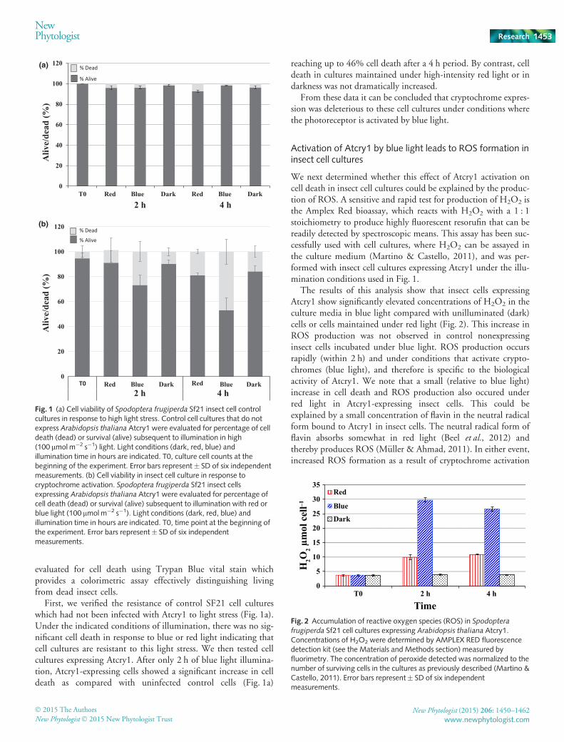
First, we verified the resistance of control SF21 cell cultureswhich had not been infected with Atcry1 to light stress (Fig. 1a).Under the indicated conditions of illumination, there was no sig-nificant cell death in response to blue or red light indicating thatcell cultures are resistant to this light stress. We then tested cellcultures expressing Atcry1. After only 2 h of blue light illumina-tion, Atcry1-expressing cells showed a significant increase in celldeath as compared with uninfected control cells (Fig. 1a)
reaching up to 46% cell death after a 4 h period. By contrast, celldeath in cultures maintained under high-intensity red light or indarkness was not dramatically increased.
From these data it can be concluded that cryptochrome expres-sion was deleterious to these cell cultures under conditions wherethe photoreceptor is activated by blue light.
Activation of Atcry1 by blue light leads to ROS formation ininsect cell cultures
We next determined whether this effect of Atcry1 activation oncell death in insect cell cultures could be explained by the produc-tion of ROS. A sensitive and rapid test for production of H2O2 isthe Amplex Red bioassay, which reacts with H2O2 with a 1 : 1stoichiometry to produce highly fluorescent resorufin that can bereadily detected by spectroscopic means. This assay has been suc-cessfully used with cell cultures, where H2O2 can be assayed inthe culture medium (Martino & Castello, 2011), and was per-formed with insect cell cultures expressing Atcry1 under the illu-mination conditions used in Fig. 1.
The results of this analysis show that insect cells expressingAtcry1 show significantly elevated concentrations of H2O2 in theculture media in blue light compared with unilluminated (dark)cells or cells maintained under red light (Fig. 2). This increase inROS production was not observed in control nonexpressinginsect cells incubated under blue light. ROS production occursrapidly (within 2 h) and under conditions that activate crypto-chromes (blue light), and therefore is specific to the biologicalactivity of Atcry1. We note that a small (relative to blue light)increase in cell death and ROS production also occured underred light in Atcry1-expressing insect cells. This could beexplained by a small concentration of flavin in the neutral radicalform bound to Atcry1 in insect cells. The neutral radical form offlavin absorbs somewhat in red light (Beel et al., 2012) andthereby produces ROS (M€uller & Ahmad, 2011). In either event,increased ROS formation as a result of cryptochrome activation
0
20
40
60
80
100
120
T0 Red Blue Dark Red Blue Dark
Aliv
e/de
ad (%
)A
live/
dead
(%)
2 h 4 h
% Dead
% Alive
0
20
40
60
80
100
120
Red Blue Dark Red Blue DarkT02 h 4 h
% Alive
% Dead
(a)
(b)
Fig. 1 (a) Cell viability of Spodoptera frugiperda Sf21 insect cell controlcultures in response to high light stress. Control cell cultures that do notexpress Arabidopsis thaliana Atcry1 were evaluated for percentage of celldeath (dead) or survival (alive) subsequent to illumination in high(100 lmol m�2 s�1) light. Light conditions (dark, red, blue) andillumination time in hours are indicated. T0, culture cell counts at thebeginning of the experiment. Error bars represent� SD of six independentmeasurements. (b) Cell viability in insect cell culture in response tocryptochrome activation. Spodoptera frugiperda Sf21 insect cellsexpressing Arabidopsis thaliana Atcry1 were evaluated for percentage ofcell death (dead) or survival (alive) subsequent to illumination with red orblue light (100 lmol m�2 s�1). Light conditions (dark, red, blue) andillumination time in hours are indicated. T0, time point at the beginning ofthe experiment. Error bars represent� SD of six independentmeasurements.
0
5
10
15
20
25
30
35
T0 2 h 4 h
H2O
2 μm
ol c
ell–1
Time
Red
Blue
Dark
Fig. 2 Accumulation of reactive oxygen species (ROS) in Spodoptera
frugiperda Sf21 cell cultures expressing Arabidopsis thaliana Atcry1.Concentrations of H2O2 were determined by AMPLEX RED fluorescencedetection kit (see the Materials and Methods section) measured byfluorimetry. The concentration of peroxide detected was normalized to thenumber of surviving cells in the cultures as previously described (Martino &Castello, 2011). Error bars represent� SD of six independentmeasurements.
� 2015 The Authors
New Phytologist� 2015 New Phytologist TrustNew Phytologist (2015) 206: 1450–1462
www.newphytologist.com
NewPhytologist Research 1453
can explain the increased cell death observed in blue light in thesecell cultures (Fig. 1b).
Oxygen concentration dependence of the Atcry1 responsein insect cell cultures
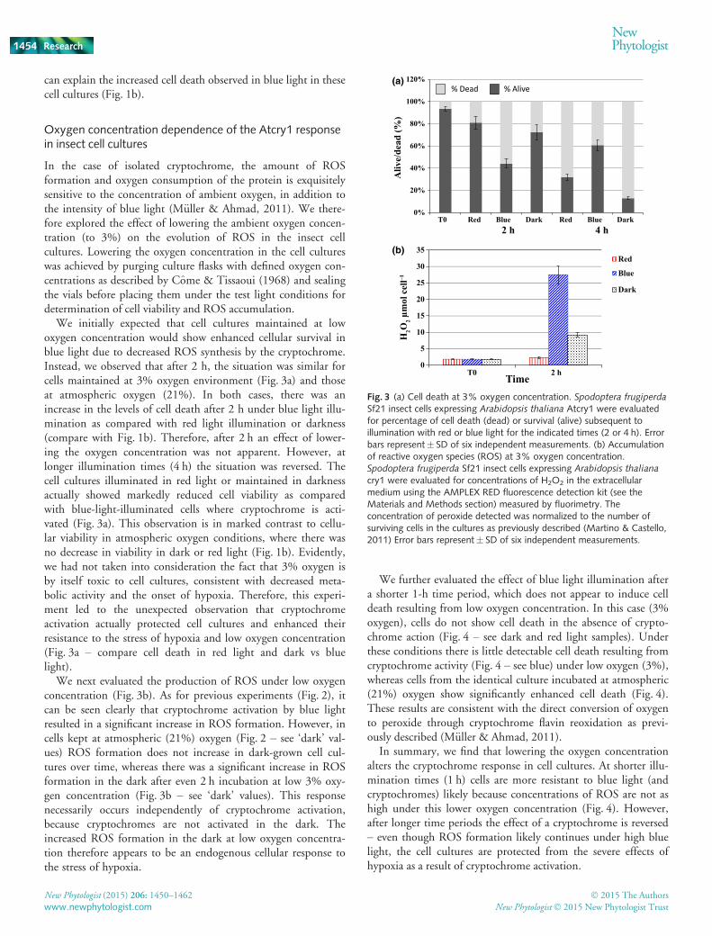
In the case of isolated cryptochrome, the amount of ROSformation and oxygen consumption of the protein is exquisitelysensitive to the concentration of ambient oxygen, in addition tothe intensity of blue light (M€uller & Ahmad, 2011). We there-fore explored the effect of lowering the ambient oxygen concen-tration (to 3%) on the evolution of ROS in the insect cellcultures. Lowering the oxygen concentration in the cell cultureswas achieved by purging culture flasks with defined oxygen con-centrations as described by Come & Tissaoui (1968) and sealingthe vials before placing them under the test light conditions fordetermination of cell viability and ROS accumulation.
We initially expected that cell cultures maintained at lowoxygen concentration would show enhanced cellular survival inblue light due to decreased ROS synthesis by the cryptochrome.Instead, we observed that after 2 h, the situation was similar forcells maintained at 3% oxygen environment (Fig. 3a) and thoseat atmospheric oxygen (21%). In both cases, there was anincrease in the levels of cell death after 2 h under blue light illu-mination as compared with red light illumination or darkness(compare with Fig. 1b). Therefore, after 2 h an effect of lower-ing the oxygen concentration was not apparent. However, atlonger illumination times (4 h) the situation was reversed. Thecell cultures illuminated in red light or maintained in darknessactually showed markedly reduced cell viability as comparedwith blue-light-illuminated cells where cryptochrome is acti-vated (Fig. 3a). This observation is in marked contrast to cellu-lar viability in atmospheric oxygen conditions, where there wasno decrease in viability in dark or red light (Fig. 1b). Evidently,we had not taken into consideration the fact that 3% oxygen isby itself toxic to cell cultures, consistent with decreased meta-bolic activity and the onset of hypoxia. Therefore, this experi-ment led to the unexpected observation that cryptochromeactivation actually protected cell cultures and enhanced theirresistance to the stress of hypoxia and low oxygen concentration(Fig. 3a – compare cell death in red light and dark vs bluelight).
We next evaluated the production of ROS under low oxygenconcentration (Fig. 3b). As for previous experiments (Fig. 2), itcan be seen clearly that cryptochrome activation by blue lightresulted in a significant increase in ROS formation. However, incells kept at atmospheric (21%) oxygen (Fig. 2 – see ‘dark’ val-ues) ROS formation does not increase in dark-grown cell cul-tures over time, whereas there was a significant increase in ROSformation in the dark after even 2 h incubation at low 3% oxy-gen concentration (Fig. 3b – see ‘dark’ values). This responsenecessarily occurs independently of cryptochrome activation,because cryptochromes are not activated in the dark. Theincreased ROS formation in the dark at low oxygen concentra-tion therefore appears to be an endogenous cellular response tothe stress of hypoxia.
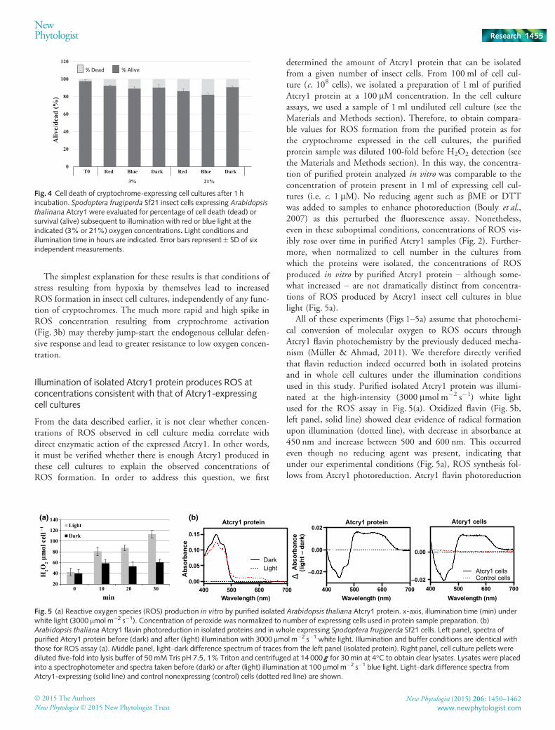
We further evaluated the effect of blue light illumination aftera shorter 1-h time period, which does not appear to induce celldeath resulting from low oxygen concentration. In this case (3%oxygen), cells do not show cell death in the absence of crypto-chrome action (Fig. 4 – see dark and red light samples). Underthese conditions there is little detectable cell death resulting fromcryptochrome activity (Fig. 4 – see blue) under low oxygen (3%),whereas cells from the identical culture incubated at atmospheric(21%) oxygen show significantly enhanced cell death (Fig. 4).These results are consistent with the direct conversion of oxygento peroxide through cryptochrome flavin reoxidation as previ-ously described (M€uller & Ahmad, 2011).
In summary, we find that lowering the oxygen concentrationalters the cryptochrome response in cell cultures. At shorter illu-mination times (1 h) cells are more resistant to blue light (andcryptochromes) likely because concentrations of ROS are not ashigh under this lower oxygen concentration (Fig. 4). However,after longer time periods the effect of a cryptochrome is reversed– even though ROS formation likely continues under high bluelight, the cell cultures are protected from the severe effects ofhypoxia as a result of cryptochrome activation.
0%
20%
40%
60%
80%
100%
120%
T0 Red Blue Dark Red Blue Dark
Aliv
e/de
ad (%
)
2 h 4 h
% Alive% Dead
0
5
10
15
20
25
30
35
T0 2 h
H2O
2 μm
ol c
ell–1
Time
Red
Blue
Dark
(a)
(b)
Fig. 3 (a) Cell death at 3% oxygen concentration. Spodoptera frugiperdaSf21 insect cells expressing Arabidopsis thaliana Atcry1 were evaluatedfor percentage of cell death (dead) or survival (alive) subsequent toillumination with red or blue light for the indicated times (2 or 4 h). Errorbars represent� SD of six independent measurements. (b) Accumulationof reactive oxygen species (ROS) at 3% oxygen concentration.Spodoptera frugiperda Sf21 insect cells expressing Arabidopsis thaliana
cry1 were evaluated for concentrations of H2O2 in the extracellularmedium using the AMPLEX RED fluorescence detection kit (see theMaterials and Methods section) measured by fluorimetry. Theconcentration of peroxide detected was normalized to the number ofsurviving cells in the cultures as previously described (Martino & Castello,2011) Error bars represent� SD of six independent measurements.
New Phytologist (2015) 206: 1450–1462 � 2015 The Authors
New Phytologist� 2015 New Phytologist Trustwww.newphytologist.com
Research
NewPhytologist1454
The simplest explanation for these results is that conditions ofstress resulting from hypoxia by themselves lead to increasedROS formation in insect cell cultures, independently of any func-tion of cryptochromes. The much more rapid and high spike inROS concentration resulting from cryptochrome activation(Fig. 3b) may thereby jump-start the endogenous cellular defen-sive response and lead to greater resistance to low oxygen concen-tration.
Illumination of isolated Atcry1 protein produces ROS atconcentrations consistent with that of Atcry1-expressingcell cultures
From the data described earlier, it is not clear whether concen-trations of ROS observed in cell culture media correlate withdirect enzymatic action of the expressed Atcry1. In other words,it must be verified whether there is enough Atcry1 produced inthese cell cultures to explain the observed concentrations ofROS formation. In order to address this question, we first
determined the amount of Atcry1 protein that can be isolatedfrom a given number of insect cells. From 100 ml of cell cul-ture (c. 108 cells), we isolated a preparation of 1 ml of purifiedAtcry1 protein at a 100 lM concentration. In the cell cultureassays, we used a sample of 1 ml undiluted cell culture (see theMaterials and Methods section). Therefore, to obtain compara-ble values for ROS formation from the purified protein as forthe cryptochrome expressed in the cell cultures, the purifiedprotein sample was diluted 100-fold before H2O2 detection (seethe Materials and Methods section). In this way, the concentra-tion of purified protein analyzed in vitro was comparable to theconcentration of protein present in 1 ml of expressing cell cul-tures (i.e. c. 1 lM). No reducing agent such as bME or DTTwas added to samples to enhance photoreduction (Bouly et al.,2007) as this perturbed the fluorescence assay. Nonetheless,even in these suboptimal conditions, concentrations of ROS vis-ibly rose over time in purified Atcry1 samples (Fig. 2). Further-more, when normalized to cell number in the cultures fromwhich the proteins were isolated, the concentrations of ROSproduced in vitro by purified Atcry1 protein – although some-what increased – are not dramatically distinct from concentra-tions of ROS produced by Atcry1 insect cell cultures in bluelight (Fig. 5a).
All of these experiments (Figs 1–5a) assume that photochemi-cal conversion of molecular oxygen to ROS occurs throughAtcry1 flavin photochemistry by the previously deduced mecha-nism (M€uller & Ahmad, 2011). We therefore directly verifiedthat flavin reduction indeed occurred both in isolated proteinsand in whole cell cultures under the illumination conditionsused in this study. Purified isolated Atcry1 protein was illumi-nated at the high-intensity (3000 lmol m�2 s�1) white lightused for the ROS assay in Fig. 5(a). Oxidized flavin (Fig. 5b,left panel, solid line) showed clear evidence of radical formationupon illumination (dotted line), with decrease in absorbance at450 nm and increase between 500 and 600 nm. This occurredeven though no reducing agent was present, indicating thatunder our experimental conditions (Fig. 5a), ROS synthesis fol-lows from Atcry1 photoreduction. Atcry1 flavin photoreduction
0
20
40
60
80
100
120
Red Blue Dark
3% 21%
Red Blue Dark
Aliv
e/de
ad (%
)
T0
% Dead % Alive
Fig. 4 Cell death of cryptochrome-expressing cell cultures after 1 hincubation. Spodoptera frugiperda Sf21 insect cells expressing Arabidopsis
thalinana Atcry1 were evaluated for percentage of cell death (dead) orsurvival (alive) subsequent to illumination with red or blue light at theindicated (3% or 21%) oxygen concentrations. Light conditions andillumination time in hours are indicated. Error bars represent� SD of sixindependent measurements.
20
40
60
80
100
120
140
0 10 20 30
H2O
2 μm
ol c
ell –1
min
Light
Dark
400 500 600 700–0.02
0.00
Wavelength (nm)Wavelength (nm)Wavelength (nm)
Atcry1 cellsControl cells
Atcry1 cells
400 500 600 700
–0.02
0.00
0.02
400 500 600 7000.00
0.05
0.10
0.15
LightDark
Atcry1 protein Atcry1 protein
Abs
orba
nce
(ligh
t –da
rk)
Abs
orba
nce
Δ
(a) (b)
Fig. 5 (a) Reactive oxygen species (ROS) production in vitro by purified isolated Arabidopsis thaliana Atcry1 protein. x-axis, illumination time (min) underwhite light (3000 lmol m�2 s�1). Concentration of peroxide was normalized to number of expressing cells used in protein sample preparation. (b)Arabidopsis thaliana Atcry1 flavin photoreduction in isolated proteins and in whole expressing Spodoptera frugiperda Sf21 cells. Left panel, spectra ofpurified Atcry1 protein before (dark) and after (light) illumination with 3000 lmol m�2 s�1 white light. Illumination and buffer conditions are identical withthose for ROS assay (a). Middle panel, light–dark difference spectrum of traces from the left panel (isolated protein). Right panel, cell culture pellets werediluted five-fold into lysis buffer of 50mM Tris pH 7.5, 1% Triton and centrifuged at 14 000 g for 30min at 4°C to obtain clear lysates. Lysates were placedinto a spectrophotometer and spectra taken before (dark) or after (light) illumination at 100 lmol m�2 s�1 blue light. Light–dark difference spectra fromAtcry1-expressing (solid line) and control nonexpressing (control) cells (dotted red line) are shown.
� 2015 The Authors
New Phytologist� 2015 New Phytologist TrustNew Phytologist (2015) 206: 1450–1462
www.newphytologist.com
NewPhytologist Research 1455
is known to occur in whole expressing insect cells using a vari-ety of spectroscopic approaches (Bouly et al., 2007; Burneyet al., 2012). To confirm that this also occurs under our illu-mination conditions, we examined extracts from whole cells inwhich Atcry1 protein can be directly visualized optically(Engelhard et al., 2014). Whole cell lysates from both Atcry1-expressing and control cells were taken either before (dark) orafter (light) illumination with 100 lmol m�2 s�1 blue light, asused in Figs 1–4. The ‘light’ scan was subtracted from the‘dark’ scan to provide a so-called ‘difference spectrum’ (light–dark) that detects any absorbance change in the sample afterillumination and therefore any photochemical reaction. As canbe seen (Fig. 5b, right panel, solid line), there is clear evidenceof flavin radical formation in Atcry1-expressing cells but nonein control cultures (Fig. 5b, right panel, red dotted line). Thedifference spectrum obtained from Atcry1-expressing cell cul-tures is moreover essentially identical to that obtained fromthe isolated purified protein (Fig. 5b, middle panel). It cantherefore be concluded that increased concentrations of ROSin cry1-expressing cell cultures is consistent with direct enzy-matic conversion of O2 by cryptochromes (M€uller & Ahmad,2011).
Colocalization of cryptochromes with sites of ROSproduction in Atcry1-expressing insect cell cultures
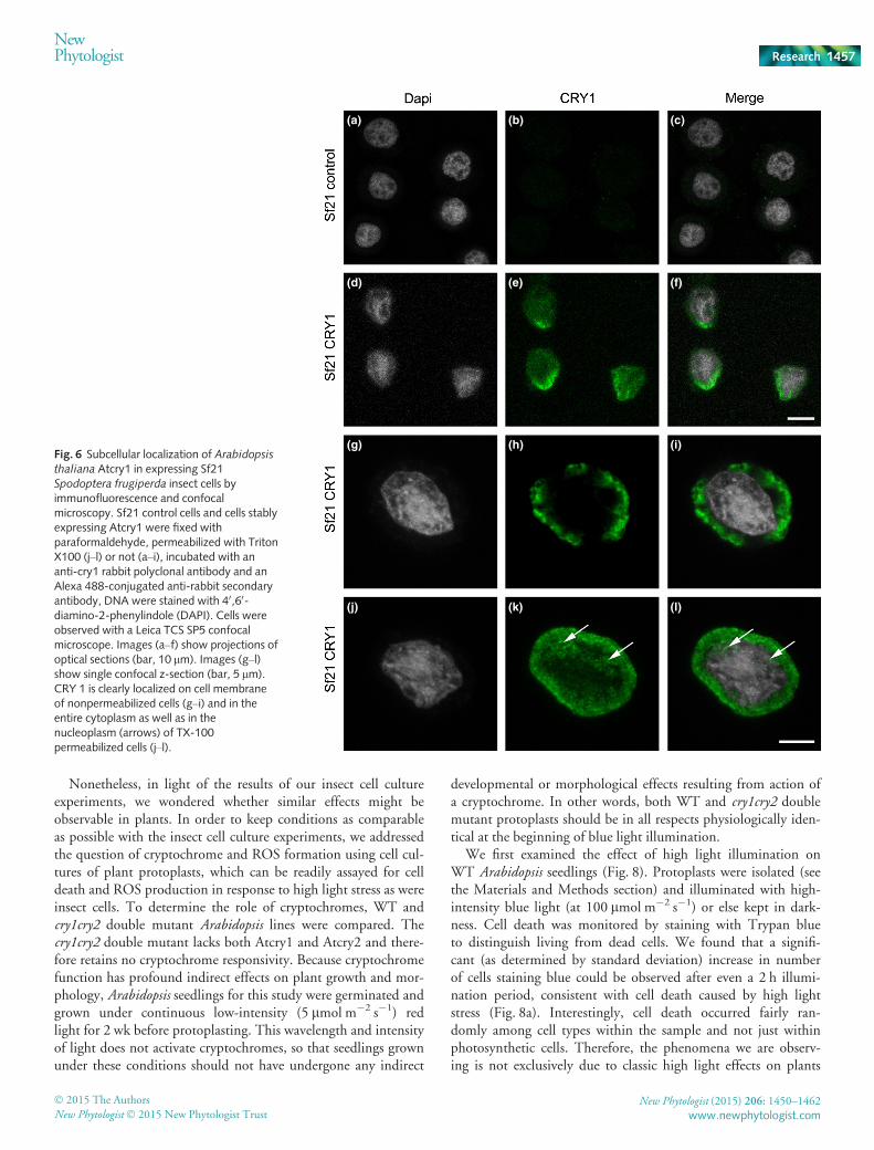
In order to obtain definitive proof linking ROS accumulatingin insect cell cultures to the enzymatic activity of crypto-chromes, it is necessary that a cryptochrome colocalizes with thesite of ROS formation in these cell cultures. To test this predic-tion, we first performed immunolocalization studies of crypto-chromes in expressing insect cells (Fig. 6), which were stainedwith DAPI in order to identify the nucleus. To unequivocallyassess the subcellular localization of Atcry1, we used a classicalmethod to label cells before or after permeabilization (Mottolaet al., 2000). Without permeabilization, the antibody crosses theplasma membrane very poorly and recognizes mainly the Atcry1protein present at or near the cell surface. After permeabilizationof cells with 0.1% Triton, antibodies can cross the cell mem-brane and label protein within the entire cell including thenucleus.
Labeling with a polyclonal anti-Atcry1 antibody showed nosignal in control uninfected cells. In Atcry1-expressing cells,immunolabeled without permeabilization, there was clear proteinsignal outside the nucleus, preferentially at the cellular membranein large concentrated zones (Fig. 6e). This is clearly seen in cellsobserved at higher magnification on the optical section across thenucleus (Fig. 6h,i). Images performed with permeabilized cells,showed the cryptochrome within the entire cytoplasm and alsowithin the nucleus itself (Fig. 6k,l). Here it is found mainlywithin the interstitial spaces within the chromatin and notapparently associated with the chromatin itself (see arrows onFig. 6k,l). This distribution is consistent with that reported inplants (Yang et al., 2000), where cryptochromes are localized inboth cytosolic and nuclear compartments. A 3D image of the im-munolocalization of the cryptochrome in insect cells is further
included in Supporting Information Video S1 (nonpermeabilizedcells) and Video S2 (permeabilized cells).
In order to better visualize Atcry1 in subnuclear structures, weperformed a 3D fluorescence imaging analysis of a cry1 positivenucleus by using TANGO, a powerful image analysis tool dedi-cated to the study of nuclear architecture (Ollion et al., 2013).With this tool we analyzed Atcry1 staining in the entire nucleusindependently from the cytoplasmic Atcry1 staining. First nucleiare segmented from the DAPI stained images, then green chan-nels corresponding to Atcry1 staining are cropped around thesegmented nuclei, and finally cry1 staining contained in subnu-clear structures can be segmented. A representative result can bevisualized by a 3D representation movie in Video S3 where acry1-positive structure can be clearly seen throw the nucleus bytransparency.
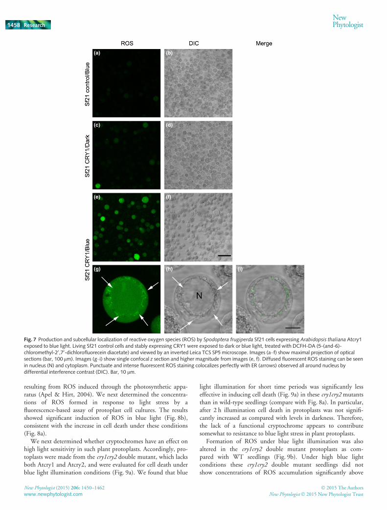
In order to evaluate the site of formation of ROS in thesecells, we used a fluorescent stain (hydroxyphenyl fluorescein)that can directly visualize H2O2 within living cells (Lariguetet al., 2013). Living insect cells were treated with fluorescentsubstrate and then either kept in the dark or illuminated withblue light (Fig. 7). Under these conditions, no ROS wasdetected in uninfected SF21 control cell cultures, and verylittle in SF21 expressing insect cells kept in darkness (Fig. 7,top panels). However, illumination with just 10 min of bluelight resulted in accumulation of ROS within cells. Further-more, the localization of the accumulated ROS was in bothcytoplasmic and nuclear compartments, consistent with theimmunolocalization studies (Fig. 6). ROS formation was partic-ularly abundant in vesicular structures surrounding the nucleusthat perfectly match endoplasmic reticulum observed by DIC(Fig. 7g,h).
In summary, ROS formation occurs on a rapid timescale con-sistent with cryptochrome activation and is colocalized within thesame cellular compartments.
Cryptochromes and ROS formation in plant protoplasts
The role of cryptochromes in plant pathogen response, highlight stress and programmed cell death (PCD) has beendocumented in numerous studies (Chaves et al., 2011). How-ever, no study has analyzed whether cryptochromes by them-selves directly synthesize ROS in living plant cells. Thisquestion has been difficult to address, because most formsof oxidative stress resulting from high light have been linkedto formation of ROS in the chloroplast. A direct involvementof cryptochrome in production of reactive oxygen is there-fore unlikely, because cryptochromes with known signalingroles such as Atcry1 and Atcry2 are not localized to the chlo-roplast. The involvement of cryptochrome in responses toROS is thought to be indirect, either through regulating thebiosynthesis of the photosynthetic apparatus (including light-absorbing antenna and photoprotective pigments such asanthocyanins), or else by interacting with defense and stressresponse signaling pathways through effects on transcriptionalactivators in the nucleus such as hyh, hy5 and so on (Chaveset al., 2011).
New Phytologist (2015) 206: 1450–1462 � 2015 The Authors
New Phytologist� 2015 New Phytologist Trustwww.newphytologist.com
Research
NewPhytologist1456
Nonetheless, in light of the results of our insect cell cultureexperiments, we wondered whether similar effects might beobservable in plants. In order to keep conditions as comparableas possible with the insect cell culture experiments, we addressedthe question of cryptochrome and ROS formation using cell cul-tures of plant protoplasts, which can be readily assayed for celldeath and ROS production in response to high light stress as wereinsect cells. To determine the role of cryptochromes, WT andcry1cry2 double mutant Arabidopsis lines were compared. Thecry1cry2 double mutant lacks both Atcry1 and Atcry2 and there-fore retains no cryptochrome responsivity. Because cryptochromefunction has profound indirect effects on plant growth and mor-phology, Arabidopsis seedlings for this study were germinated andgrown under continuous low-intensity (5 lmol m�2 s�1) redlight for 2 wk before protoplasting. This wavelength and intensityof light does not activate cryptochromes, so that seedlings grownunder these conditions should not have undergone any indirect
developmental or morphological effects resulting from action ofa cryptochrome. In other words, both WT and cry1cry2 doublemutant protoplasts should be in all respects physiologically iden-tical at the beginning of blue light illumination.
We first examined the effect of high light illumination onWT Arabidopsis seedlings (Fig. 8). Protoplasts were isolated (seethe Materials and Methods section) and illuminated with high-intensity blue light (at 100 lmol m�2 s�1) or else kept in dark-ness. Cell death was monitored by staining with Trypan blueto distinguish living from dead cells. We found that a signifi-cant (as determined by standard deviation) increase in numberof cells staining blue could be observed after even a 2 h illumi-nation period, consistent with cell death caused by high lightstress (Fig. 8a). Interestingly, cell death occurred fairly ran-domly among cell types within the sample and not just withinphotosynthetic cells. Therefore, the phenomena we are observ-ing is not exclusively due to classic high light effects on plants
(a) (b) (c)
(d) (e) (f)
(g) (h) (i)
(j) (k) (l)
Fig. 6 Subcellular localization of Arabidopsisthaliana Atcry1 in expressing Sf21Spodoptera frugiperda insect cells byimmunofluorescence and confocalmicroscopy. Sf21 control cells and cells stablyexpressing Atcry1 were fixed withparaformaldehyde, permeabilized with TritonX100 (j–l) or not (a–i), incubated with ananti-cry1 rabbit polyclonal antibody and anAlexa 488-conjugated anti-rabbit secondaryantibody, DNA were stained with 40,60-diamino-2-phenylindole (DAPI). Cells wereobserved with a Leica TCS SP5 confocalmicroscope. Images (a–f) show projections ofoptical sections (bar, 10 lm). Images (g–l)show single confocal z-section (bar, 5 lm).CRY 1 is clearly localized on cell membraneof nonpermeabilized cells (g–i) and in theentire cytoplasm as well as in thenucleoplasm (arrows) of TX-100permeabilized cells (j–l).
� 2015 The Authors
New Phytologist� 2015 New Phytologist TrustNew Phytologist (2015) 206: 1450–1462
www.newphytologist.com
NewPhytologist Research 1457
resulting from ROS induced through the photosynthetic appa-ratus (Apel & Hirt, 2004). We next determined the concentra-tions of ROS formed in response to light stress by afluorescence-based assay of protoplast cell cultures. The resultsshowed significant induction of ROS in blue light (Fig. 8b),consistent with the increase in cell death under these conditions(Fig. 8a).
We next determined whether cryptochromes have an effect onhigh light sensitivity in such plant protoplasts. Accordingly, pro-toplasts were made from the cry1cry2 double mutant, which lacksboth Atcry1 and Atcry2, and were evaluated for cell death underblue light illumination conditions (Fig. 9a). We found that blue
light illumination for short time periods was significantly lesseffective in inducing cell death (Fig. 9a) in these cry1cry2 mutantsthan in wild-type seedlings (compare with Fig. 8a). In particular,after 2 h illumination cell death in protoplasts was not signifi-cantly increased as compared with levels in darkness. Therefore,the lack of a functional cryptochrome appears to contributesomewhat to resistance to blue light stress in plant protoplasts.
Formation of ROS under blue light illumination was alsoaltered in the cry1cry2 double mutant protoplasts as com-pared with WT seedlings (Fig. 9b). Under high blue lightconditions these cry1cry2 double mutant seedlings did notshow concentrations of ROS accumulation significantly above
(a) (b)
(c) (d)
(e)
(g) (h) (i)
(f)
Fig. 7 Production and subcellular localization of reactive oxygen species (ROS) by Spodoptera frugiperda Sf21 cells expressing Arabidopsis thaliana Atcry1exposed to blue light. Living Sf21 control cells and stably expressing CRY1 were exposed to dark or blue light, treated with DCFH-DA (5-(and-6)-chloromethyl-20,70-dichlorofluorecein diacetate) and viewed by an inverted Leica TCS SP5 microscope. Images (a–f) show maximal projection of opticalsections (bar, 100 lm). Images (g–i) show single confocal z section and higher magnitude from images (e, f). Diffused fluorescent ROS staining can be seenin nucleus (N) and cytoplasm. Punctuate and intense fluorescent ROS staining colocalizes perfectly with ER (arrows) observed all around nucleus bydifferential interference contrast (DIC). Bar, 10 lm.
New Phytologist (2015) 206: 1450–1462 � 2015 The Authors
New Phytologist� 2015 New Phytologist Trustwww.newphytologist.com
Research
NewPhytologist1458
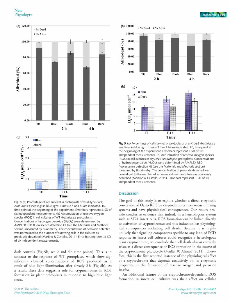
dark controls (Fig. 9b, see 2 and 4 h time points). This is incontrast to the response of WT protoplasts, which show sig-nificantly elevated concentrations of ROS produced as aresult of blue light illumination after already 2 h (Fig. 8b). Asa result, these data suggest a role for cryptochromes in ROSformation in plant protoplasts in response to high blue lightstress.
Discussion
The goal of this study is to explore whether a direct enzymaticconversion of O2 to ROS by cryptochromes may occur in livingsystems and have physiological consequences. Our results pro-vide conclusive evidence that indeed, in a heterologous systemsuch as SF21 insect cells, ROS formation can be linked directlyto activation of cryptochromes and this induction has physiolog-ical consequences including cell death. Because it is highlyunlikely that signaling components specific to any kind of PCDresponse in insect cell cultures could recognize a heterologousplant cryptochrome, we conclude that cell death almost certainlyarises as a direct consequence of ROS formation in the course ofthe cryptochrome photocycle (M€uller & Ahmad, 2011). There-fore, this is the first reported instance of the physiological effectof a cryptochrome that depends exclusively on its enzymaticproperties in the formation of reactive oxygen and peroxidein vivo.
An additional feature of the cryptochrome-dependent ROSformation in insect cell cultures was their effect on cellular
0.00
20.00
40.00
60.00
80.00
100.00
120.00
T0 Blue Dark Blue Dark
Aliv
e/de
ad (%
)
2 h 4 h
% Dead% Alive
1
1.5
2
2.5
3
3.5
T0 T 2 h T 4 h
H2O
2 μm
ol c
ell–1
(%)
BlueDark
Time
(a)
(b)
Fig. 8 (a) Percentage of cell survival in protoplasts of wild-type (WT)Arabidopsis seedlings in blue light. Times (2 h or 4 h) are indicated. T0,time point at the beginning of the experiment. Error bars represent� SD ofsix independent measurements. (b) Accumulation of reactive oxygenspecies (ROS) in cell cultures of WT Arabidopsis protoplasts.Concentrations of hydrogen peroxide (H2O2) were determined byAMPLEX RED fluorescence detection kit (see the Materials and Methodssection) measured by fluorimetry. The concentration of peroxide detectedwas normalized to the number of surviving cells in the cultures aspreviously described (Martino & Castello, 2011). Error bars represent� SDof six independent measurements.
0.00
20.00
40.00
60.00
80.00
100.00
120.00
T0 Blue Dark Blue Dark
Aliv
e/de
ad (%
)
2 h 4 h
% Dead % Alive
5
7
9
11
13
15
17
T0 T 2 h T 4 hH
2O2 μ
mol
cel
l–1
BlueDark
Time
(a)
(b)
Fig. 9 (a) Percentage of cell survival of protoplasts of cry1cry2 Arabidopsis
seedlings in blue light. Times (2 h or 4 h) are indicated. T0, time point atthe beginning of the experiment. Error bars represent� SD of sixindependent measurements. (b) Accumulation of reactive oxygen species(ROS) in cell cultures of cry1cry2 Arabidopsis protoplasts. Concentrationsof hydrogen peroxide (H2O2) were determined by AMPLEX REDfluorescence detection kit (see the Materials and Methods section)measured by fluorimetry. The concentration of peroxide detected wasnormalized to the number of surviving cells in the cultures as previouslydescribed (Martino & Castello, 2011). Error bars represent� SD of sixindependent measurements.
� 2015 The Authors
New Phytologist� 2015 New Phytologist TrustNew Phytologist (2015) 206: 1450–1462
www.newphytologist.com
NewPhytologist Research 1459
survival under conditions of hypoxia (low oxygen concentration).These experiments were initially undertaken in order to deter-mine whether low oxygen concentration results in reduced ROSformation by cryptochromes and thereby reduced cell death. Thisprediction arises from the enzymatic properties of cryptochromesand their (reduced) activity at low oxygen concentration (M€uller& Ahmad, 2011). In keeping with this prediction, after a shortillumination time (1 h) there was indeed reduced cell death underlow oxygen concentration (3%) as compared with atmosphericoxygen (21%) (Fig. 4). A direct enzymatic role for cryptochromesin cell death by means of formation of H2O2 and ROS couldtherefore be further strengthened.
However, an unexpected finding of the effect of low oxygenconcentration was that at longer illumination times (4 h), crypto-chromes actually played a protective role. Under conditions oflow oxygen, cell cultures die as a result of hypoxia, as can be seenfrom the high percentage of cell death after 4 h continuous dark-ness (Fig. 3a). Under blue light illumination (and therefore acti-vation of Atcry1) there was significantly less cell death after 4 hincubation in low oxygen than in dark controls. This indicatesthat that activation of the cryptochrome resulted in induction ofresistance (defensive mechanisms) to hypoxia. Consistent withthis interpretation, we noted elevated concentrations of ROS incell cultures subjected to low oxygen concentrations, even in thedark (Fig. 3b). This indicates that hypoxia leads to the onset ofoxidative stress and ROS formation independently of any actionof cryptochromes; however, the concentrations of ROS are lowerthan under blue light illumination. The likely explanation of theprotective effect of Atcry1 on insect cell cultures under conditionsof hypoxia is to provide a large early burst of ROS compared withcell cultures maintained in darkness. This burst of ROS thenreleases downstream signaling pathways that lead to protectivemechanisms. Examples of both protective or toxic effects of ROSabound in the literature (Mittler et al., 2011) and in the case ofinsect cells ROS signaling may play a part in the endogenousresponse to hypoxia. These results are therefore further consistentwith a physiological role resulting from enzymatic ROS forma-tion by cryptochrome photoreceptors.
Production of ROS in animal cells is thought to occur primar-ily as a result of metabolic activity in the mitochondria (Holm-str€om & Finkel, 2014). By contrast, plant cryptochromes havebeen reported to be localized to cytosolic and nuclear cellularcompartments (Guo et al., 1999; Kleiner et al., 1999; Yang et al.,2000). Consistent with this data, immunolocalization studies forAtcry1 performed in insect cell cultures show that Atcry is local-ized to both nuclear and cytosolic compartments. Fluorescentstaining showed that H2O2 is localized to the same compart-ments as the Atcry1 protein. Moreover, that formation of ROS isclearly apparent only 10 min after the onset of illumination, atimescale too short for significant diffusion across membranecompartments. These results suggest that increased ROS forma-tion in insect cells is most likely to result directly from enzymaticaction of Atcry1, and not involve any endogenous signalingmechanisms.
Amounts of cryptochromes in plant cells are at least 100-foldless concentrated than in insect cells (M. Ahmad, unpublished)
and therefore plant cells should not form as large concentrationsof ROS as in insect cell cultures in response to Atcry enzymaticconversion of O2. Nonetheless, illumination of plant cell protop-lasts obtained from Arabidopsis seedlings revealed increased celldeath and ROS formation induced after even 2 h illumination inhigh-intensity blue light. Significantly, both cell death and ROSformation in blue light was somewhat reduced in cryptochromedouble mutant seedlings (cry1cry2 mutants). Furthermore, celldeath resulting from the action of cryptochromes occurred inboth photosynthetic and nonphotosynthetic protoplasts at appar-ently equal efficiency, and also in protoplasts from dark grownetiolated seedlings (not shown), suggesting that evolution ofROS did not require the photosynthetic apparatus. These effects,although significant, are rather weak, and need to be confirmedby additional studies and also in other systems.
It cannot be concluded that the elevated ROS concentrationsmeasured in the cell cultures result directly from enzymaticconversion of oxygen to ROS by cryptochromes in this mate-rial. The concentrations of cryptochrome are simply too low(almost 1009 lower than in transfected insect cell cultures –M. Ahmad, unpublished) to account for the concentrations ofROS being synthesized in plant protoplasts, unless enzymaticconversion is in some way dramatically enhanced. It is never-theless possible that an initial burst of ROS formation resultingfrom the cryptochrome – which occurs within both cytosolicand nuclear cellular compartments – provides sufficient stressto the plant protoplasts to release further ROS signaling and/orsynthetic cascades.
This role for cryptochromes in plant cell protoplasts appears tooccur by a mechanism that is distinct from the well-studied PCDand apoptosis response (Danon et al., 2006; Kim & Apel, 2013).In these prior studies, cryptochromes did not affect ROS forma-tion per se (singlet oxygen accumulates in the plastids as a resultof the flu sensitizer mutation). Instead, the role of Atcry1 wasdetermined to be as a downstream transcriptional activator forexpression of singlet-oxygen induced genes required for the PCDresponse. In fact, no effect of Atcry1 on PCD was documented inthe absence of the flu mutation, which results in increased pro-duction of singlet oxygen in the plastid and is required for thesubsequent effect of Atcry1 (Danon et al., 2006; Kim & Apel,2013). By contrast, under the much higher blue light illumina-tion used in our present study (100 lmol m�2 s�1), the crypto-chrome effect on ROS accumulation occurs in the absence of theflu mutation and appears to be modulated by Atcry1 by someother means. A more recent study has shown reduced ROS accu-mulation and cell death in response to high light stress in cry1and cry2 mutants after a shorter illumination in high white light(Chen et al., 2013), more consistent with our present results.These authors proposed that the downstream transcriptionaleffects of cryptochrome on induction of stress-regulated geneswere responsible for the reduced ROS production and cell deaththey observed in cryptochrome mutants. However, in our find-ings we see effects on cell viability already after 2 h, which do notappear readily explainable by mechanisms that require de novotranscription/protein synthesis. Further experiments will beneeded to resolve what effects a direct enzymatic conversion of
New Phytologist (2015) 206: 1450–1462 � 2015 The Authors
New Phytologist� 2015 New Phytologist Trustwww.newphytologist.com
Research
NewPhytologist1460
molecular oxygen to ROS by cryptochromes may have on theseresponses.
In conclusion, cryptochromess are involved in numerous cellu-lar processes in multiple organisms that implicate formation ofROS. In flies, circadian clock function has been linked to burstsof oxidative stress; whereas blue-light dependent seed germina-tion, cell death and de-etiolation is known to be under the con-trol both of ROS and cryptochromes (Chen et al., 2013; Barreroet al., 2014; Hoang et al., 2014). A role for ROS in photoprotec-tive and stress responses in mammalian cell cultures has also beenextensively documented (Holmstr€om & Finkel, 2014). The cur-rent widely accepted paradigm for cryptochrome activationinvolves light-induced conformational change followed by inter-action with downstream signaling proteins to provide a versatilelight activated switch with many different output functions. Herewe present an additional possibility of cryptochromes playing adirect enzymatic role in the generation of ROS signals whichmay have impacted on responsivity to stress in multiple systemsthrough the course of evolution.
Acknowledgements
We thank the ‘Imagerie Paris Seine’ imaging platform for confo-cal microscopy analyses. We acknowledge the contribution HFSPaward #RGP0045 (to M.A) and AFOSR: FA-9550-14-1-0409(to C.M and M.A). M.E-E is funded by a scholarship from theMinist�ere de l’�education Nationale, France.
References
Apel K, Hirt H. 2004. Reactive oxygen species: metabolism, oxidative stress, and
signal transduction. Annual Review of Plant Biology 55: 373–399.Bailly C, El-Maarouf-Bouteau H, Corbineau F. 2008. From intracellular
signaling networks to cell death: the dual role of reactive oxygen species in seed
physiology. Comptes Rendus Biologies 331: 806–814.Barrero JM, Downie AB, Xu Q, Gubler F. 2014. A role for barley
CRYPTOCHROME1 in light regulation of grain dormancy and germination.
Plant Cell 26: 1094–1104.Beel B, Prager K, Spexard M, Sasso S, Weiss D, M€uller N, Heinnickel M,
Dewez D, Ikoma D, Grossman AR et al. 2012. A flavin binding cryptochrome
photoreceptor responds to both blue and red light in Chlamydomonasreinhardtii. Plant Cell 24: 2992–3008.
Bouly JP, Schleicher E, Dionisio-Sese M, Vandenbussche F, Van Der Straeten
D, Bakrim N, Meier S, Batschauer A, Galland P, Bittl R et al. 2007.Cryptochrome blue light photoreceptors are activated through interconversion
of flavin redox states. Journal of Biological Chemistry 282: 9383–9391.Burney S, Wenzel R, Kottke T, Roussel T, Hoang N, Bouly JP, Bittl R, Heberle
J, Ahmad M. 2012. Single amino acid substitution reveals latent photolyase
activity in Arabidopsis cry1. Angewandte Chemie International Edition 51:9356–9360.
Chaves I, Pokorny R, Byrdin M, Hoang N, Ritz T, Brettel K, Essen LO, van der
Horst GT, Batschauer A, Ahmad M. 2011. The cryptochromes: blue light
photoreceptors in plants and animals. Annual Review of Plant Biology 62: 335–364.
Chen D, Xu G, Tang W, Jing Y, Ji Q, Fei Z, Lin R. 2013. Antagonistic basic
helix-loop-helix/bZIP transcription factors form transcriptional modules that
integrate light and reactive oxygen species signaling in Arabidopsis. Plant Cell25: 1657–1673.
Come D, Tissaoui T. 1968. Induction d’une dormance embryonnaire secondaire
chez le pommier (Pirus malus L.) par des atmospheres tr�es appauvries enoxygene. Comptes Rendus de l’Acad�emie des Sciences, Paris 266: 444–479.
Danon A, Coll NS, Apel K. 2006. Cryptochrome-1-dependent execution of
programmed cell death induced by singlet oxygen in Arabidopsis thaliana.Proceedings of the National Academy of Sciences, USA 103: 17 036–17 041.
Engelhard C, Wang X, Robles D, Moldt J, Essen L-O, Batschauer A, Bittl R,
Ahmad M. 2014. Cellular metabolites enhance light sensitivity through
alternate electron transfer pathways in Arabidopsis cryptochrome. Plant Cell 26:4519–4531.
Gechev TS, Van Breusegem F, Stone JM, Denev I, Laloi C. 2006. Reactive
oxygen species as signals that modulate plant stress responses and programmed
cell death. BioEssays 28: 1091–1101.Guo H, Duong H, Ma N, Lin C. 1999. The Arabidopsis blue light receptor
cryptochrome 2 is a nuclear protein regulated by a blue light-dependent post-
transcriptional mechanism. Plant Journal 19: 279–287.Hoang HH, Sechet J, Bailly C, Leymarie J, Corbineau F. 2014. Inhibition of
germination of dormant barley (Hordeum vulgare L.) grains by blue light asrelated to oxygen and hormonal regulation. Plant, Cell & Environment 37:1393–1403.
Holmstr€om KM, Finkel T. 2014. Cellular mechanisms and physiological
consequences of redox-dependent signalling. Nature Reviews Molecular CellBiology 15: 411–421.
Jeong RD, Chandra-Shekara AC, Barman SR, Navarre D, Klessig DF, Kachroo
A, Kachroo P. 2010. Cryptochrome 2 and phototropin 2 regulate resistance
protein-mediated viral defense by negatively regulating an E3 ubiquitin ligase.
Proceedings of the National Academy of Sciences, USA 107: 13 538–13 543.Kim C, Apel K. 2013. Singlet oxygen-mediated signaling in plants: moving from
flu to wild type reveals an increasing complexity. Photosynthesis Research 116:455–464.
Kleine T, Kindgren P, Benedict C, Hendrickson L, Strand A. 2007. Genome-
wide gene expression analysis reveals a critical role for CRYPTOCHROME1 inthe response of Arabidopsis to high irradiance. Plant Physiology 144: 1391–1406.
Kleiner O, Kircher S, Harter K, Batschauer A. 1999. Nuclear localization of the
Arabidopsis blue light receptor cryptochrome 2. Plant Journal 19: 289–296.Lariguet P, Ranocha P, De Meyer M, Barbier O, Penel C, Dunand C. 2013.
Identification of a hydrogen peroxide signalling pathway in the control of light-
dependent germination in Arabidopsis. Planta 238: 381–395.Liu H, Yu X, Li K, Klejnot J, Yang H, Lisiero D, Lin C. 2008. Photoexcited
CRY2 interacts with CIB1 to regulate transcription and floral initiation in
Arabidopsis. Science 322: 1535–1539.Lopez L, Carbone F, Bianco L, Giuliano G, Facella P, Perrotta G. 2012.
Tomato plants overexpressing cryptochrome 2 reveal altered expression of
energy and stress-related gene products in response to diurnal cues. Plant, Cell& Environment 35: 994–1012.
Martino CF, Castello P. 2011.Modulation of hydrogen peroxide production in
cellular systems by low level magnetic fields. PLoS ONE 6: 1.
Miller G, Shulaev V, Mittler R. 2008. Reactive oxygen signaling and abiotic
stress. Physiologia Plantarum 133: 481–489.Mittler R, Vanderauwera S, Gollery M, Van Breusegem F. 2004. Reactive
oxygen gene network of plants. Trends in Plant Science 9: 490–498.Mittler R, Vanderauwera S, Suzuki N, Miller G, Tognetti VB, Vandepoele K,
Gollery M, Shulaev V, Van Breusegem F. 2011. ROS signaling: the new wave?
Trends in Plant Science 16: 300–309.Mottola G, Jourdan N, Castaldo G, Malagolini N, Lahm A, Serafini-Cessi F,
Migliaccio G, Bonatti S. 2000. A new determinant of endoplasmic reticulum
localization is contained in the juxtamembrane region of the ectodomain of
hepatitis C virus glycoprotein E1. Journal of Biological Chemistry 275: 24 070–24 079.
M€uller P, Ahmad M. 2011. Light-activated cryptochrome reacts with molecular
oxygen to form a flavin-superoxide radical pair consistent with
magnetoreception. Journal of Biological Chemistry 286: 21 033–21 040.Ollion J, Cochennec J, Loll F, Escud�e C, Boudier T. 2013. TANGO: a generic
tool for high throughput 3D image analysis for studying nuclear organization.
Bioinformatics 29: 1840–1841.Orozco-Cardenas M, Ryan CA. 1999. Hydrogen peroxide is generated
systemically in plant leaves by wounding and systemin via the octadecanoid
pathway. Proceedings of the National Academy of Sciences, USA 96: 6553–6557.
� 2015 The Authors
New Phytologist� 2015 New Phytologist TrustNew Phytologist (2015) 206: 1450–1462
www.newphytologist.com
NewPhytologist Research 1461
Quan LJ, Zhang B, Shi WW, Li HY. 2008.Hydrogen peroxide in plants: a
versatile molecule of the reactive oxygen species network. Journal of IntegrativePlant Biology 50: 2–18.
Sanchez A, Shin J, Davis SJ. 2011. Abiotic stress and the plant circadian clock.
Plant Signaling & Behavior 6: 223–231.Sharma P, Chatterjee M, Burman N, Khurana JP. 2014. Cryptochrome 1
regulates growth and development in Brassica through alteration in the
expression of genes involved in light, phytohormone and stress signalling.
Plant, Cell & Environment 37: 961–977.Wu L, Yang HQ. 2010. CRYPTOCHROME 1 is implicated in promoting R
protein-mediated plant resistance to Pseudomonas syringae in Arabidopsis.Molecular Plant 3: 539–548.
Xu P, Ma Z. 2009. Plant cryptochromes employ complicated mechanisms for
subcellular localization and are involved in pathways apart from
photomorphogenesis. Plant Signaling & Behavior 4: 200–201.Xu P, Xiang Y, Zhu HL, Xu HB, Zhang ZZ, Zhang CQ, Zhang L, Ma Z. 2009.
Wheat cryptochromes: subcellular localization and involvement in
photomorphogenesis and osmotic stress responses. Plant Physiology 149: 760–774.Yang HQ, Wu YJ, Tang RH, Liu D, Liu Y, Cashmore AR. 2000. The C-termini
of Arabidopsis cryptochromes mediate a constitutive light response. Cell 103:815–827.
Zuo Z, Liu H, Liu B, Liu X, Lin C. 2011. Blue light-dependent interaction of
CRY2 with SPA1 regulates COP1 activity and floral initiation in Arabidopsis.Current Biology 21: 841–847.
Supporting Information
Additional supporting information may be found in the onlineversion of this article.
Video S1 Three-dimensional (3D) localization of cell membraneCRY1.
Video S2 Three-dimensional (3D) localization of intracellularCRY1.
Video S3 Three-dimensional (3D) subnuclear localization ofCRY1.
Please note: Wiley Blackwell are not responsible for the contentor functionality of any supporting information supplied by theauthors. Any queries (other than missing material) should bedirected to the New Phytologist Central Office.
New Phytologist is an electronic (online-only) journal owned by the New Phytologist Trust, a not-for-profit organization dedicatedto the promotion of plant science, facilitating projects from symposia to free access for our Tansley reviews.
Regular papers, Letters, Research reviews, Rapid reports and both Modelling/Theory and Methods papers are encouraged. We are committed to rapid processing, from online submission through to publication ‘as ready’ via Early View – our average timeto decision is <26 days. There are no page or colour charges and a PDF version will be provided for each article.
The journal is available online at Wiley Online Library. Visit www.newphytologist.com to search the articles and register for tableof contents email alerts.
If you have any questions, do get in touch with Central Office ([email protected]) or, if it is more convenient,our USA Office ([email protected])
For submission instructions, subscription and all the latest information visit www.newphytologist.com
New Phytologist (2015) 206: 1450–1462 � 2015 The Authors
New Phytologist� 2015 New Phytologist Trustwww.newphytologist.com
Research
NewPhytologist1462