Embed Size (px)
Citation preview
BOLD fMRI
Cheryl Olman
4th year student
Department of Neuroscience and
Center for Magnetic Resonance Research
Lecture ‘series’
• Week 1: Biological basis: where’s the signal coming from?
• Week 2: Physical basis: what is the signal, how is it measured?
• Week 3: Imaging basics: imaging sequences, noise and artifacts.
• Week 4: The specific case of BOLD fMRI.
• Week 5: BOLD analysis: what’s significant and what’s not?
• Week 6: Spikes vs. BOLD: neural activity in visual areas
Biological basis
• fMRI measures blood oxygenation and/or flow
• How are blood oxygenation and flow related to neural
activity?
– Oxygen consumption – Blood flow
– Metabolism – Energy budgets
• Things to consider:
– Spatial resolution – Spatial specificity
– Temporal resolution – Neural specificity
Terms
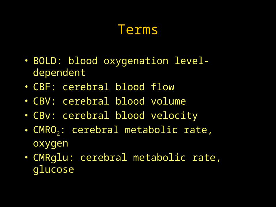
• BOLD: blood oxygenation level-dependent• CBF: cerebral blood flow• CBV: cerebral blood volume• CBv: cerebral blood velocity
• CMRO2: cerebral metabolic rate, oxygen
• CMRglu: cerebral metabolic rate, glucose
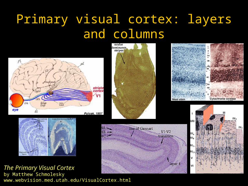
Primary visual cortex: layers and columns
The Primary Visual Cortexby Matthew Schmoleskywww.webvision.med.utah.edu/VisualCortex.html
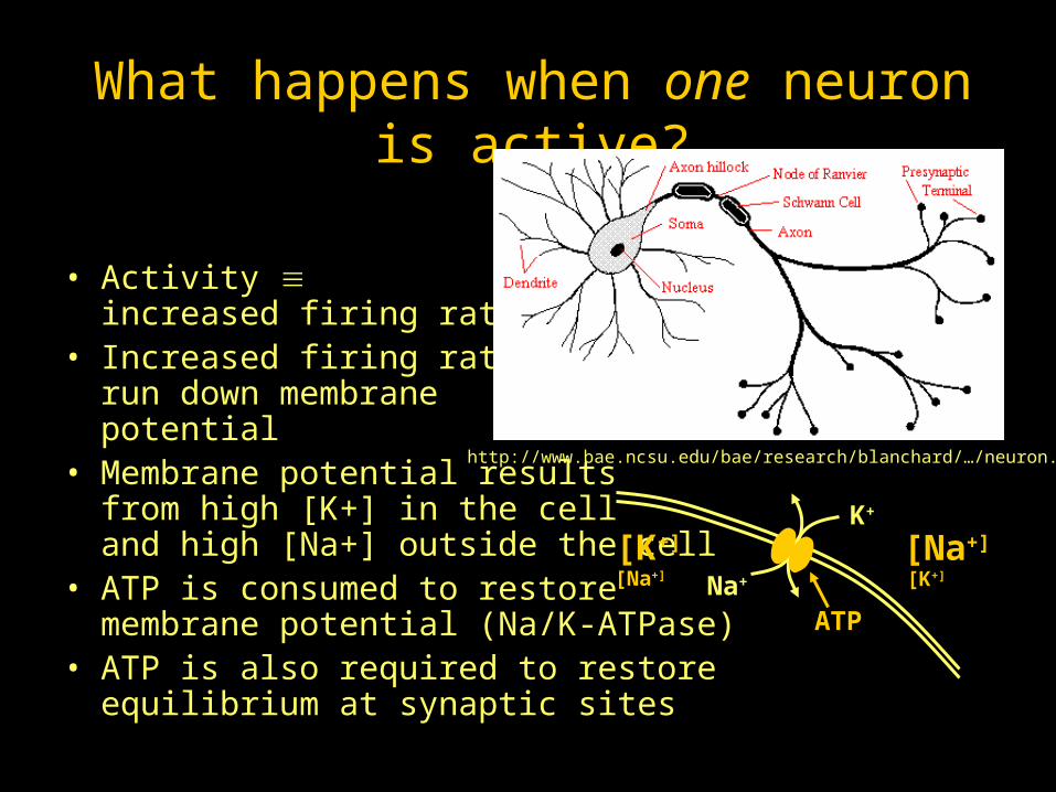
What happens when one neuron is active?
• Activity increased firing rate
• Increased firing rates run down membrane potential
• Membrane potential resultsfrom high [K+] in the celland high [Na+] outside the cell
• ATP is consumed to restore membrane potential (Na/K-ATPase)
• ATP is also required to restore equilibrium at synaptic sites
http://www.bae.ncsu.edu/bae/research/blanchard/…/neuron.gif
ATPNa+
K+
[Na+]
[K+]
[K+]
[Na+]
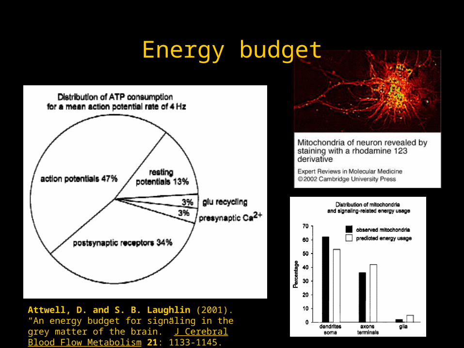
Energy budget
Attwell, D. and S. B. Laughlin (2001). “An energy budget for signaling in the grey matter of the brain.” J Cerebral Blood Flow Metabolism 21: 1133-1145.
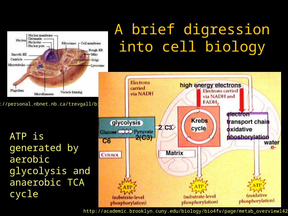
A brief digression into cell biology
http://personal.nbnet.nb.ca/trevgall/biology/
http://academic.brooklyn.cuny.edu/biology/bio4fv/page/metab_overview1424.JPG
ATP is generated by aerobic glycolysis and anaerobic TCA cycle
What happens when many neurons are active?
• The population needs increased CBF to provide glucose and oxygen– Excitatory vs. inhibitory activity
• 90% of neurons are glutamatergic/excitatory• 10% GABAergic/inhibitory
– It’s not just neurons doing the signaling (neurons and glia exist in ~1:1 ratio)
• Possible signals for increased blood flow:– Increased extracellular potassium (direct or indirect effect)– NO: range and timing match well– Other signals transmitted along capillaries or glia?
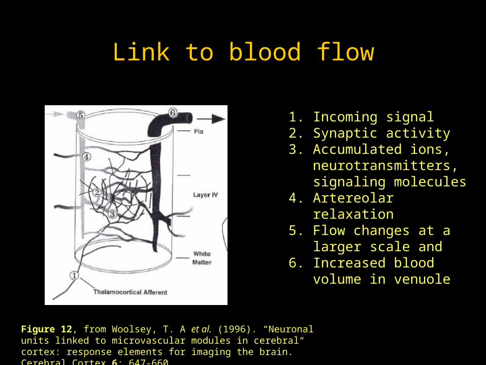
Link to blood flow
Figure 12, from Woolsey, T. A et al. (1996). “Neuronal units linked to microvascular modules in cerebral cortex: response elements for imaging the brain.” Cerebral Cortex 6: 647-660.
1. Incoming signal 2. Synaptic activity3. Accumulated ions,
neurotransmitters, signaling molecules
4. Artereolar relaxation5. Flow changes at a larger
scale and6. Increased blood volume in
venuole
Decoupling of CBF, CMRglu and CMRO2
• PET studies by Fox and Raichle demonstrated 40% increase
in CBF and CMRglu, but only 5% increase in CMRO2
– Fox, P. T. and M. E. Raichle (1986). “Focal physiological uncoupling of cerebral blood flow and
oxidative metabolism during somatosensory stimulation in human subjects.” Proc Natl Acad Sci
USA 83: 1140-1144.
– Fox, P. T., M. E. Raichle, M. A. Mintun and C. Dence (1988). “Nonoxidative glucose consumption
during focal physiologic neural activity.” Science 241: 462-464.
• Positive BOLD signal confirms this!
• Is neural activity anaerobic? Is oxygen consumption
delayed? Is CBF much more widespread than CMRO2?
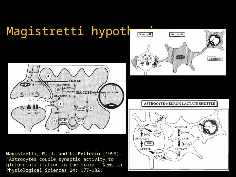
Magistretti hypothesis
Magistretti, P. J. and L. Pellerin (1999). “Astrocytes couple synaptic activity to glucose utilization in the brain.” News in Physiological Sciences 14: 177-182.
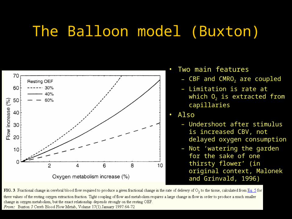
The Balloon model (Buxton)
• Two main features– CBF and CMRO2 are coupled
– Limitation is rate at which O2 is extracted from capillaries
• Also – Undershoot after stimulus is
increased CBV, not delayed oxygen consumption
– Not ‘watering the garden for the sake of one thirsty flower’ (in original context, Malonek and Grinvald, 1996)

Spatial specificity
Figure 10, from Woolsey, T. A et al. (1996). “Neuronal units linked to microvascular modules in cerebral cortex: response elements for imaging the brain.” Cerebral Cortex 6: 647-660.
Cortical territory for a large venuole is about the size of a barrel, but …… not in register with barrels… not in register with feeding arterioles, where CBF is regulated