Embed Size (px)
Citation preview
Kidney International, Vol. 31 (1987), pp. 1344—1350
CLINICAL INVESTIGATION
Bone histologic response to deferoxamine in aluminum—relatedbone disease
DENNIS L. ANDRESS, HENRY G. NEBEKER, SUSAN M. OTT, DAVID B. ENDRES,ALLEN C. ALFREY, EDUARDO A. SLATOPOLSKY, JACK W. COBURN,
and DONALD J. SHERRARD
Departments of Medicine and Nephrology at the Seattle Veterans Administration Medical Center and the University of Washington, Seattle,Washington; University of Southern Cal(fornia Medical Center, and Veterans Administration Medical Center, Wadsworth Division, Los
Angeles, California; University of Colorado, Denver, Colorado; and Washington University,St. Louis, Missouri, USA
Bone histologic response to deferoxamine in aluminum—related bonedisease. We have examined the changes in bone histology in 28 uremicpatients after long—term treatment with the aluminum chelator,deferoxamine. Marked declines in stainable bone—surface aluminumwere associated with increases in bone formation rate and osteoblasticosteoid following deferoxamine. The increased bone formation resultedfrom increases in bone apposition and length of double—tetracyclinelabels, the latter being highly correlated with the increase inosteoblastic osteoid (r = 0.85). While bone surface aluminum washighly correlated with bone formation rate (r = .69, p < .001), bonealuminum content did not correlate with bone formation (r = 0.13) andwas often elevated after treatment despite an improvement in bonehistology. Patients who had undergone prior parathyroidectomy wereless likely to have improved bone histology than those with intactparathyroid glands. We conclude that aluminum chelation therapy withdeferoxamine is effective in ameliorating the bone histology of patientswith chronic renal failure and bone aluminum accumulation, and thatthe change in stainable bone—surface aluminum is a more sensitiveindicator than the change in bone aluminum content in assessingadequacy of chelation therapy. Patients who need deferoxamine treat-ment but have undergone a prior parathyroidectomy will probablyrequire a more intensive treatment schedule than those who have intactparathyroid glands.
Bone aluminum accumulation in chronic renal failure wasfirst identified in groups of uremic patients who underwentdialysis with aluminum contaminated water [1—4]. More re-cently, aluminum—related bone disease has developed in pa-tients with chronic renal failure not yet receiving dialysis [5, 6],in long—term dialysis patients [71 and in diabetic dialysis pa-tients [8] not exposed to water contaminated with aluminum,presumably from intestinal absorption of aluminum fromaluminum—containing phosphate binders.
Successful removal of aluminum during dialysis has beenrecently demonstrated using the aluminum chelating agent,deferoxamine [9—12]. While reports have suggested thatdeferoxamine may be effective in the treatment of bone alumi-
Received for publication August 21, 1986and in revised form January 12, 1987
© 1987 by the International Society of Nephrology
num accumulation [13, 14], long—term studies in a large popu-lation have not been reported. In the present study, we reportthe histologic response of bone to long—term aluminum chela-tion with deferoxamine in uremic patients with increasedsurface—aluminum staining of bone.
Methods
Patients
Twenty—eight patients (23 males and 5 females) underwentiliac crest bone—biopsies (after double tetracycline labeling)both before and after intravenous therapy with deferoxamine.Twenty—one patients were symptomatic with bone pain and/ornontraumatic fractures and seven were asymptomatic for bonedisease at the start of the study. Patients were included forstudy if aluminum staining occupied at least 30% or more of themineralized bone surface. Seven patients had undergone a priorparathyroidectomy, and one patient had insulin—dependentdiabetes mellitus.
Maintenance deferoxamine was administered as an intrave-nous infusion during the last two hours of dialysis, except in onepatient with chronic renal failure not on dialysis who receivedweekly deferoxamine subcutaneously by infusion pump. Theindividual weekly dosage of deferoxamine ranged from 2 to 6grams and the duration of treatment ranged from 6 to 18 months(mean SD, 11.7 2.7 months). In the dialysis patients,deferoxamine was given either as a single dose or as two dosesdivided by at least a three—day interval while dialyzing on athrice weekly schedule. Eleven patients dialyzed in the Seattlearea where the aluminum content of the water was consistentlybelow 15 jig/liter during deferoxamine treatment; all but one ofthe remainder dialyzed with water treated by deionization andreverse osmosis either at home or in regional dialysis centers.The one patient without water treatment consistently haddialysate aluminum levels that ranged from 50 to 70 jig/literthroughout the treatment period. Each patient continued toreceive aluminum-containing phosphate binders throughout thestudy period, in doses adjusted to maintain serum phosphoruslevels of 4.5 to 6.0 mg/l00 ml. Baseline serum biochemistriesare given in Table 1.
1344
Treatment of aluminum bone disease 1345
Table 1. Baseline serum biochemistries
Patientsa Normal range
Calcium, mg/dl 10.3 0.7 8.5—10.5
Phosphorus, mg/dl 5.7 1.1 2.5—4.5Alkaline phosphatase, U/liter 193 132 30—115
Aluminum jig/liter 206 130 <10
Values are mean SD.a N = 27 except for phosphorus and aluminum where N = 23
Bone biopsy
Iliac crest bone—specimens were taken before and aftertreatment with deferoxamine, fixed in iced neutral formalin andprocessed as previously described [15]. Histomorphometricanalysis of undecalcified, Goldner—stained sections was doneusing a computerized digitizer. Preliminary data in 10 of thepatients have been reported previously [14].
Static bone histologic—measurements included osteoid sur-face (as percent of total surface), osteoblastic osteoid (cuboidalor "plump" osteoblasts as percent of total surface), total andmineralized bone area and endosteal fibrosis (as percent oftissue area), osteoid area (as percent of total bone area), andosteoid width (jim). Acid phosphatase staining was utilized forthe identification of osteoclasts [16], which were expressed asthe number of osteoclasts per millimeter of total bone surface.Sections were also stained with aurin—tricarboxylic acid for thedetection of aluminum on the mineralized bone surface andwithin cement lines [17]. The Prussian blue stain was applied forthe detection of iron. The amount of aluminum present wasexpressed as: stainable bone—surface aluminum (as percent oftotal bone surface), stainable aluminum on the cement—line (aspercent of total stainable aluminum), and total stainablebone—aluminum (surface and cement lines stained, as mm/mm2tissue area). In 13 patients, two separate bone samples wereavailable for measurement of total aluminum content usingflameless atomic absorption spectroscopy as previously de-scribed [18]. Normal bone aluminum content is 2.4 1.2 mg/kgdry weight.
Dynamic bone parameters were quantitated on unstainedsections. The bone apposition rate (BAR) was determined bydividing the distance between the two tetracycline labels by thenumber of days between tetracycline administration. The boneformation rate (BFR) was calculated by multiplying the BAR bythe length of bone surface occupied by double tetracyclinelabels. Data from bone in biopsies of 19 normal males (age 21 to69) from the Seattle area were used for comparisons. The BARin the normals ranged from 0.51 to 0.78 jim/day and the BFRranged from 106 to 602 jim2/mm2/day.
Biopsies were classified histologically, as previously de-scribed [19], as mild (osteold area < 15%, fibrosis <0.5% andBFR � 106 jim2/mm2/day), osteitis fibrosa (osteoid area < 15%,fibrosis � 0.5% and BFR � 106 jim2/mm2/day), osteomalacia(osteoid area � 15%, fibrosis < 0.5% and BFR < 106jim2/mm2/day), aplastic (osteoid area <15%, fibrosis < 0.5%and BFR < 106 jim2/mm2/day), or mixed (osteoid area � 15%,fihrncic � 0 5tr,).
Serum parathyroid hormone (PTH)A radioimmunoassay with an antiserum (CH 9) specific to the
mid-region and carboxy-terminal portions of the PTH molecule[20] was employed for PTH determinations before and afterdeferoxamine treatment in 13 patients. The normal limit for thisassay is < 10 jilEq/ml. To assess acute PTH secretion in sevenother patients, antiserum specific for the amino—terminal por-tion of PTH was used to measure the PTH response during anacute hypocalcemic challenge [21] before and after therapy withdeferoxamine. This antiserum does not cross—react with biolog-ically inactive mid-region/carboxy-terminal P1'H fragments[22]. The normal range is 11 to 24 pg/mI.
StatisticsAll results are expressed as the mean SEM. The Wilcoxon
sign—rank test was used for the comparisons of all paired datawith P < 0.05 as the minimum level of significance. Linearregression analysis was used for the comparisons of bonehistology.
Results
Bone biopsiesSelected parameters of bone histology are displayed in Table
2 for each type of renal bone disease. Before treatment withdeferoxamine, 11 patients had osteomalacia, 12 had aplasticdisease, four had mild disease, and one patient had mixed bonedisease. Stainable bone—surface aluminum was significantlygreater in the patients with osteomalacia than in the aplastic andmild groups (P < 0.01). Osteoblastic osteoid was less in boththe osteomalacic and aplastic groups when compared to thegroup with mild disease (P < 0.01). Osteoclast number was notstatistically different among the groups. Tetracycline labelsfailed to separate in eight patients with osteomalacia and in fivepatients with aplastic disease. The mean BAR was lower in theosteomalacic group than in the mild group (P < 0.01) but notsignificantly lower than in the aplastic group (P = 0.057). Therewas no difference in bone apposition rate between the mild andaplastic groups.
As shown in Table 3, stainable bone—surface aluminum was61 3% before and 21 4% after deferoxamine treatment (P <0.001). Total stainable bone—aluminum also decreased signifi-cantly following deferoxamine therapy (2.57 0.19 vs. 1.320.25 mm/mm2 tissue area; P < 0.001). Concomitantly, stainablealuminum on the cement—line increased from 11 2% to 395% of the total stainable aluminum (P < 0.001). No iron wasdetected on the mineralized bone surface. Among the staticbone histologic—parameters, there was a significant increase inosteoblastic osteoid and osteoclast number and among thedynamic bone parameters there were increases in BFR andBAR following deferoxamine treatment. The increase indouble—tetracycline label length from 0.11 0.04 to 0.40 0.09mm/mm2 (P < 0.001) represents a greater than 250% change.There was a negative correlation of stainable bone—surfacealuminum with BFR (r = —0.47, P < 0.001), BAR (r = —0.61,P < 0.001), and double—tetracycline labeled surfaces (r = —0.54,P < 0.001). BFR correlated with osteoblastic osteoid (r = 0.85,P < 0.001) primarily because of the high correlation betweendouble—tetracycline label length and osteoblastic osteoid (r =0.85. P <0.001). Osteoblastic osteoid also correlated with BAR
1346 Andress et a!
Table 2. Surface bone aluminum, osteoid width, osteoblastic osteoid, osteoclast number, and bone apposition rate in28 dialysis patients before treatment with deferoxamine
Bone
Type ofhistology
Bone surfacealuminum
%
Osteoidwidthun
Osteoblasticosteoid
%Osteoclast
number
appositionrate
stun/day
Osteomalacia (N = 11) 75 17 26 7 0.9 Ø9b 0.27 0.27 0.14 0.28Aplastic (N = 12) 56 12 11 4 0.8 0,5b 0.38 0.21 0.47 0.39Mild (N = 4) 44 10 12 5 3.0 1.6 0.92 0.83 0.84 0.30cMixed (N = 1) 42 29 19.8 1,25 0.71
a P < 0.01 vs. aplastic and mildb P < 0.01 vs. mildP < 0.01 vs. osteomalacia
NormalBefore Aftera value&'
Stainable bone—surfacealuminum, % 61 3 21 4 0
Total stainable bonealuminum, mm/mm2 2.57 0.19 1.32 0.25 0
Stainable cement—linealuminum, % of total 11 2 39 5 0
Osteoblastic osteoid %of total surface 2.0 0.7 4.6 1.1 3.4 0.6
Osteoclasts, no. permm bone surface 0.47 0.09 0.89 0.14 0.4 0.1
Bone apposition ratewn/day 0.39 0.08 0.72 0.14 0.63 0.2
Double tetracycline labellength, mm/mm2 0.11 0.04 0.40 0.09 0.41 0.05
Bone formation rate,wn2/mm2/day 80 27 388 104 311 39
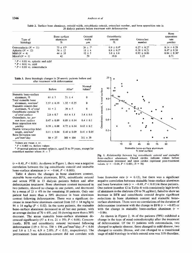
(r = 0.41, P <0.01). As shown in Figure 1, there was a negativecorrelation between the log osteoblastic osteoid and stainablebone—surface aluminum (r = —0.64, P < 0.001).
Table 4 shows the changes in bone aluminum content,stainable bone—surface aluminum, BFR, osteoblastic osteoidand serum PTH in 13 dialysis patients before and afterdeferoxamine treatment. Bone aluminum content increased intwo patients, showed no change in one patient, and decreasedby a mean of 23 4% in the remaining 10 patients. Only onepatient had more than a 50% decrease in bone aluminumcontent following deferoxamine. There was a significant de-crease in mean bone aluminum—content from 115 14 mg/kg to94 14 mg/kg (P <0.02). In the same patients, the stainablebone—surface aluminum decreased in all but one patient, withan average decline of 70 6%, and 10 showing more than a 50%decrease. The mean stainable bone—surface aluminum de-creased significantly (57 4 vs. 21 5%; P < 0.01). Concom-itantly, BFR and osteoblastic osteoid increased followingdeferoxamine (119 54 vs. 530 196 m2/mm2/day; P < 0.01and 3.0 1.5 vs. 6.9 2.0%; P < 0.01, respectively). Thepretreatment bone aluminum-content did not correlate with
bone formation rate (r = 0.13), but there was a significantnegative correlation between stainable bone—surface aluminumand bone formation rate (r = —0.69, P <0.01) in these patients.One patient (number 12 in Table 4) with consistently high levelsof aluminum in the dialysate (50 to 70 pg/liter), failed to show anincrease in BFR and osteoblastic osteoid despite significantreductions in bone aluminum content and stainable bone—surface aluminum. There were no correlations of the duration ofdeferoxamine treatment with the change in BFR (r = —0.05) orwith the change in stainable bone—surface aluminum (r =—0.06).
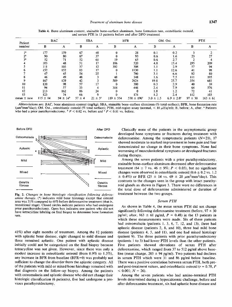
As shown in Figure 2, 16 of the patients (59%) exhibited achange in the type of renal osteodystrophy after the treatmentof deferoxamine. Of the 11 patients with osteomalacia, twochanged to aplastic disease, three changed to mild disease, twochanged to osteitis fibrosa, and one changed to a transitionalstage of mild histology in which osteoid area was 31% (baseline,
Table 3. Bone histologic changes in 28 uremic patients before andafter treatment with deferoxamine
25
10
.00
0
00SS
S0
0.6
S.S .
0.2
.
Values are mean SE.P < 0.005 vs. Before values
b 19 normal patients normal subjects, aged 21 to 59 years, except forosteoblast number where N = 7
tE
Stainable bone—surface aluminum% total surface
Fig. 1. Relationship between log osteoblastic osteoid and stainablebone—surface aluminum. Closed circles indicate values beforedeferoxamine treatment and open circles represent post-treatmentvalues. (r = —0.64; P < 0.001; N = 56)
15 30 45 60 75
Treatment of aluminum bone disease 1347
Table 4. Bone aluminum content, stainable bone—surface aluminum, bone formation rate, osteoblastic osteoid,and serum PTH in 13 patients before and after DFO treatment
.Patientnumber
BAC
B
SBA
A
BFR 0bl. Ost.
B
PTH
AB A B A B A
ia 177 159 67 48 0 28 0.1 0.2 3 22 90 80 67 37 0 93 0.6 1.6 23 273 52 71 52 61 19 65 0.6 2.7 2 44 103 48 51 17 186 520 4.8 15.4 295 2095 111 103 37 12 392 308 1.1 2.9 57 946 152 152 83 17 0 1130 1.1 12.6 41 987 47 45 54 25 I 780 3.1 6.6 82 808 46 49 48 3 48 348 1.6 7.5 111 1959 167 129 42 3 589 2624 19.8 25.7 354 681
10 120 98 57 17 0 380 0.3 2.9 40 3911 94 57 33 4 314 446 2.4 7.9 64 57612 213 162 86 24 0 0 1.8 1.2 72 4113 123 74 66 9 0 174 1.2 2.4 112 103
mean SEM 115 14 94 14b 57 4 21 5C 119 554 530 196C 3.0 1.5 6.9 2.0c 97 30 163 61
Abbreviations are: BAC, bone aluminum content (mg/kg); SBA, stainable bone—surface aluminum (% total surface); BFR, bone formation rate(sm2/mm2/day); ObI. Ost,, osteoblastic osteoid (% total surface); PTH, mid-region assay (normal, < 10 dEq/ml); B, before; A, after. a Patientswho had a prior parathyroidectomy. b P < 0,02 vs. before and C P < 0.01 vs. before.
Fig. 2. Changes in bone histologic classification following deferox-amine therapy. (*) indicates mild bone histology except that osteoidarea was 31% compared to 41% before deferoxamine treatment (that is,transitional stage). Closed circles indicate patients who had undergoneprior parathyroidectomy. Open box indicates one patient who did nothave tetracycline labeling on final biopsy to determine bone formationrate.
41%) after eight months of treatment. Among the 12 patientswith aplastic bone disease, eight changed to mild disease andthree remained aplastic. One patient with aplastic diseaseinitially could not be categorized on the final biopsy becausetetracycline was not given. However, since there was only amodest increase in osteoblastic osteoid (from 0.9% to 1.3%),any increase in BFR from baseline (BFR=0) was probably notsufficient to change the disorder from the aplastic category. Allof the patients with mild or mixed bone histology remained withthat diagnosis on the follow—up biopsy. Among the patientswith osteomalacia and aplastic disease who did not change theirhistologic classification (6 patients), five had undergone a pre-vious parathyroidectomy.
Serum PTHAs shown in Table 4, the mean serum PTH did not change
significantly following deferoxamine treatment (before, 97 30
pg/mi; after, 163 61 pg/mI, P = 0.48) in the 13 patients inwhich these measurements were made. Six of these patientshad osteomalacia (patients 1, 3, 6, 7, 12, and 13), three hadaplastic disease (patients 2, 8, and 10), three had mild bonedisease (patients 4, 5, and 11), and one had mixed histology(patient 9). The three patients with prior parathyroidectomy(patients 1 to 3) had lower PTH levels than the other patients.Five patients showed elevations of serum PTH afterdeferoxamine, which ranged from 37 to 512 pg/mI above base-line (mean change, 203 94 pg/mI). Two patients had declinesin serum PTH which were 31 and 86 pg/mI below baseline.There was a positive correlation between serum PTH, both pre-and post-treatment values, and osteoblastic osteoid (r =0.78, P<0.001; N = 26).
Among the seven patients who had amino—terminal PTHlevels determined during a hypocalcemic challenge, before andafter deferoxamine treatment, six had aplastic bone disease and
Before DFO
Osteomalacia
Aplastic
Mild
Mixed
Osteitis
fibrosa
After DFO Clinically none of the patients in the asymptomatic groupdeveloped bone symptoms or fractures during treatment with
Osteomalacia deferoxamine. Among the symptomatic patients (N=21), 17showed moderate to marked improvement inbone pain and four
A Idemonstrated no change in their bone symptoms. None had
P asticworsening ofmusculoskeletal symptoms or developed fracturesduring therapy.
Mild Among the seven patients with a prior parathyroidectomy,stainable bone—surface aluminum decreasedafter deferoxaminetreatment (64 7 vs. 46 9%; P < 0.05), but no significant
Mixed changes were observed in osteoblastic osteoid (0.6 0.2 vs. 1.2_________ 0.4%) or BFR (25 14 vs. 69 20 m2/mm2/day). This
Osteitiscontrasts to the changes seen in the group with intact parathy-
fibrosa roid glands as shown in Figure 3. There were no differences inthe total dose of deferoxamine administered or duration oftreatment between the two groups.
1348 Andress et a!
Rffore After
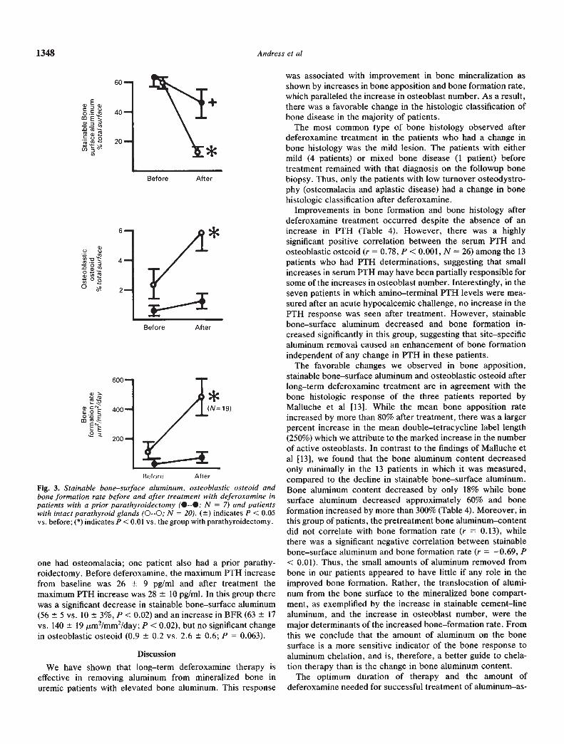
Fig. 3. Stainable bone—surface aluminu,n, osteoblastic osteoid andbone formation rate before and after treatment with deferoxamine inpatients with a prior parathyroidectomy (•—I; N = 7) and patientswith intact parathyroid glands (O--O; N = 20). (±) indicates P < 0.05vs. before; (*) indicates P <0.01 vs. the group with parathyroidectomy.
one had osteomalacia; one patient also had a prior parathy-roidectomy. Before deferoxamine, the maximum PTH increasefrom baseline was 26 9 pg/mi and after treatment themaximum PTH increase was 28 10 pg/mI. In this group therewas a significant decrease in stainable bone—surface aluminum(56 5 vs. 10 3%, P < 0.02) and an increase in BFR (63 17
vs. 140 19 m2/mm2/day; P < 0.02), but no significant changein osteoblastic osteoid (0.9 0.2 vs. 2.6 0.6; P = 0.063).
Discussion
We have shown that long—term deferoxamine therapy iseffective in removing aluminum from mineralized bone inuremic patients with elevated bone aluminum. This response
was associated with improvement in bone mineralization asshown by increases in bone apposition and bone formation rate,which paralleled the increase in osteoblast number. As a result,there was a favorable change in the histologic classification ofbone disease in the majority of patients.
The most common type of bone histology observed afterdeferoxamine treatment in the patients who had a change inbone histology was the mild lesion. The patients with eithermild (4 patients) or mixed bone disease (1 patient) beforetreatment remained with that diagnosis on the followup bonebiopsy. Thus, only the patients with low turnover osteodystro-phy (osteomalacia and aplastic disease) had a change in bonehistologic classification after deferoxamine.
Improvements in bone formation and bone histology afterdeferoxamine treatment occurred despite the absence of anincrease in PTH (Table 4). However, there was a highlysignificant positive correlation between the serum PTH andosteoblastic osteoid (r = 0.78, P <0.001, N = 26) among the 13patients who had PTH determinations, suggesting that smallincreases in serum PTH may have been partially responsible forsome of the increases in osteoblast number. Interestingly, in theseven patients in which amino—terminal PTH levels were mea-sured after an acute hypocalcemic challenge, no increase in thePTH response was seen after treatment. However, stainablebone—surface aluminum decreased and bone formation in-creased significantly in this group, suggesting that site—specificaluminum removal caused an enhancement of bone formationindependent of any change in PTH in these patients.
The favorable changes we observed in bone apposition,stainable bone—surface aluminum and osteoblastic osteoid afterlong—term deferoxamine treatment are in agreement with thebone histologic response of the three patients reported byMalluche et al [13]. While the mean bone apposition rateincreased by more than 80% after treatment, there was a largerpercent increase in the mean double—tetracycline label length(250%) which we attribute to the marked increase in the numberof active osteoblasts. In contrast to the findings of Malluche etal [13], we found that the bone aluminum content decreasedonly minimally in the 13 patients in which it was measured,compared to the decline in stainable bone—surface aluminum.Bone aluminum content decreased by only 18% while bonesurface aluminum decreased approximately 60% and boneformation increased by more than 300% (Table 4). Moreover, inthis group of patients, the pretreatment bone aluminum—contentdid not correlate with bone formation rate (r = 0.13), whilethere was a significant negative correlation between stainablebone—surface aluminum and bone formation rate (r = —0.69, P
<0.01). Thus, the small amounts of aluminum removed frombone in our patients appeared to have little if any role in theimproved bone formation. Rather, the translocation of alumi-num from the bone surface to the mineralized bone compart-ment, as exemplified by the increase in stainable cement—linealuminum, and the increase in osteoblast number, were themajor determinants of the increased bone—formation rate. Fromthis we conclude that the amount of aluminum on the bonesurface is a more sensitive indicator of the bone response toaluminum chelation, and is, therefore, a better guide to chela-tion therapy than is the change in bone aluminum content.
The optimum duration of therapy and the amount ofdeferoxamine needed for successful treatment of aluminum—as-
60—
40
20—
Before After
6
4,
C0.— 'E'.0 m—C
Q 0(oc.-
Q ,..0.n.0
o
w.coSon
2
Before After
Treatment of aluminum bone disease 1349
sociated bone disease cannot be determined from this study. Itappears, however, that a rather prolonged treatment schedulemay be necessary in the majority of patients. It is possible thatthe concomitant administration of aluminum—containing phos-phate binders in our patients may have contributed to this longtreatment period. Perhaps the use of calcium carbonate as aphosphate binder[23] would adequately control serum phospho-rus levels and allow for the discontinuation of aluminum bindersduring chelation therapy. Whether the use of newer methods toenhance aluminum removal during dialysis [24] can also makechronic deferoxamine treatment more efficient remains to bedetermined.
Patients who have had a parathyroidectomy prior todeferoxamine therapy may require more intensive treatmentthan patients with intact parathyroid glands. Among the sixpatients with low turnover bone disease (3 with osteomalaciaand 3 with aplastic disease) who failed to change their bonehistologic classification (Fig. 2), five had undergone a para-thyroidectomy prior to receiving deferoxamine. When all sevenpatients with parathyroidectomy were analyzed together, avery modest decrease in bone surface aluminum was noted inthe group, although osteoblastic osteoid and bone formation didnot improve (Fig. 3). The reason for their poor response totreatment is unclear but may be related to a requirement for anoptimum level of circulating PTH for the maintenance of normalbone formation. Thus, a greater dosage of deferoxamine may berequired in patients with prior parathyroidectomy. This dosageadjustment should probably also include the discontinuation ofaluminum-containing phosphate binders. These data and thefinding that bone aluminum accumulation is enhanced afterparathyroidectomy [25—271 emphasize the need to developbetter treatments for secondary hyperparathyroidism.
In summary, we have shown that long—term aluminum chela-tion therapy with deferoxamine effectively ameliorates alumi-num associated, low—turnover renal osteodystrophy in patientswith intact parathyroid glands. Removal of aluminum from thebone surface is associated with marked increases in osteoblastnumber and bone formation, resulting in improved bone histol-ogy. While small increases in serum PTH following aluminumchelation may have an anabolic effect on bone function, alumi-num removal alone can result in increased bone formationwithout changes in PTH.
AcknowledgmentsThis study was supported in part by General Medical Research funds
from the Veterans Administration.
Reprint requests to Dennis L. Andress, M.D., Dialysis Unit (11/A),VA Medical Center, /660 S. Columbian Way, Seattle, Washington98108, USA.
References
I. WARD WK, FEEST TG, ELLIS HA, PARKINSON IS, KERR DNS:Osteomalacic dialysis osteodystrophy: Evidence for a water—borneaetiological agent, probably aluminum. Lancet 1:841—845, 1978
2. PARKINSON IS, FEEST TG, WARD MK, FAWCETT RWP, KERR
DNS: Fracturing dialysis osteodystrophy and dialysis encephalop-athy: An epidemiological survey. Lancet 1:406—409, 1979
3. ELLIS HA, MCCARTHY JH, HERRINGTON J: Bone aluminum inhaemodialysed patients and in rats injected with aluminum chlo-
ride: Relationship to impaired bone mineralization. J Clin Pathol32:832—844, 1979
4. Pierides AM, Edwards WG, Cullum UX, McCall iT, Ellis HA:Hemodialysis encephalopathy with osteomalacia fractures andmuscle weakness. Kidney mt 18:115—124, 1980
5. FELSENFELD AJ, GUTMAN RA, LLACH F, HARRELSON JMOsteomalacia in chronic renal failure: A syndrome previouslyreported only with maintenance dialysis. Am J Nephrol 2:147—154,1982
6. ANDREOLI SP, BEROSTEIN JM, SHERRARD Di: Aluminum intoxi-cation from aluminum—containing phosphate binders in childrenwith azotemia not undergoing dialysis. N Engl J Med 310:1079—1084, 1984
7. ANDRESs DL, MALONEY NA, ENDRE5 DB, SHERRARD Di:Aluminum—associated bone disease in chronic renal failure: Highprevalence in a long—term dialysis population. J Bone Miner Res1:391—398, 1986
8. ANDRESS DL, Ko JB, MALONEY NA, COBURN iW, SHERRARDDi: Early deposition of aluminum in bone in patients with diabeteson hemodialysis. N Engi J Med 316:292—296, 1987
9. ACKRILL P. RALSTON Ai, DAY iP, HODGE KC: Successful removalof aluminum from a patient with dialysis encephalopathy. Lancet2:692—693, 1980
10. BROWN Di, HAM KN, DAWBORN iK, XIPPEL iM: Treatment ofdialysis osteomalacia with desferrioxamine, Lancet 2:343—345, 1982
11. IHLE BU, BUCHANAN MRC, STEVENS B, BECKER GJ,KINCAID—SMITH P: The efficacy of various treatment modalities onaluminum associated bone disease. Proc EDTA 19:195—201, 1982
12. ACKRILL P, DAY JP, GARSTANG FM, HODGE KC, METCALFE Pi,BENZO Z, HILL K, RALSTON Ai, DENTON i: Treatment of fractur-ing renal osteodystrophy by desferrioxamine. Proc EDTA19:203—207, 1982
13. MALLUCHE HH, SMITH AJ, ABREO K, FAIJGERE MC: The use ofdeferoxamine in the management of aluminum accumulation inbone in patients with renal failure. N Engi J Med 311:140—144, 1984
14. OTT SM, ANDRESS DL, NEBEKER HG, MILLINER DS, MALONEYNA, COBURN JW, SHERRARD Di: Changes in bone histology aftertreatment with deferrioxamine. Kidney mt 29(Suppl 18): 108—113,1986
15. SHERRARD Di, BAYLINK Di, WEGEDAL JE, MALONEY NA: Quan-titative histological studies on the pathogenesis of uremic bonedisease. J Clin Endocrinol Metab 39:119—135, 1974
16. EVANS RA, DUNSTAN CR, BAYLINK Di: Histochemical identifica-tion of osteoclasts in undecalcified sections of human bone. MinerElectrol Metab 2:179—185, 1979
17. MALONEY NA, O-rr S, ALFREY AC, Cosu JW, SHERRARD Di:Histologic quantitation of aluminum in iliac bone from patients withrenal failure. J Lab Gun Med 99:206—216, 1982
18. LEGENDRE GR, ALFREY AC: Measuring picogram amounts ofaluminum in biological tissue by flameless atomic absorption anal-ysis of a chelate. Clin Chem 22:53—56, 1976
19. ANDRESS DL, ENDRES DB, MALONEY NA, KoP iB, COBLJRN JW,SHERRARD Di: Comparison of parathyroid hormone assays withbone histomorphometry in renal osteodystrophy. J Clin EndocrinolMetab 63:1163—1 169, 1986
20. HRUSKA KA, KOPELMAN R, RUTHERFORD WE, KLAHR S,SLATOPOLSKY E: Metabolism of immunoreactive parathyroid hor-mone in the dog: The role of the kidney and the effects of chronicrenal disease. J Gun Invest 56:39—48, 1975
21. ANDRESS D, FELSENFELD Ai, VOIGTS A, LLACH F: Parathyroidhormone response to hypocalcemia in hemodialysis patients withosteomalacia. Kidney mt 24:364—370, 1983
22. SEGRE GV: Amino—terminal radioimmunoassays for human para-thyroid hormone, in Clinical Disorders of Bone and MineralMetabolism, edited by FRAME B, POTTS iT iR. Amsterdam,Excerpta Medica 14-7, 1983
23. SLATOPOLSKY E, WEERTS C, LOPEz—HILKER S, NoRwooD K,ZINK M, WINDUS D, DELMEZ i: Calcium carbonate as a phosphatebinder in patients with chronic renal failure undergoing hemo-dialysis. NEnglJMed3l5:157—16l, 1986
24. SLATOPOL5KY E, WEERTS C, FINCH J, LEE W: The use ofmicroencapsulated carbon in the removal of aluminum in dialysis
1350 Andress et a!
patients. (abstract) Kidney In: 29:226, 198625. ANDaESS DL, Orr SM, MALONEY NA, SHERRARD DJ: Effect of
parathyroidectomy on bone aluminum accumulation in chronicrenal failure. N Engi J Med 312:468—473, 1985
26. CHARHON SA, BERLANDYF, OLMER MJ, DELAWARI E, TRAGAR J,MEUNIER P: Effects of parathyroidectomy on bone formation and
mineralization in hemodialyzed patients. Kidney In: 27:426—435,1985
27. DE VERNEJOIJL MC, MARCHAIS S. LONDON 0, MORIEUX C,BIELAKOFF J, MIRAVET L: Increased bone aluminum depositionafter subtotal parathyroidectomy in dialyzed patients. Kidney In:27:785—791, 1985