Embed Size (px)
Citation preview

Bones – Test Review
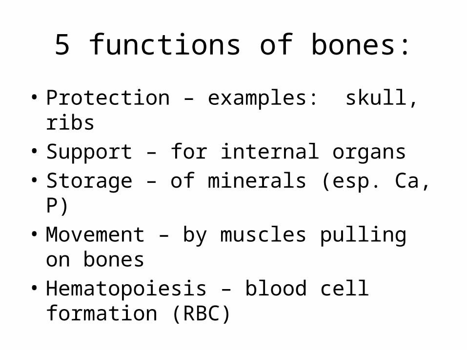
5 functions of bones:
• Protection – examples: skull, ribs• Support – for internal organs• Storage – of minerals (esp. Ca, P)• Movement – by muscles pulling on bones• Hematopoiesis – blood cell formation (RBC)

2 subdivisions of the skeletal system
• Axial skeleton – skull, thorax, vertebrae
• Appendicular skeleton – limbs, girdles

4 Classifications of bones• Long– Ex. Upper and lower limbs
• Short– Ex. Carpals and tarsals
• Flat– Ex. Ribs, sternum, skull, scapulae
• Irregular– Ex. Vertebrae, facial
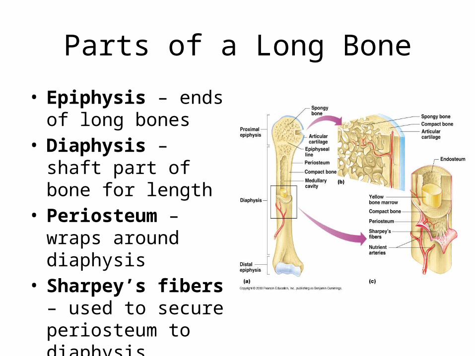
Parts of a Long Bone
• Epiphysis – ends of long bones
• Diaphysis – shaft part of bone for length
• Periosteum – wraps around diaphysis
• Sharpey’s fibers – used to secure periosteum to diaphysis

Spongy vs. Compact Bone
• Spongy bone – found on epiphysis
· Small needle-like pieces of bone
· Many open spaces
• Compact bone - Dense and smooth

Microanatomy of Bone
• Osteons (Haversian system)
• Central (Haversian) Canals
• Lamellae – Rings around the central canal
• Canaliculi – Tiny canals• Lacunae - Cavities
containing bone cells

Bone Formation, Growth, Remodeling
Bone Formation
· In embryos, the skeleton is primarily hyaline cartilage
· During development, much of this cartilage is replaced by bone
· Cartilage remains in isolated areas
· Bridge of the nose
· Parts of ribs
· Joints
Bone Growth and Remodeling Cartilage is broken down, bone
replaces cartilage, epiphyseal plates allow for growth of long bones during childhood
· Bones are remodeled and lengthened until growth stops
· Bones change shape somewhat
· Bones grow in width

PTH / Calcitonin
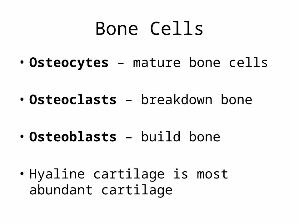
Bone Cells
• Osteocytes – mature bone cells
• Osteoclasts – breakdown bone
• Osteoblasts – build bone
• Hyaline cartilage is most abundant cartilage
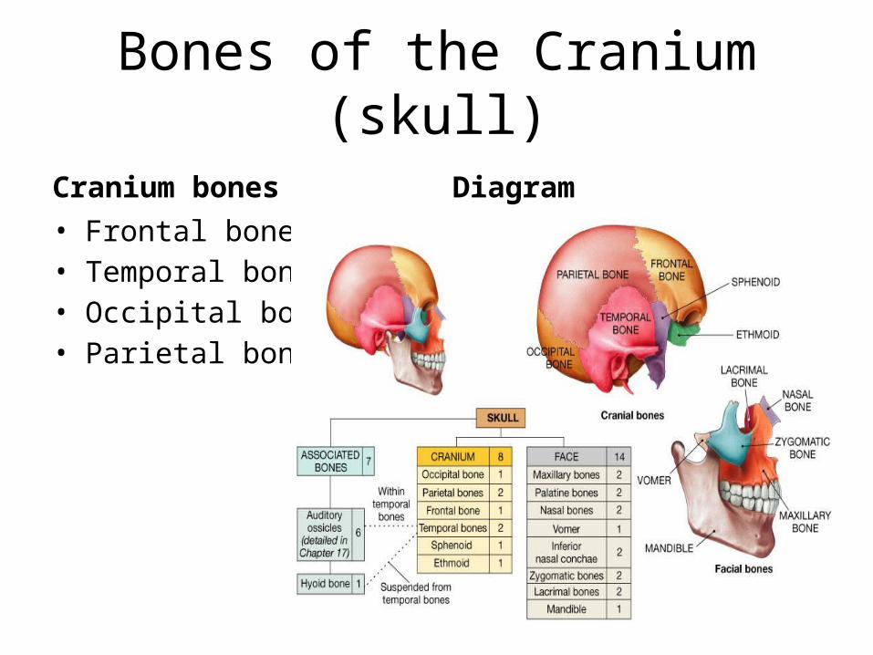
Bones of the Cranium (skull)
Cranium bones• Frontal bone• Temporal bone• Occipital bone• Parietal bone
Diagram

Bones of the skull – facial bones
Facial bones• Zygomatic• Nasal• Maxilla• Lacrimal • Sphenoid• Mandible
Diagram

Bone Markings (skull)
• Foramen magnum – on occipital bone
• Styloid process – temporal bone
• Mastoid process – temporal bone
• Zygomatic process – temporal bone
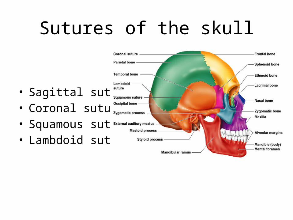
Sutures of the skull
• Sagittal suture• Coronal suture• Squamous suture• Lambdoid suture

Fractures• Treatment is reduction
open or closed• Types of fractures:• Simple• Compound• Comminuted• Impacted• Epiphyseal• Greenstick• Osteomyelitis (problem)
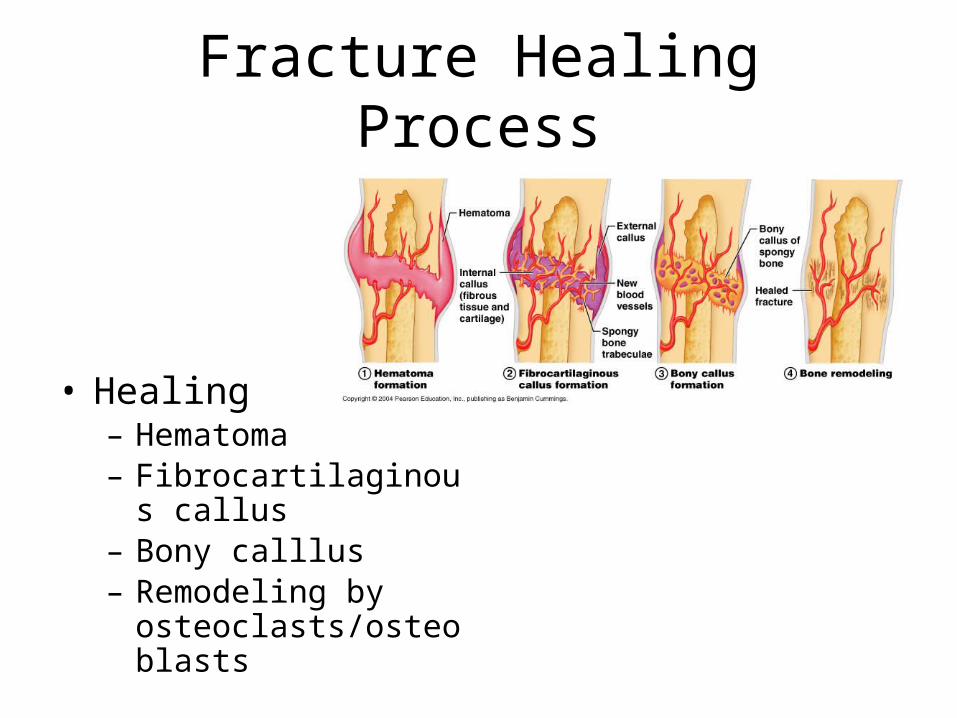
Fracture Healing Process
• Healing– Hematoma– Fibrocartilaginous callus– Bony calllus– Remodeling by
osteoclasts/osteoblasts
Pectoral Girdle (bones, joints)
• Clavicle, Scapula make up the girdle• Joints: S/C (sternoclavicular)
A/C (acromioclavicluar) - separation
Glenohumeral joint - dislocation• Glenoid of the scapula• Coracoid process
Bone of the Arm
• HumerusHead of the humerus (proximal) articulates with the glenoid of the scapulaDistal: condyle, trochlea, capitulum to help form the elbow joint
• Fossa – Ant. Coronoid fossaPost. Olecranon fossa
• Deltoid tuberosity – for the deltoid muscle
Humerus
• Anatomical Neck – site of fractures
Interosseous membrane
• Surgical neck
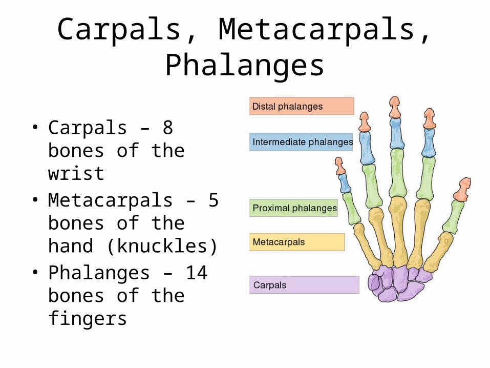
Carpals, Metacarpals, Phalanges
• Carpals – 8 bones of the wrist
• Metacarpals – 5 bones of the hand (knuckles)
• Phalanges – 14 bones of the fingers
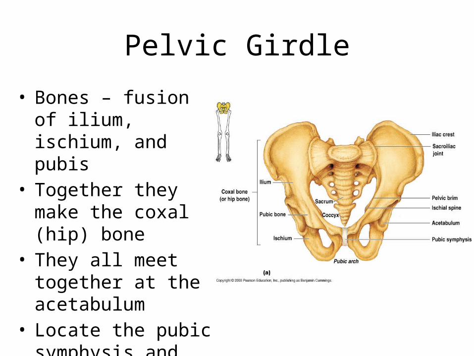
Pelvic Girdle
• Bones – fusion of ilium, ischium, and pubis
• Together they make the coxal (hip) bone
• They all meet together at the acetabulum
• Locate the pubic symphysis and the obturator foramen

Bones of the Thigh and Leg
• Thigh – head of the femur articulates with the acetabulum superiorly and inferiorly at the knee with the tibia/fibula
• Longest, strongest bone in the body
• Bones of the leg - Tibia/fibula
• Tibia bears the weight of the leg; fibula is non-weight bearing
• Distal end of tibia is medial malleolus and lateral malleolus is the distal end of the fibula

Arches of the Foot