Embed Size (px)
Citation preview

bowel obstruction
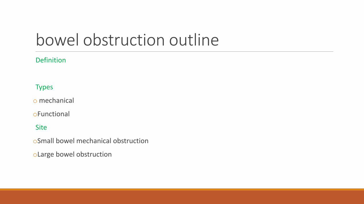
bowel obstruction outlineDefinition
Types
o mechanical
oFunctional
Site
oSmall bowel mechanical obstruction
oLarge bowel obstruction
DefinitionsInterruption in the normal flow of intestinal contents along the intestinal tract
Ileus= paralytic= adynamic; when obstruction is functional

Mechanical Small bowel obstruction
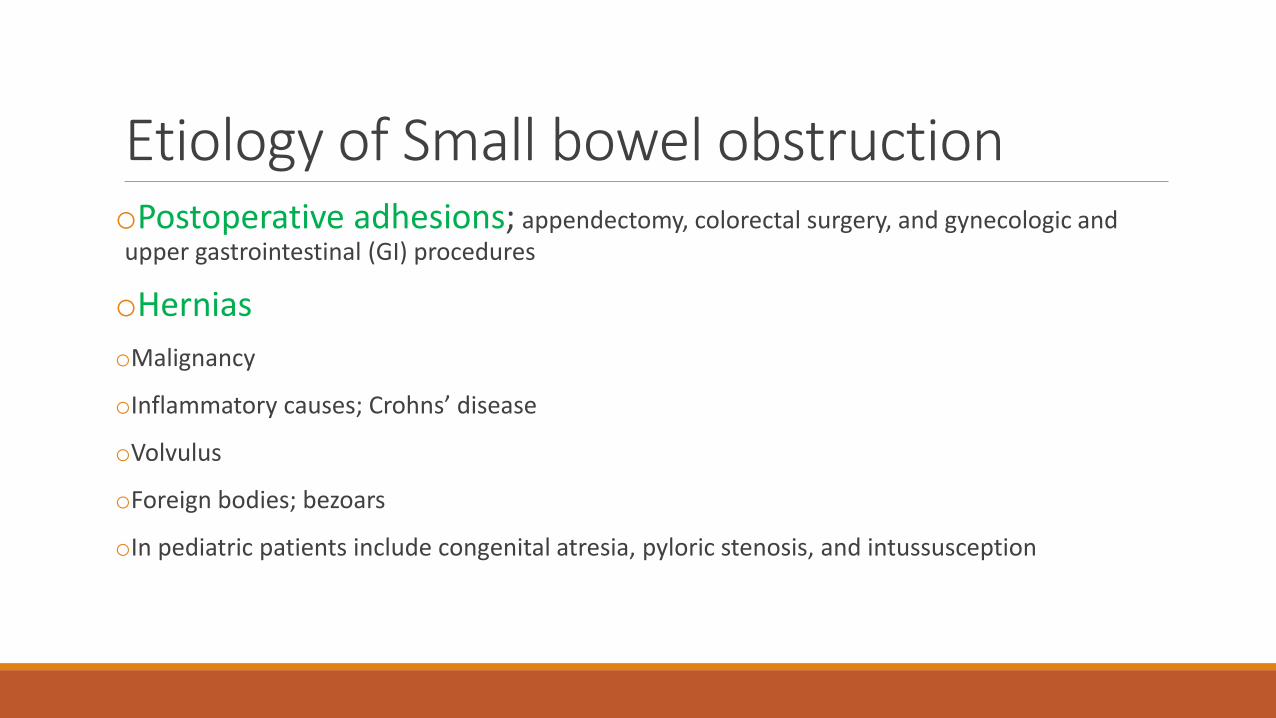
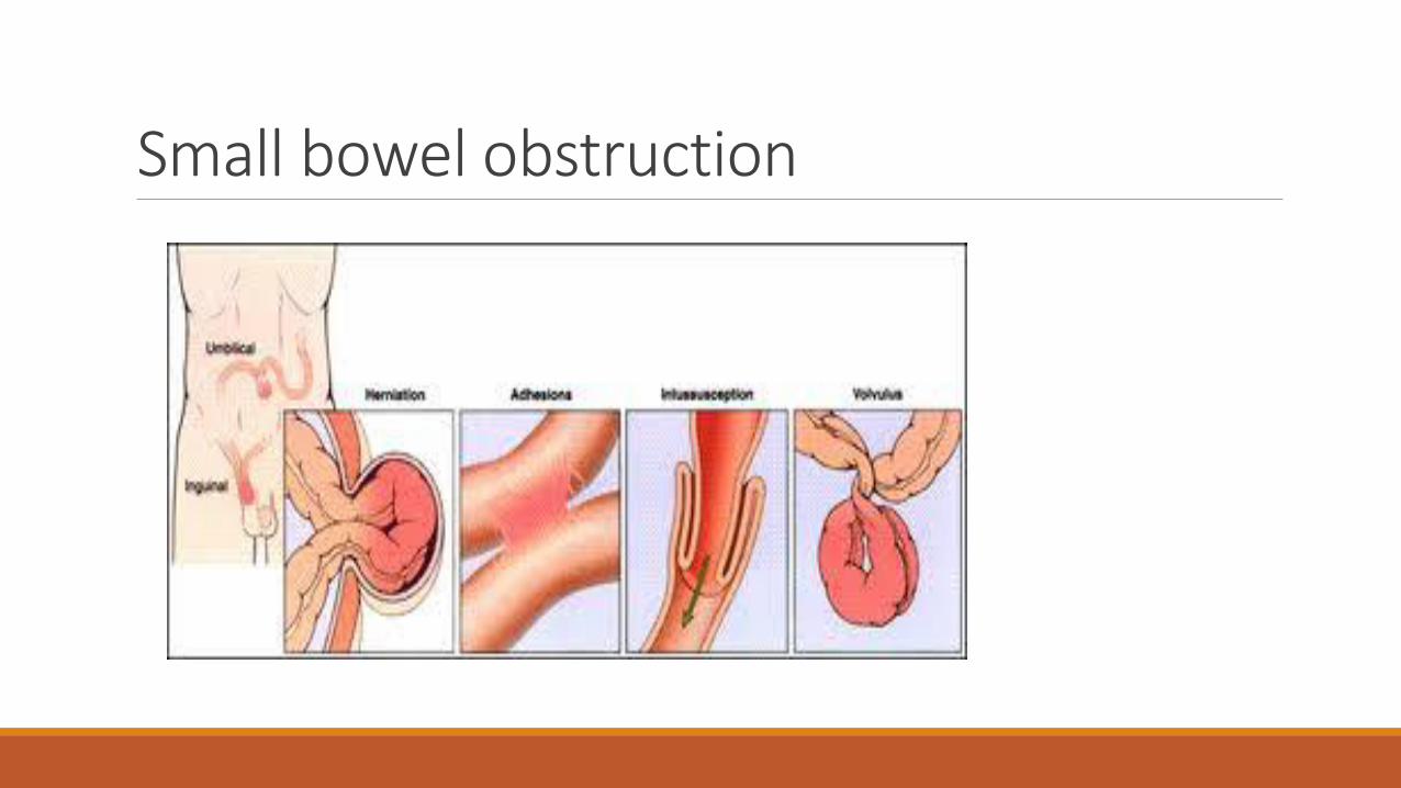
Etiology of Small bowel obstructionoPostoperative adhesions; appendectomy, colorectal surgery, and gynecologic and upper gastrointestinal (GI) procedures
oHernias
oMalignancy
oInflammatory causes; Crohns’ disease
oVolvulus
oForeign bodies; bezoars
oIn pediatric patients include congenital atresia, pyloric stenosis, and intussusception

Closed loop obstruction1. Hernia
2. Volvulus
3. Colonic obstruction with a competent ileocecal valve
4. intussusception
5. Some adhesive obstructions
Small bowel obstruction
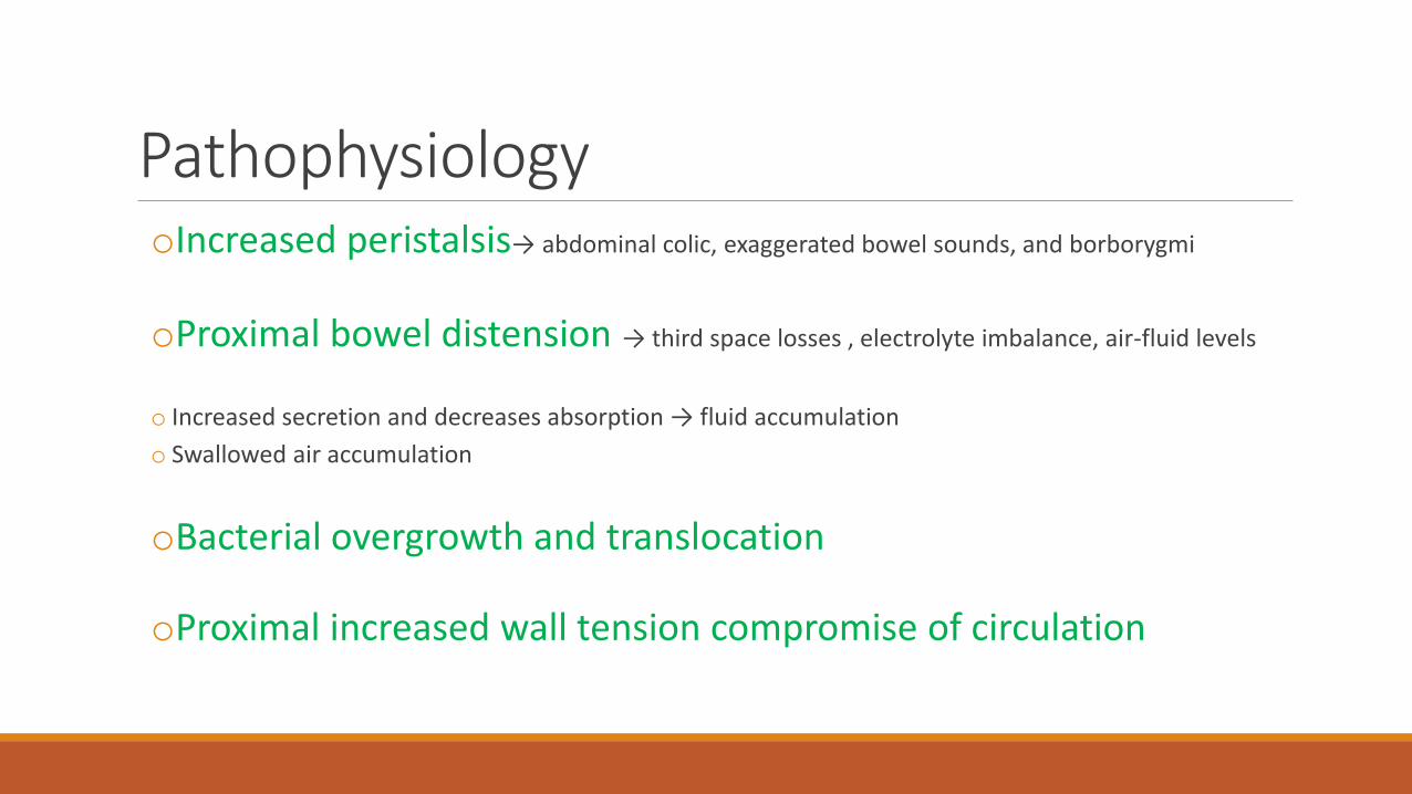
PathophysiologyoIncreased peristalsis→ abdominal colic, exaggerated bowel sounds, and borborygmi
oProximal bowel distension → third space losses , electrolyte imbalance, air-fluid levels
o Increased secretion and decreases absorption → fluid accumulation
o Swallowed air accumulation
oBacterial overgrowth and translocation
oProximal increased wall tension compromise of circulation
History
Abdominal pain
Crampy and intermittent, is more prevalent in simple obstruction.
Central
Changes in the character of the pain may indicate the development of a more serious complication (i.e., constant pain of a strangulated or ischemic bowel).
History❑Nausea
❑ Vomiting; reflex and reflux
❑ constipation or obstipation; more than 24 hours
❑ Diarrhea; in partial and intermittent obstruction like volvulus and gallstone ileus
History▪Fever and tachycardia - Occur late and may be associated with strangulation
▪ Previous abdominal or pelvic surgery, previous radiation therapy, or both
▪ History of malignancy - Particularly ovarian and colonic malignancy
Physical Examination
1. Abdominal distention; The proximal small bowel has less distention when obstructed than the distal bowel has when obstructed.
2. Hyperactive bowel sounds occur early as GI contents attempt to overcome the obstruction and typically related to the colic
3. Visible peristalsis
4. Borborygmi; audible peristalsis
5. Abdominal scars
6. Abdominal hernias
Rectal examination:• Gross or occult blood, which suggests late strangulation or malignancy
• Masses, which suggest obturator hernia

Clinical typeso Partial vs complete
o Simple vs strangulated
SBO accounts for 20% of all acute surgical admissions
Strangulated SBOsCheck for findings commonly believed to be more diagnostic of intestinal ischemia, including the following:
• Fever
• Tachycardia
• Peritoneal signs
No reliable way exists to differentiate simple from early strangulated obstruction on physical examination.
Serial abdominal examinations are important and may detect changes early.
Terms▪Bowel fatigue; ileus complicating mechanical obstruction
▪Feculent vomiting
Both are indicative of prolonged obstruction and the need for surgery

LabsoBlood urea nitrogen (BUN) level
oElectrolytes
oCreatinine
oComplete blood count (CBC)
oUrinalysis
oType and cross match
Imaging tests
1. Plain radiographs first for patients in whom SBO is suspected. At least 2 views, supine or flat and upright, are required. Plain radiographs are diagnostically more accurate in cases of simple obstruction.
2. Enteroclysis is valuable in detecting the presence of obstruction and in differentiating partial from complete blockages. This study is useful when plain radiographic findings are normal in the presence of clinical signs of SBO or when plain radiographic findings are nonspecific.
3. Computed tomography (CT) scanning is the study of choice if the patient has fever, tachycardia, localized abdominal pain, and/or leukocytosis.
4. Ultrasonography is less costly and invasive than CT scanning and may reliably exclude SBO in as many as 89% of patients; specificity is reportedly 100%.
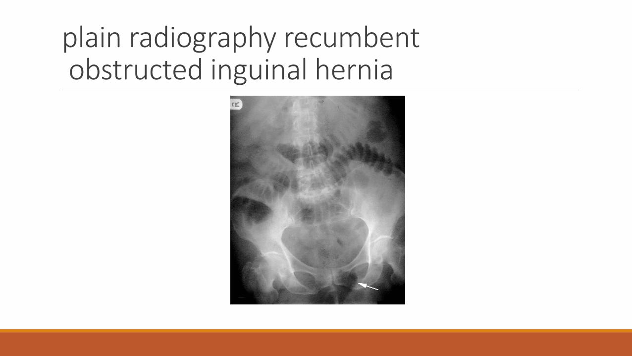
plain radiography recumbent obstructed inguinal hernia
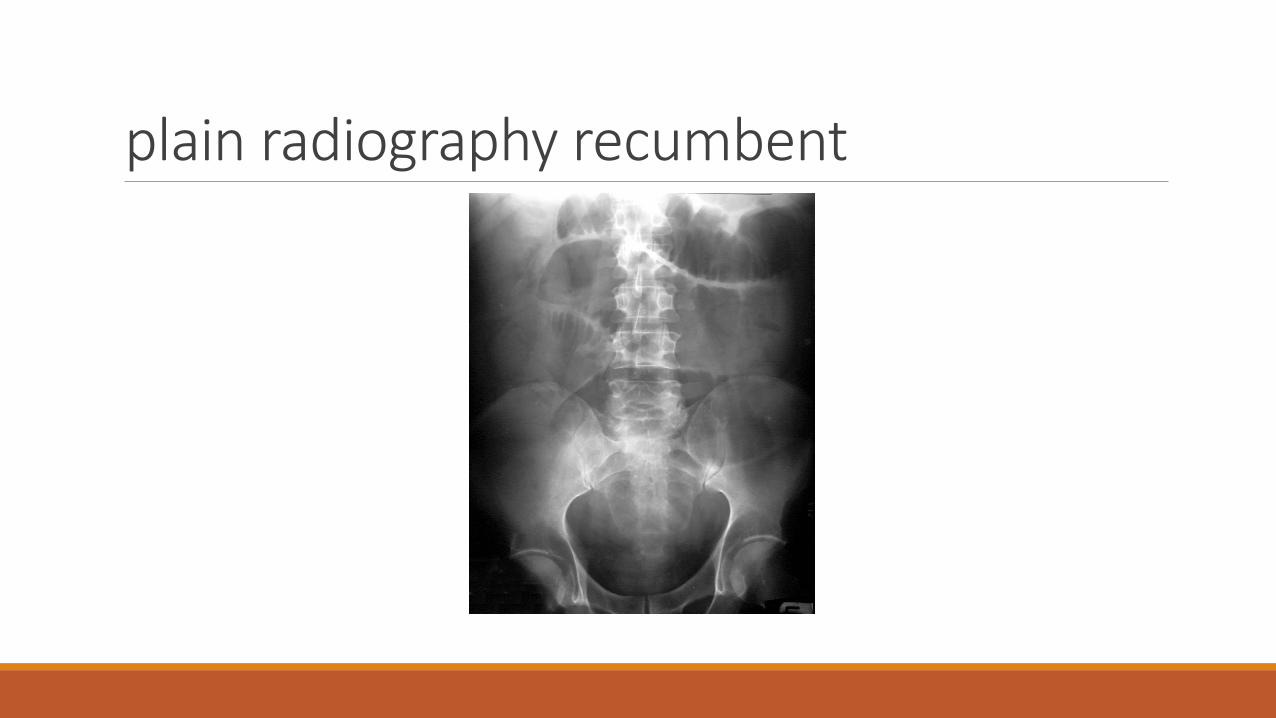
plain radiography recumbent
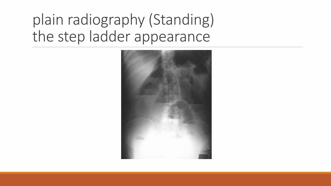
plain radiography (Standing)the step ladder appearance
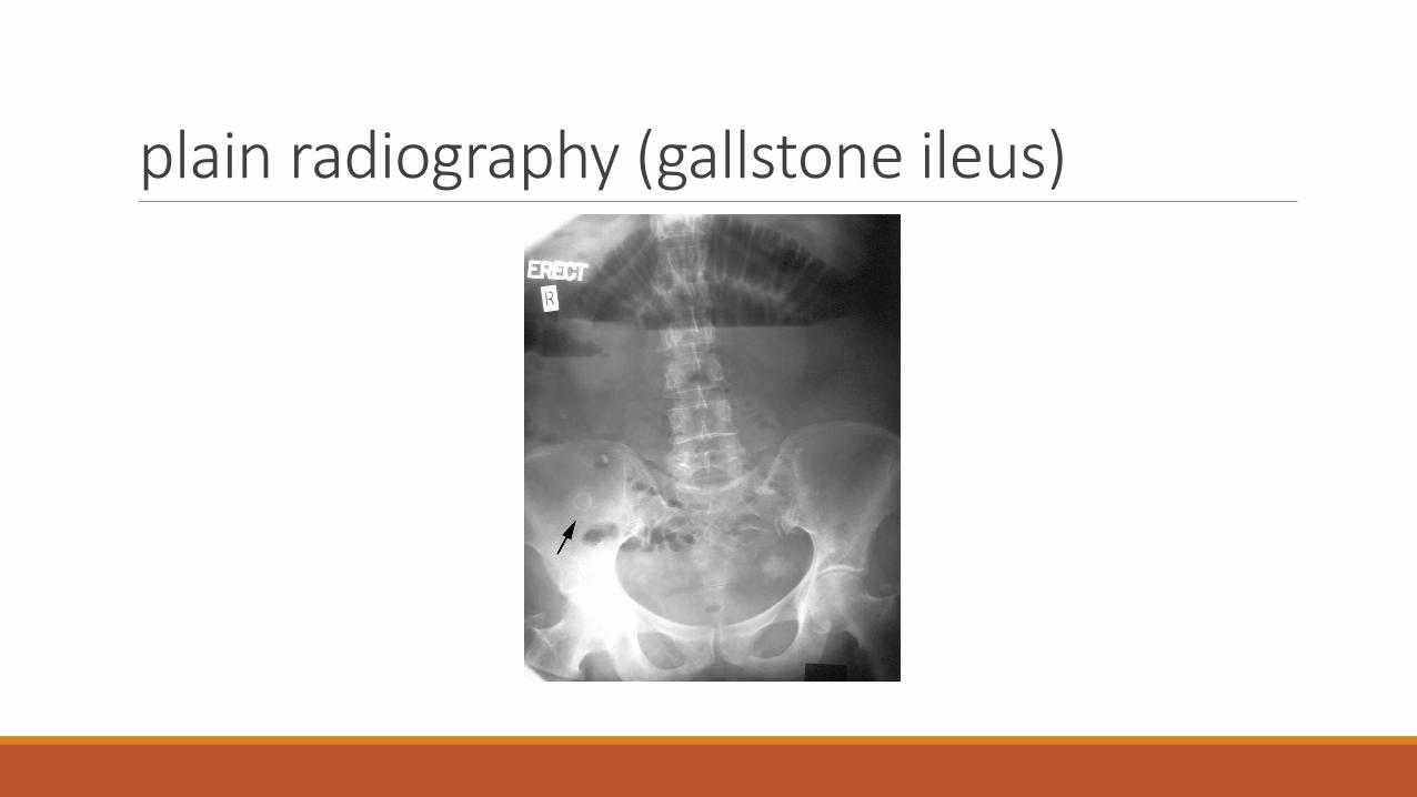
plain radiography (gallstone ileus)
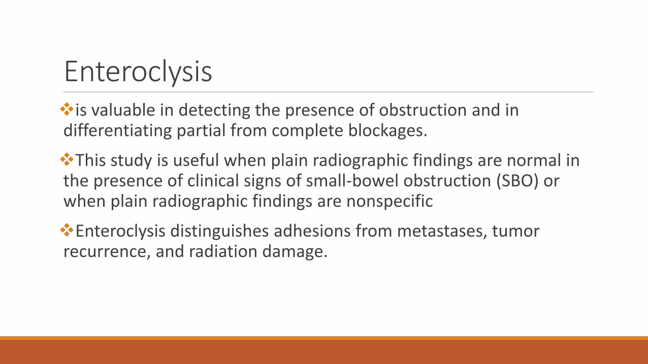
Enteroclysis❖is valuable in detecting the presence of obstruction and in differentiating partial from complete blockages.
❖This study is useful when plain radiographic findings are normal in the presence of clinical signs of small-bowel obstruction (SBO) or when plain radiographic findings are nonspecific
❖Enteroclysis distinguishes adhesions from metastases, tumor recurrence, and radiation damage.
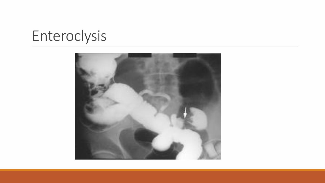
Enteroclysis
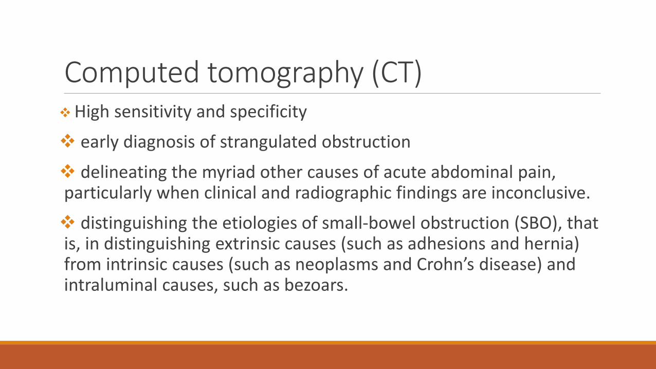
Computed tomography (CT) ❖High sensitivity and specificity
❖ early diagnosis of strangulated obstruction
❖ delineating the myriad other causes of acute abdominal pain, particularly when clinical and radiographic findings are inconclusive.
❖ distinguishing the etiologies of small-bowel obstruction (SBO), that is, in distinguishing extrinsic causes (such as adhesions and hernia) from intrinsic causes (such as neoplasms and Crohn’s disease) and intraluminal causes, such as bezoars.
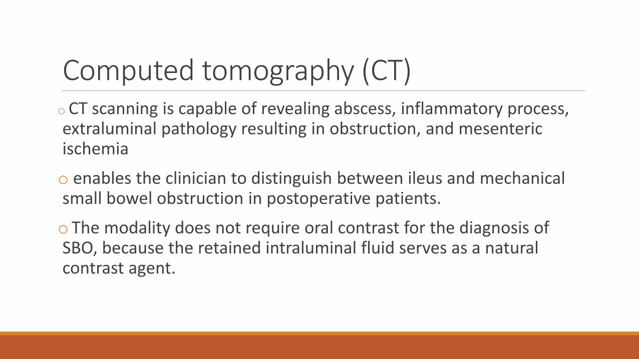
Computed tomography (CT) oCT scanning is capable of revealing abscess, inflammatory process, extraluminal pathology resulting in obstruction, and mesenteric ischemia
o enables the clinician to distinguish between ileus and mechanical small bowel obstruction in postoperative patients.
o The modality does not require oral contrast for the diagnosis of SBO, because the retained intraluminal fluid serves as a natural contrast agent.
Computed tomography (CT)
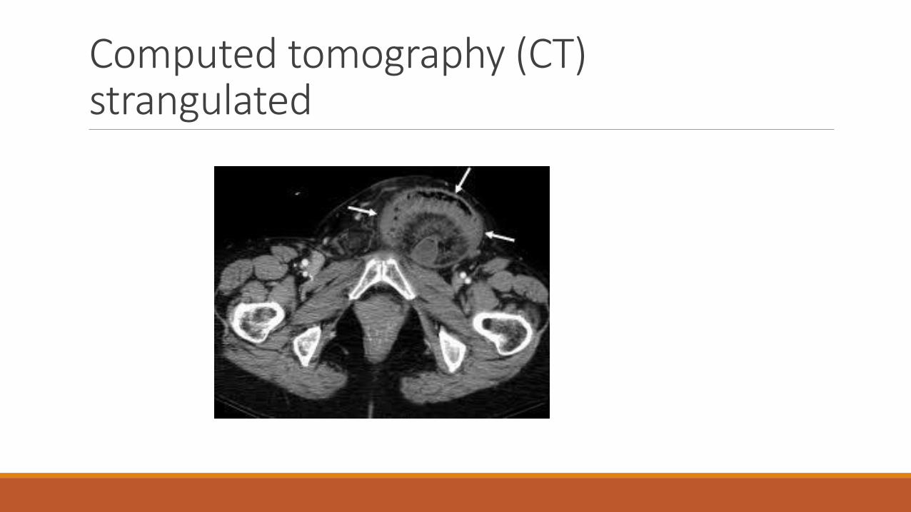
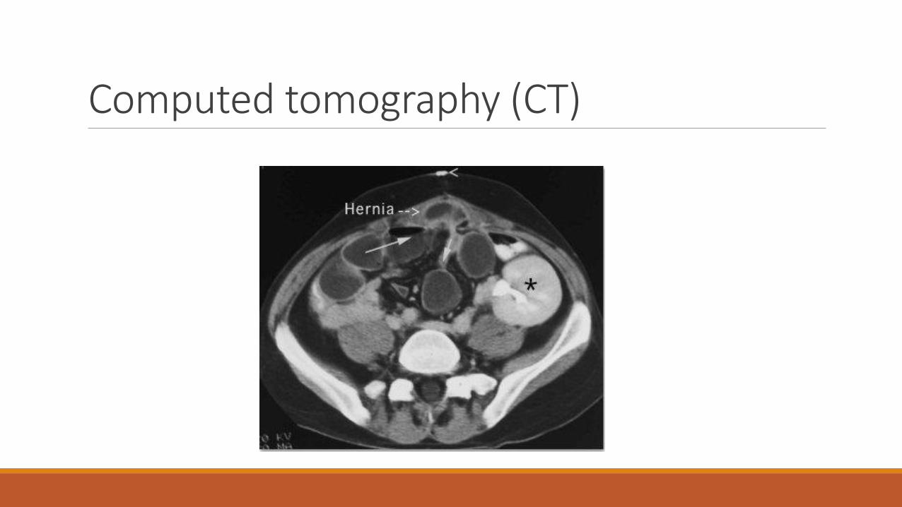
Computed tomography (CT)strangulated
Ultrasonography Is less costly and invasive than CT scanning and may reliably exclude SBO in as many as 89% of patients; specificity is reportedly 100%.
Emergency physician ̶ performed ultrasonography compared favorably with radiography.
Indications of Nonoperative treatmentof SBO ❑ Adhesions
❑Malignant tumor - Obstruction by tumor is usually caused by metastasis; initial treatment should be nonoperative (surgical resection is recommended when feasible)
❑Pediatric obstructed hernia - Initially use manual reduction and observation; advise elective hernia repair as soon as possible after reduction
❑ Inflammatory bowel disease - high-dose steroids; consider parenteral treatment for prolonged periods of bowel rest, and undertake surgical treatment, bowel resection, and/or stricturoplasty if nonoperative treatment fails.
Indications of Nonoperative treatmentof SBO (con…)❑Intra-abdominal abscess - CT scan ̶ guided drainage is usually sufficient to relieve obstruction
❑ Radiation enteritis - acutely, nonoperative treatment accompanied by steroids is usually sufficient; if the obstruction is a chronic sequela of radiation therapy, surgical treatment is indicated
❑Acute postoperative obstruction - This is difficult to diagnose due to postoperative ileus
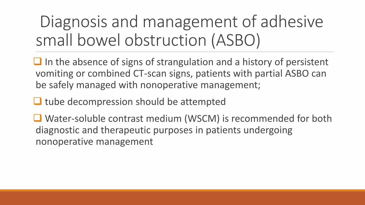
Diagnosis and management of adhesive small bowel obstruction (ASBO)❑ In the absence of signs of strangulation and a history of persistent vomiting or combined CT-scan signs, patients with partial ASBO can be safely managed with nonoperative management;
❑ tube decompression should be attempted
❑ Water-soluble contrast medium (WSCM) is recommended for both diagnostic and therapeutic purposes in patients undergoing nonoperative management
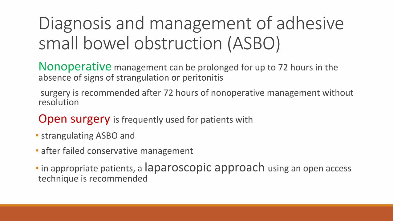
Diagnosis and management of adhesive small bowel obstruction (ASBO)Nonoperative management can be prolonged for up to 72 hours in the absence of signs of strangulation or peritonitis
surgery is recommended after 72 hours of nonoperative management without resolution
Open surgery is frequently used for patients with
• strangulating ASBO and
• after failed conservative management
• in appropriate patients, a laparoscopic approach using an open access technique is recommended
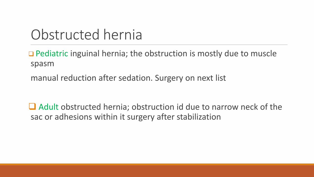
Obstructed hernia❑ Pediatric inguinal hernia; the obstruction is mostly due to muscle spasm
manual reduction after sedation. Surgery on next list
❑ Adult obstructed hernia; obstruction id due to narrow neck of the sac or adhesions within it surgery after stabilization
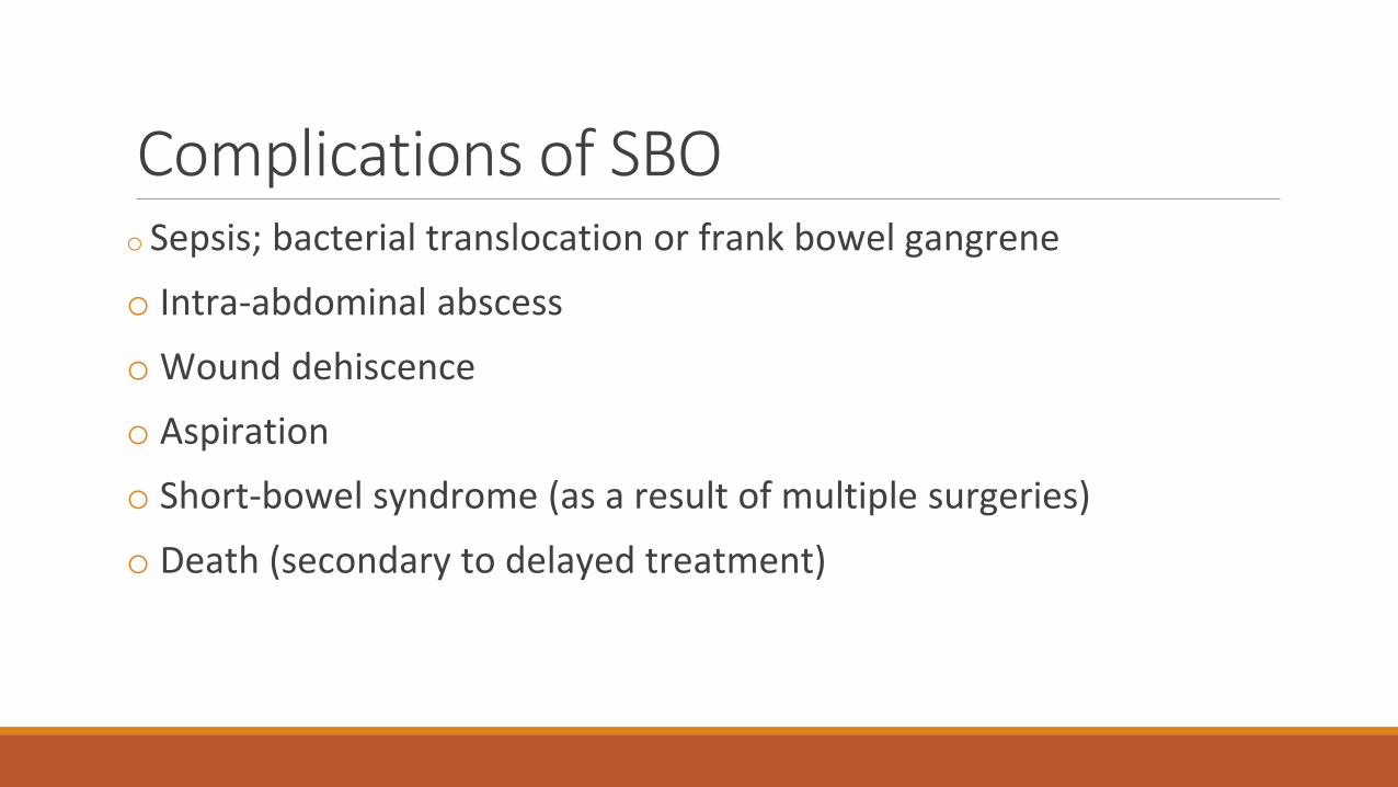
Complications of SBO o Sepsis; bacterial translocation or frank bowel gangrene
o Intra-abdominal abscess
o Wound dehiscence
o Aspiration
o Short-bowel syndrome (as a result of multiple surgeries)
o Death (secondary to delayed treatment)
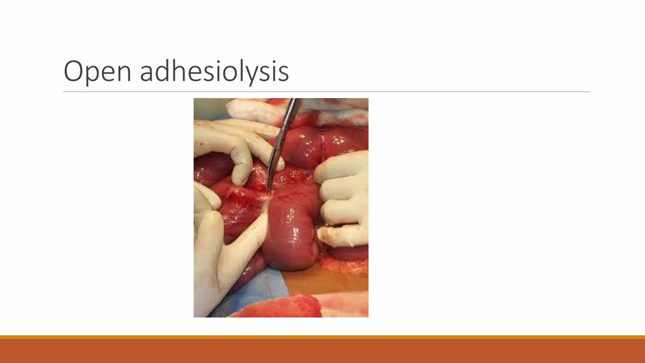
Open adhesiolysis
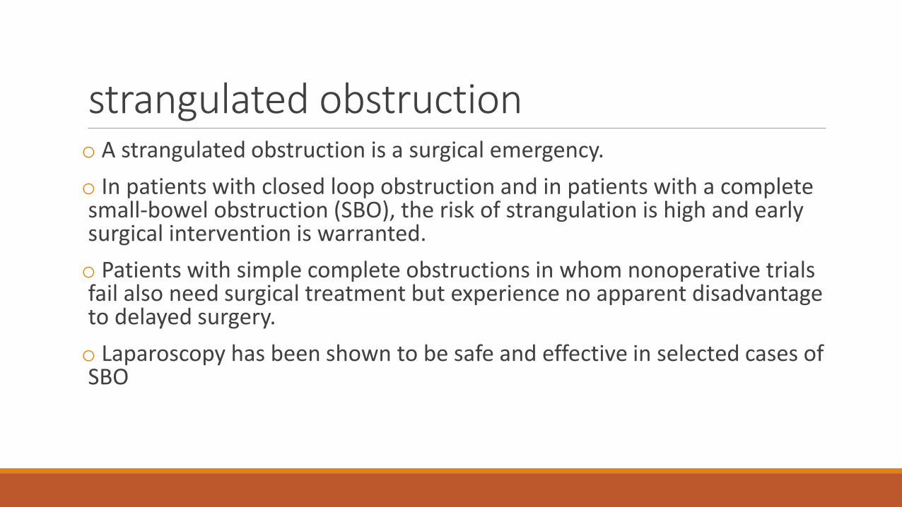
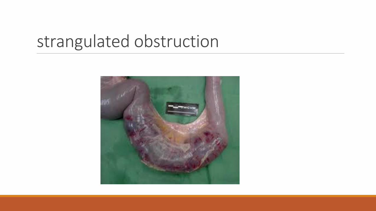
strangulated obstructiono A strangulated obstruction is a surgical emergency.
o In patients with closed loop obstruction and in patients with a complete small-bowel obstruction (SBO), the risk of strangulation is high and early surgical intervention is warranted.
o Patients with simple complete obstructions in whom nonoperative trials fail also need surgical treatment but experience no apparent disadvantage to delayed surgery.
o Laparoscopy has been shown to be safe and effective in selected cases of SBO
strangulated obstruction
strangulated obstructionmortalityoIf untreated, strangulated obstructions cause death in 100% of patients.
oIf surgery is performed within 36 hours, the mortality rate decreases to 8%.
oThe mortality rate is 25% if the surgery is postponed beyond 36 hours in these patients.

Ileus
Definitions Interruption of the normal propagation of intestinal contents due to decreased motor activity
Synonyms; 1. functional
2. Paralytic
3. adynamic
Etiology1. Peritonitis
2. Postoperative
3. Stress, sepsis, hypoperfusion, hypoxia
4. Trauma
5. Drugs, narcotics, anticholinergics, sedatives,… etc.
6. Metabolic, electrolyte disturbances, DKA, organ failures
7. Idiopathic
Distribution ❑Generalized
❑ Localized◦ Small bowel as in pancreatitis
◦ Large bowel as acute appendicitis
Diagnosisclinicalo The predisposing factor
o Constipation or obstipation
o Abdominal distention
o Vomiting or regurge
o Diminished bowel sounds
o Minimal or no abdominal pain
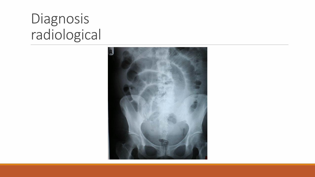
Diagnosisradiological

Postoperative ileusoAffects small and large bowel
o Small bowel regains activity before the large (usually within hours)
o May last few days
o CT scan is the best modality to distinguish postop ileus from postop mechanical obstruction
General Management
1. NPO
2. Nasogastric intubation
3. Fluid and electrolyte resuscitation
4. Reverse the primary cause
5. Use of prokinetic drugs ?