Embed Size (px)
Citation preview
Hindawi Publishing CorporationThe Scientific World JournalVolume 2013, Article ID 298392, 6 pageshttp://dx.doi.org/10.1155/2013/298392
Clinical StudyCT Findings of Patients with Small Bowel Obstruction due toBezoar: A Descriptive Study
Fatih Altintoprak,1 Bumin Degirmenci,2,3 Enis Dikicier,4 Guner Cakmak,4 Taner Kivilcim,4
Gokhan Akbulut,1 Osman Nuri Dilek,1 and Yasemin Gunduz2
1 Department of General Surgery, Faculty of Medicine, Sakarya University, Turkey2Department of Radiology, Faculty of Medicine, Sakarya University, Turkey3 Department of Radiology, Faculty of Medicine, Suleyman Demirel University, Turkey4Department of General Surgery, Sakarya University Research and Educational Hospital, Turkey
Correspondence should be addressed to Fatih Altintoprak; [email protected]
Received 19 February 2013; Accepted 1 April 2013
Academic Editors: A. P. Kypson and A. Shamiyeh
Copyright © 2013 Fatih Altintoprak et al. This is an open access article distributed under the Creative Commons AttributionLicense, which permits unrestricted use, distribution, and reproduction in any medium, provided the original work is properlycited.
Purpose.The aim of this study was to present the computed tomography (CT) findings of bezoars that cause obstruction in the smallbowel and to emphasize that some CT findings can be considered specific to some bezoar types.Materials andMethods.The recordsof 39 patients who underwent preoperative abdominal CT and subsequent operation with a diagnosis of intestinal obstruction dueto bezoars were retrospectively analyzed. Results. In total, 56 bezoars were surgically removed from 39 patients. Bezoars were mostcommonly located in the jejunum (𝑛 = 26/56, 46.4%). Sixteen (41.0%) patients hadmultiple bezoar locations in the gastrointestinaltract. Common CT findings in all patients were a mottled gas pattern and a focal ovoid or round intraluminal mass with regularmargins and a heterogeneous internal structure. Furthermore, some CT findings were determined to be specific to bezoars causedby persimmons. Conclusions. Preoperative CT is valuable in patients admitted with signs of intestinal obstruction in geographicregions with a high bezoar prevalence. We believe that the correct diagnosis of bezoars and the identification of their number andlocation provide a great advantage for all physicians and surgeons. In addition, some types of bezoars have unique CT findings, andwe believe that these findings may help to establish a diagnosis.
1. Introduction
Bezoar is a mass of swallowed foreign indigestible materialfound within the gastrointestinal tract (GIT). Despite the factthat bezoars are a rare cause of intestinal obstruction, thisemergency pathology is a frequently encountered problemworldwide [1]. Predisposing factors in bezoar formationinclude systemic diseases that reduce gastrointestinalmotilityand previous peptic ulcer surgery [2].
Radiologic findings are very valuable for bezoar diagno-sis, because clinical and laboratory findings are similar forbezoars and other causes. Radiographic andultrasonographicfindings have been defined for bezoars [3, 4]. However, bothmethods have disadvantages. Computed tomography (CT) issuperior to other radiologic tools for bezoar diagnosis anddifferential diagnosis in patients with intestinal obstruction.
In this paper, we evaluated the preoperative CT findingsof bezoars that cause obstructions in the small intestines, andhighlight some special CT appearances that may be useful fordifferential diagnoses in the preoperative period.
2. Materials and Methods
The records of 39 patients who underwent preoperativeabdominal CT and subsequent operation with a diagnosisof intestinal obstruction due to bezoars were retrospectivelyanalyzed between January 2004 and December 2012 at singlecenter.The diagnosis of intestinal obstructionwas establishedon the basis of clinical presentation (vomiting and abdominalpain and/or distention) and radiologic findings (plain X-rayor CT). The diagnosis of intestinal bezoar was confirmedeither by intraoperative findings or on CT findings.
2 The Scientific World Journal
Table 1: Total bezoars and multiple bezoar locations.
Total bezoar locations 𝑛 = 56, (100%) Multiple bezoars locations 𝑛 = 39, (100%)Stomach 12 (21.4%) Stomach and jejunum 6 (15.3%)Duodenum 1 (1.7%) Stomach and Ileum 5 (12.8%)Jejunum 26 (46.4%) Jejenum and ileum 2 (5.1%)Ileum 17 (30.3%) #Stomach and jejenum 1 (2.5%)
∗Jejenum 2 (5.1%)#One bezoar in stomach and two bezoars in jejunum.∗Two bezoars in jejenum.
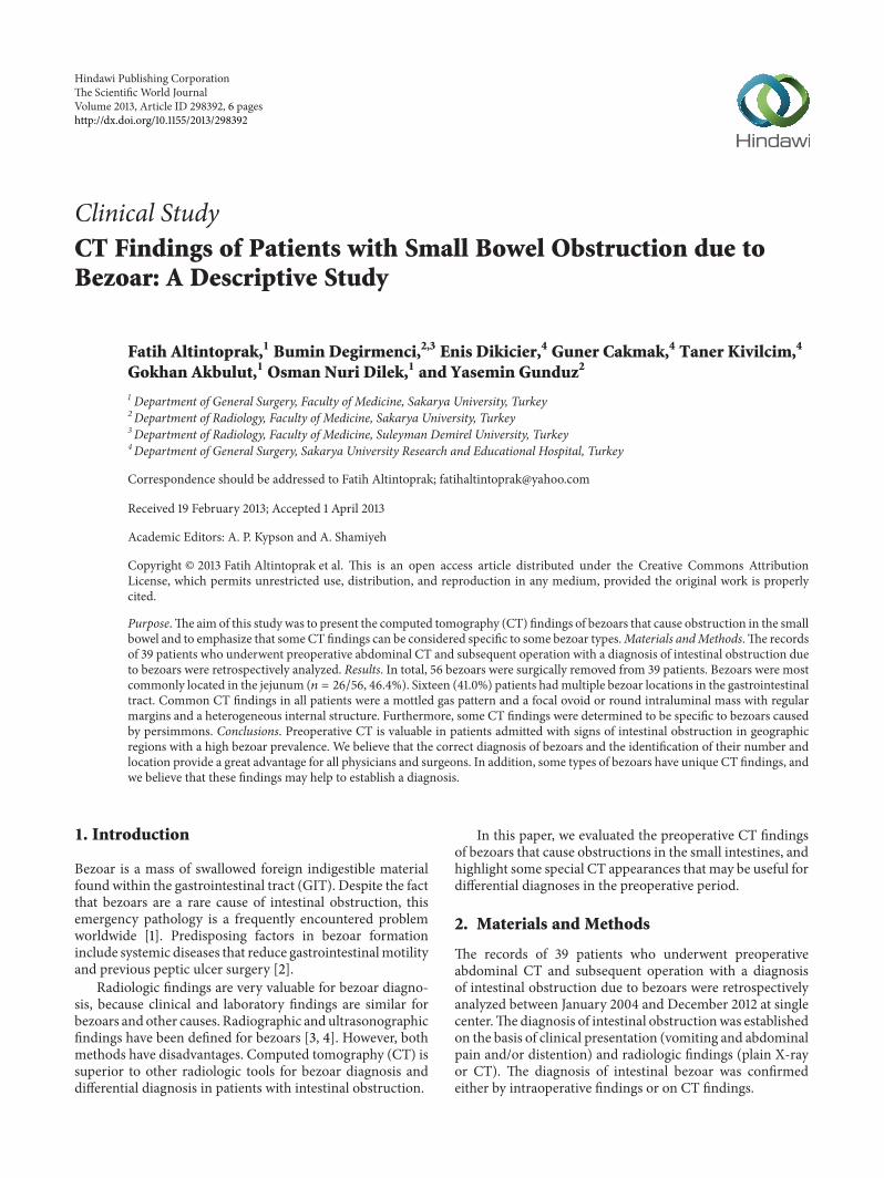
(a) (b)
Figure 1: 61-year-old female patient. (a) CT findings: an intraluminal ovoid bezoar. Fusiform-shaped seeds were seen in the mass, andwall thickening was seen in the duodenum (arrow). (b) Intraoperative findings: an ovoid-shaped bezoar containing seeds was removed viaenterotomy.
All patients’ CT examinations were performed with a4-multidetector computed tomography (4-MDCT) scanner(Asteion; Toshiba, Kobe, Japan). The following parameterswere used in the CT examination protocols: 4 × 5mm colli-mation, 5mm slice thickness, 2.5mm scan interval, 120 kVp,and 250mAs. Approximately 125mL of intravenous (IV)iohexol (Omnipaque 300; GE Healthcare, Little Chalfont,United Kingdom), iopromide (Ultravist 300; Bayer-Schering,Berlin, Germany), or iomeprol (Iomeron 350; Bracco,Milano,Italy) was given to patients who have no contraindicationsfor IV contrast use. Contrast-enhanced examinations usinga routine portal venous phase (60–70 s) were performed.
When considering the diameter and shape of bezoars, theaxial CT sequences of bezoars which were located only inobstruction region were evaluated.
3. Results
The female :male ratio was 24 : 15 (1.6 : 1), and the mean agewas 62.0 (range 28–82) years. Eighteen patients were >65years of age. A total of 56 bezoars were found in 39 patients.
Bezoarsweremost commonly located in the jejunum (𝑛 =26/56, 46.4%). Other bezoar locations include ileum (𝑛 =17/56, 30.3%), stomach (𝑛 = 12/56, 21.4%), and duodenum(𝑛 = 1/56, 1.7%). Sixteen (41.0%) patients had multiplebezoars in different GIT locations stomach and jejunum, oneeach in six patients (15.3%); stomach and ileum, one each in
five patients (12.8%); jejunum and ileum, one each in twopatients (5.1%); jejunum and stomach, two and one in onepatient (2.5%); two bezoars in jejenum, two patients (5.1%)(Table 1).
Twenty-nine (74.3%) patients had a history of previousabdominal surgery, twenty-one (21/29, 72.4%) of them due topeptic ulcers. The surgery had been performed an average of13 (range 10–26) years previously. Nine of twenty-one (42.8%)patients who underwent previous abdominal surgery due topeptic ulcers had multiple bezoars. Six (15.3%) patients haddiabetes mellitus (DM; type I, 𝑛 = 2; type II, 𝑛 = 4).
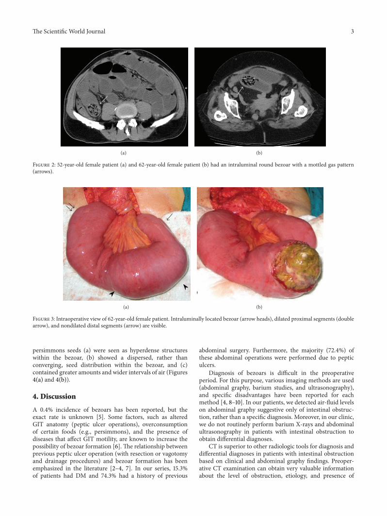
Computed tomography findings of bezoars in the smallintestine included amottled gas pattern and a focal ovoid (𝑛 =31/44, 70.4%; Figure 1) or round (𝑛 = 13/44, 29.6%) intra-luminal mass located in the obstruction region with regularmargins and a heterogeneous internal structure. In addition,while the small intestine proximal to themass was dilated, thedistal intestines had a normal diameter, suggesting intestinalobstruction (Figures 2 and 3). Contrast changes caused byinflammation at and proximal to the obstruction site werealso detected.
Patients with bezoars due to seedy type persimmon(removed bezoars contained persimmon seeds). Round orovoid intraluminal masses with a heterogeneous internalstructure and mottled gas pattern were detected at differentlevels of the small intestines and the stomach. As a resultof our examines on CT images, we have also noticed that
The Scientific World Journal 3
(a) (b)
Figure 2: 52-year-old female patient (a) and 62-year-old female patient (b) had an intraluminal round bezoar with a mottled gas pattern(arrows).
(a) (b)
Figure 3: Intraoperative view of 62-year-old female patient. Intraluminally located bezoar (arrow heads), dilated proximal segments (doublearrow), and nondilated distal segments (arrow) are visible.
persimmons seeds (a) were seen as hyperdense structureswithin the bezoar, (b) showed a dispersed, rather thanconverging, seed distribution within the bezoar, and (c)contained greater amounts and wider intervals of air (Figures4(a) and 4(b)).
4. Discussion
A 0.4% incidence of bezoars has been reported, but theexact rate is unknown [5]. Some factors, such as alteredGIT anatomy (peptic ulcer operations), overconsumptionof certain foods (e.g., persimmons), and the presence ofdiseases that affect GIT motility, are known to increase thepossibility of bezoar formation [6]. The relationship betweenprevious peptic ulcer operation (with resection or vagotomyand drainage procedures) and bezoar formation has beenemphasized in the literature [2–4, 7]. In our series, 15.3%of patients had DM and 74.3% had a history of previous
abdominal surgery. Furthermore, the majority (72.4%) ofthese abdominal operations were performed due to pepticulcers.
Diagnosis of bezoars is difficult in the preoperativeperiod. For this purpose, various imaging methods are used(abdominal graphy, barium studies, and ultrasonography),and specific disadvantages have been reported for eachmethod [4, 8–10]. In our patients, we detected air-fluid levelson abdominal graphy suggestive only of intestinal obstruc-tion, rather than a specific diagnosis. Moreover, in our clinic,we do not routinely perform barium X-rays and abdominalultrasonography in patients with intestinal obstruction toobtain differential diagnoses.
CT is superior to other radiologic tools for diagnosis anddifferential diagnoses in patients with intestinal obstructionbased on clinical and abdominal graphy findings. Preoper-ative CT examination can obtain very valuable informationabout the level of obstruction, etiology, and presence of
4 The Scientific World Journal
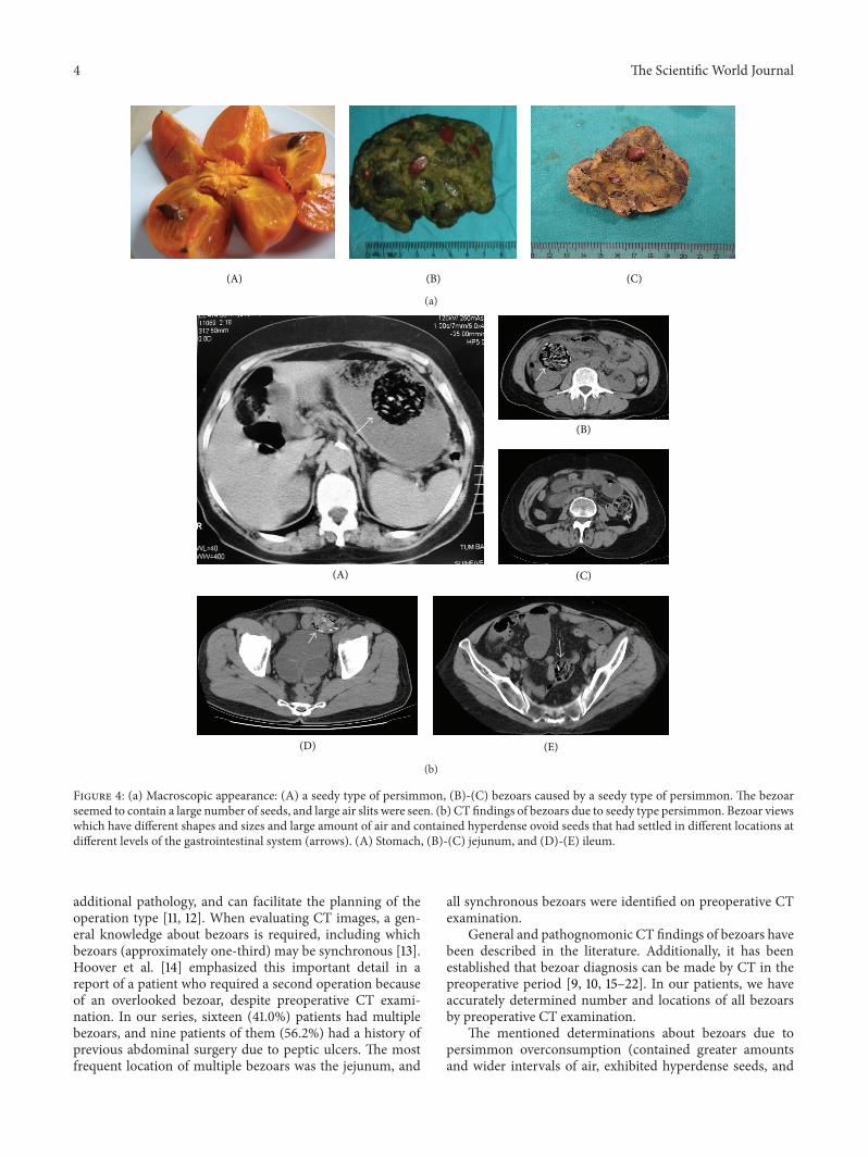
(A) (B) (C)
(a)
(A)
(B)
(C)
(D) (E)
(b)
Figure 4: (a) Macroscopic appearance: (A) a seedy type of persimmon, (B)-(C) bezoars caused by a seedy type of persimmon. The bezoarseemed to contain a large number of seeds, and large air slits were seen. (b) CT findings of bezoars due to seedy type persimmon. Bezoar viewswhich have different shapes and sizes and large amount of air and contained hyperdense ovoid seeds that had settled in different locations atdifferent levels of the gastrointestinal system (arrows). (A) Stomach, (B)-(C) jejunum, and (D)-(E) ileum.
additional pathology, and can facilitate the planning of theoperation type [11, 12]. When evaluating CT images, a gen-eral knowledge about bezoars is required, including whichbezoars (approximately one-third) may be synchronous [13].Hoover et al. [14] emphasized this important detail in areport of a patient who required a second operation becauseof an overlooked bezoar, despite preoperative CT exami-nation. In our series, sixteen (41.0%) patients had multiplebezoars, and nine patients of them (56.2%) had a history ofprevious abdominal surgery due to peptic ulcers. The mostfrequent location of multiple bezoars was the jejunum, and
all synchronous bezoars were identified on preoperative CTexamination.
General and pathognomonic CT findings of bezoars havebeen described in the literature. Additionally, it has beenestablished that bezoar diagnosis can be made by CT in thepreoperative period [9, 10, 15–22]. In our patients, we haveaccurately determined number and locations of all bezoarsby preoperative CT examination.
The mentioned determinations about bezoars due topersimmon overconsumption (contained greater amountsand wider intervals of air, exhibited hyperdense seeds, and
The Scientific World Journal 5
showed a dispersed, rather than converging, seed distributionwithin the bezoar) are subjective and may not be unique topersimmon seeds. But these findings may be typical featuresfor this specific type of bezoar, andwe believe that they shouldbe kept in mind when evaluating abdominal CT findings insome geographic regions.
The appearances about small bowel bezoars may besimilar to small bowel feces (SBF). SBF is frequently foundwithin the lumen of a relatively long segment of a dilatedsmall bowel loop, and the incidence is approximately 8% insmall bowel obstruction cases [15]. SBF is placed proximal tothe obstruction site as distinct from bezoar and longer thanbezoar. Kim et al. [13] defined that SBF ismore tubular shapedthan bezoar, and Zissin et al. [15] indicated that the length ofthe feces-likematerial foundwithin the dilated loop proximalto the transition zone is the key for differentiating SBF signfrom a bezoar. We did not think SBF diagnosis in our casesdue to the fact that the intraluminal gas-containing massimagings with round or ovoid-shaped bezoar are located atthe obstruction site.
Carrying out only the axial section CT findings’ evalua-tion is the restrictive aspect of our study. By themultidetectorrow CT (MDCT), more detailed assessments can be made bytaking axial, coronal, sagittal, and oblique images in severalsequences. Hodel et al. [23] found that multiplanar refor-mations (MPRs) can increase both accuracy and confidencein the location of the transition zone in CT of mechan-ical small bowel obstruction. If our study was designedby MPR, probably different results could be achieved(especially about shape of the bezoar at the obstructionregion).
In conclusion, although bezoars are a rare cause ofintestinal obstruction, the possible presence of bezoarsshould not be forgotten in patients admitted with signs ofintestinal obstruction, especially patients with a history ofprevious abdominal surgery for peptic ulcer. In addition,we believe that some bezoars, especially those caused bypersimmons, have specific CT appearances that may help toestablish a preoperative diagnosis in some patients admit-ted with signs of intestinal obstruction and who live ingeographic regions where persimmon overconsumption iscommon.
Conflict of Interests
The authors declare that they have no conflict of interests.
References
[1] D. Menzies and H. Ellis, “Intestinal obstruction fromadhesions—how big is the problem?” Annals of the RoyalCollege of Surgeons of England, vol. 72, no. 1, pp. 60–63, 1990.
[2] C. Escamilla, R. Robles-Campos, P. Parrilla-Paricio, J. Lujan-Mompean, R. Liron-Ruiz, and J. A. Torralba-Martinez, “Intesti-nal obstruction and bezoars,” Journal of the American College ofSurgeons, vol. 179, no. 3, pp. 285–288, 1994.
[3] Y. T. Ko, J. H. Lim, D. H. Lee, and Y. Yoon, “Small intestinalphytobezoars: sonographic detection,” Abdominal Imaging, vol.18, no. 3, pp. 271–273, 1993.
[4] A. G. Verstandig, B. Klin, R. A. Bloom, I. Hadas, and E. Libson,“Small bowel phytobezoars: detection with radiography,” Radi-ology, vol. 172, no. 3, pp. 705–707, 1989.
[5] R. S. Kadian, J. F. Rose, and N. S. Mann, “Gastric bezoars.Spontaneous resolution,” American Journal of Gastroenterology,vol. 70, no. 1, pp. 79–82, 1978.
[6] C. H. Andrus and J. L. Ponsky, “Bezoars: classification, patho-physiology, and treatment,” American Journal of Gastroenterol-ogy, vol. 83, no. 5, pp. 476–478, 1988.
[7] K. Erzurumlu, Z. Malazgirt, A. Bektas et al., “Gastrointestinalbezoars: a retrospective analysis of 34 cases,” World Journal ofGastroenterology, vol. 11, no. 12, pp. 1813–1817, 2005.
[8] E. J. Balthazar, “CT of small-bowel obstruction,” AmericanJournal of Roentgenology, vol. 162, no. 2, pp. 255–261, 1994.
[9] T. Ripolles, J. Garcıa-Aguayo, M. J. Martınez, and P. Gill,“Gastrointestinal bezoars: sonographic and CT characteristics,”American Journal of Roentgenology, vol. 177, no. 1, pp. 65–69,2001.
[10] S. F. Ko, T. Y. Lee, and S. H. Ng, “Small bowel obstruction dueto phytobezoar: CT diagnosis,” Abdominal Imaging, vol. 22, no.5, pp. 471–473, 1997.
[11] D. Frager, S. W. Medwid, J. W. Baer, B. Mollinelli, and M. Fried-man, “CT of small-bowel obstruction: value in establishing thediagnosis and determining the degree and cause,” AmericanJournal of Roentgenology, vol. 162, no. 1, pp. 37–41, 1994.
[12] G. Burkill, J. Bell, and J. Healy, “Small bowel obstruction: therole of computed tomography in its diagnosis and managementwith reference to other imaging modalities,” European Radiol-ogy, vol. 11, no. 8, pp. 1405–1422, 2001.
[13] J. H. Kim, H. K. Ha, M. J. Sohn et al., “CT findings of phy-tobezoar associated with small bowel obstruction,” EuropeanRadiology, vol. 13, no. 2, pp. 299–304, 2003.
[14] K. Hoover, J. Piotrowski, K. S. Pierre, A. Katz, and A. M.Goldstein, “Simultaneous gastric and small intestinal trichobe-zoars—a hairy problem,” Journal of Pediatric Surgery, vol. 41, no.8, pp. 1495–1497, 2006.
[15] R. Zissin, A. Osadchy, V. Gutman, V. Rathaus, M. Shapiro-Feinberg, and G. Gayer, “CT findings in patients with smallbowel obstruction due to phytobezoar,” Emergency Radiology,vol. 10, no. 4, pp. 197–200, 2004.
[16] S. Quiroga, A. Alvarez-Castells, M. C. Sebastia, E. Pallisa, and E.Barluenga, “Small bowel obstruction secondary to bezoar: CTdiagnosis,” Abdominal Imaging, vol. 22, no. 3, pp. 315–317, 1997.
[17] M. Licht, B. M. Gold, and D. S. Katz, “Obstructing small-bowelbezoar: diagnosis usingCT,”American Journal of Roentgenology,vol. 173, no. 2, pp. 500–501, 1999.
[18] S. Ulusan, Z. Koc, and N. Torer, “Small bowel obstructionssecondary to bezoars,” Ulusal Travma ve Acil Cerrahi Dergisi,vol. 13, no. 3, pp. 217–221, 2007.
[19] H. Bedioui, A. Daghfous, M. Ayadi et al., “A report of 15 casesof small-bowel obstruction secondary to phytobezoars: pre-disposing factors and diagnostic difficulties,” GastroenterologieClinique et Biologique, vol. 32, no. 6-7, pp. 596–600, 2008.
[20] G. Gayer, T. Jonas, S. Apter et al., “Bezoars in the stomach andsmall bowel—CT appearance,” Clinical Radiology, vol. 54, no. 4,pp. 228–232, 1999.
[21] E. Delabrousse, S. Brunelle, O. Saguet, N. Destrumelle, G.Landecy, and B. Kastler, “Small bowel obstruction secondary tophytobezoar CT findings,” Clinical Imaging, vol. 25, no. 1, pp.44–46, 2001.

6 The Scientific World Journal
[22] J. Tamminen and D. Rosenfeld, “CT diagnosis of a gastrictrichobezoar,”ComputerizedMedical Imaging andGraphics, vol.12, no. 6, pp. 339–341, 1988.
[23] J. Hodel,M. Zins, L. Desmottes et al., “Location of the transitionzone inCTof small-bowel obstruction: added value ofmultipla-nar reformations,” Abdominal Imaging, vol. 34, no. 1, pp. 35–41,2009.

Submit your manuscripts athttp://www.hindawi.com
Stem CellsInternational
Hindawi Publishing Corporationhttp://www.hindawi.com Volume 2014
Hindawi Publishing Corporationhttp://www.hindawi.com Volume 2014
MEDIATORSINFLAMMATION
of
Hindawi Publishing Corporationhttp://www.hindawi.com Volume 2014
Behavioural Neurology
EndocrinologyInternational Journal of
Hindawi Publishing Corporationhttp://www.hindawi.com Volume 2014
Hindawi Publishing Corporationhttp://www.hindawi.com Volume 2014
Disease Markers
Hindawi Publishing Corporationhttp://www.hindawi.com Volume 2014
BioMed Research International
OncologyJournal of
Hindawi Publishing Corporationhttp://www.hindawi.com Volume 2014
Hindawi Publishing Corporationhttp://www.hindawi.com Volume 2014
Oxidative Medicine and Cellular Longevity
Hindawi Publishing Corporationhttp://www.hindawi.com Volume 2014
PPAR Research
The Scientific World JournalHindawi Publishing Corporation http://www.hindawi.com Volume 2014
Immunology ResearchHindawi Publishing Corporationhttp://www.hindawi.com Volume 2014
Journal of
ObesityJournal of
Hindawi Publishing Corporationhttp://www.hindawi.com Volume 2014
Hindawi Publishing Corporationhttp://www.hindawi.com Volume 2014
Computational and Mathematical Methods in Medicine
OphthalmologyJournal of
Hindawi Publishing Corporationhttp://www.hindawi.com Volume 2014
Diabetes ResearchJournal of
Hindawi Publishing Corporationhttp://www.hindawi.com Volume 2014
Hindawi Publishing Corporationhttp://www.hindawi.com Volume 2014
Research and TreatmentAIDS
Hindawi Publishing Corporationhttp://www.hindawi.com Volume 2014
Gastroenterology Research and Practice
Hindawi Publishing Corporationhttp://www.hindawi.com Volume 2014
Parkinson’s Disease
Evidence-Based Complementary and Alternative Medicine
Volume 2014Hindawi Publishing Corporationhttp://www.hindawi.com

![Intrapulmonary Lymph Nodes:Thin-Section CT … Lymph Nodes:Thin-Section CT Findings, Pathological Findings,and CT Differential Diagnosis from Pulmonary Metastatic ... shadows[3-10],and](https://img.pdfslide.net/doc/110x75/5acbaebf7f8b9a73128bf3e4/intrapulmonary-lymph-nodesthin-section-ct-lymph-nodesthin-section-ct-findings.jpg)