Embed Size (px)
Citation preview
BRAIN ABSCESS
MANAGEMENT
Presenter : PANKAJ AILAWADHI
Moderators : PROF. BS SHARMA
DR. SUMIT SINHA


LAKSHMI V, R. R. RAO RR, DINAKAR I: Bacteriology of brain abscess - observations
on 50 cases. J. Med. Microbiol. -Vol. 38 (1993), 187-190

INCIDENCE
• VARIES ACCORDING TO GEOGRAPHY
• CONSTITUTES 8 % OF ALL INTRACRANIAL LESIONS IN INDIA TO 1-2% IN DEVELOPED LESIONS IN INDIA TO 1-2% IN DEVELOPED NATIONS.*
*Osenbach RK, Loftus CM: Diagnosis and management of brain abscess.
Neurosurg Clin N Am 1992; 3: 403-420.
Bhatia R, Tandon PN, Banerji AK: Brain abscess - an analysis of 55 cases. Int Surg 1973, 58: 565-568

ETILOGICAL AGENTS
• In about one third of patients, more than one organism found.
• Organisms most frequently isolated include streptococci (both aerobic and anaerobic), anaerobes and staphylococci
• Microbiological profile is also changing
• Gram-negative organisms/ anaerobes - increasing cause of cerebral abscess.
• In neonates, more common organisms include Citrobacter,
Proteus, Pseudomonas, and Serratia species
• These abscesses are often large with poorly formed capsules
• Organisms other than pyogenic bacteria include
Mycobacterium tuberculosisNontuberculous mycobacteriaFungiFungiParasitesActinomyces and Nocardia speciesrarely salmonella


ETIOLOGICAL AGENTS
• Most abscesses are monomicrobial
• 1/3 of all abscesses are polymicrobial
(especially otogenic)
Incidence of negative cultures is 25-30% Incidence of negative cultures is 25-30%

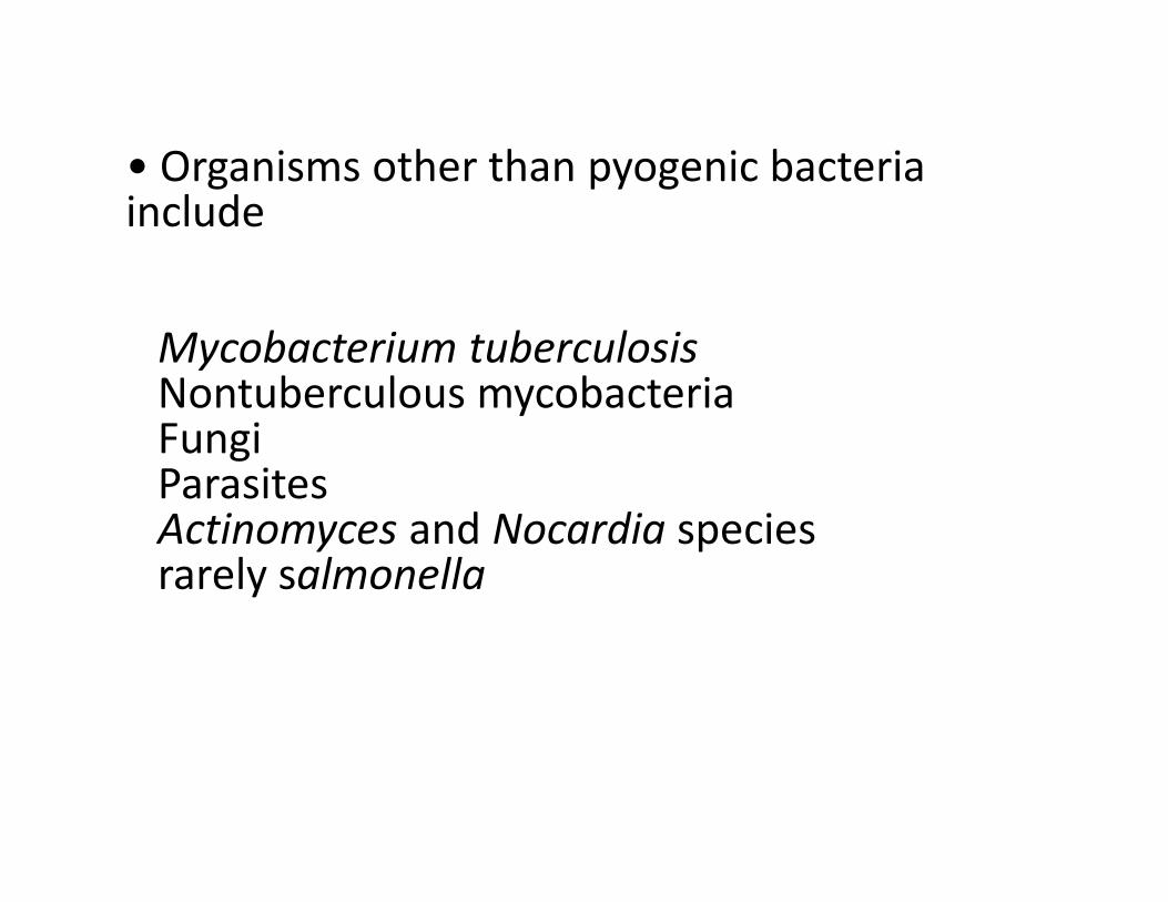
Location
• OTOGENIC 24(48%)
• CONGENITAL HEART 09(18%)
DISEASE
• PULMONARY DISEASE 06(12%)• PULMONARY DISEASE 06(12%)
• POST TRAUMATIC 01
TOTAL 50LAKSHMI V, R. R. RAO RR, DINAKAR I: Bacteriology of brain abscess - observations on 50
cases. J. Med. Microbiol. -Vol. 38 (1993), 187-190
• MECHANISM:RETROGRADE VENOUS THROMBO PHLEBITIS


•TOF → CHRONIC HYPOXEMIA→ POLYCYTHEMIA/ INCREASED
VISCOSITY→MULTIPLE INFARCTS AT GREYWHITE JUNCTION
→→MILIEU FOR BACTERIAL GROWTH

3. TRAUMA
• Open cranial fracture with dural breach
• Neurosurgery or foreign body
4. CRYPTOGENIC


CAPSULE formation influenced by:
•Type of organism
•Metastatic abscesses poorly capsulated•Metastatic abscesses poorly capsulated
•Use of corticosteroids


CLINICAL PRESENTATION Depends on:
•Location of the lesion
•Size of the lesion
•Multiplicity•Multiplicity
• virulence of the organism
•Host response
•Severity of edema

Classical triad of headache, neurol deficits,
fever seen in 25%*fever seen in 25%*
*Loftus CM, Osenbach RK, Biller J: Diagnosis and management of brain abscess. In: Neurosurgery, second edition, Vol. III, Wilkins
RH, Rengachary SS (eds) McGraw-Hill, New York 1996; 3285-3298

HEADACHE


• CYSTIC GLIOMAS
• RESOLVING HEMATOMAS
• DEMYELINTING DISEASE

LAB INVESTIGATIONS
• NOT MUCH HELPFUL
• TLC > 20000 SEEN IN ONLY 10%
• ESR IS INCREASED IN > 90%• ESR IS INCREASED IN > 90%
• CRP levels - Help in following response to treatment
• L.P. IS INCONCLUSIVE AND DANGEROUS – TO BE AVOIDED

Plain X-Rays
• Radiographic findings usually limited to paranasal or mastoid sinus opacification
• Gas bubbles or air-fluid levels within cranium indicate gas-producing organism or communication indicate gas-producing organism or communication with paranasal sinuses
• Occasionally, foreign bodies or osteomyelitis of maxillary bone indicate source
• Bone destruction of roof, floor, or lateral wall of sinuses indicates aggressive osteomyelitis


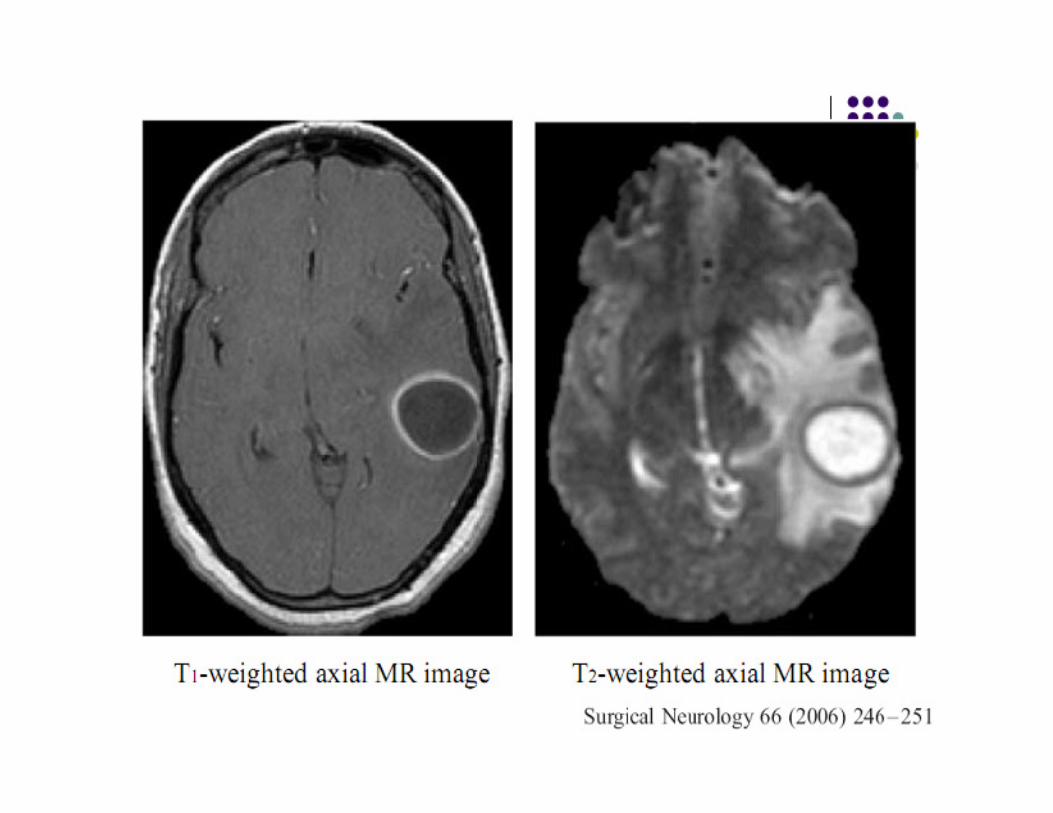
MRI


(However, fever, meningism, raised ESR, multilocularity,
leptomeningeal or ependymal enhancement, reduction of ring
enhancement in delayed scan and finding of gas within the lesion
favour a diagnosis of abscess.)


Noninvasively by localized in vivo


LEUKOCYTE LABELLED SCANS
• Finding of uptake of labeled leukocytes exceeding the normal physiological uptake of the base of skull*
• Sensitivity approaches 100%,Specifity 94%• Sensitivity approaches 100%,Specifity 94%
• Non invasive
• However , optimal images takes 24 hrs, can be used only in pts. with good neurological condition.
*Rehncrona S, Brismar J, Holtas 5: Diagnosisof brain abscesses with indium-i 1i-labeled
leukocytes. Neuro surgery16:23-26,1985.

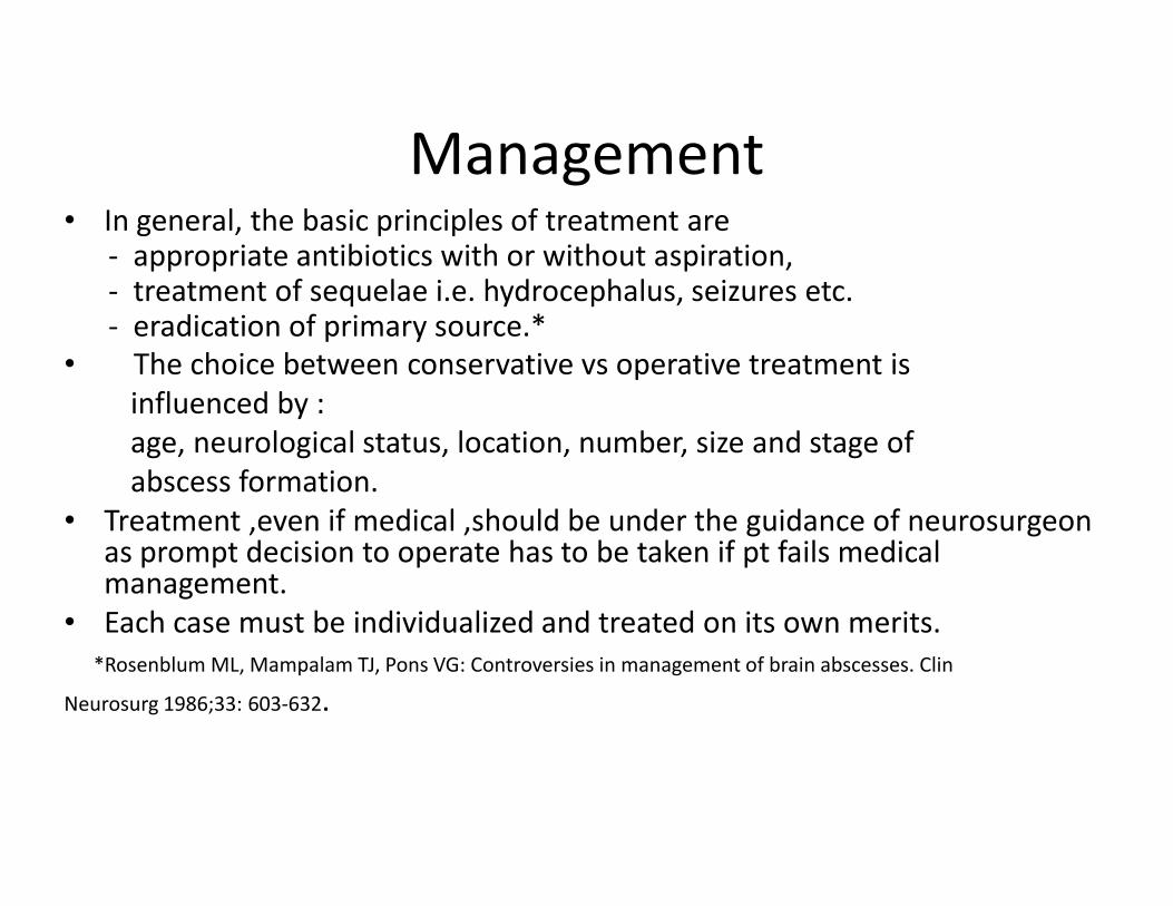
Management • In general, the basic principles of treatment are
- appropriate antibiotics with or without aspiration, - treatment of sequelae i.e. hydrocephalus, seizures etc. - eradication of primary source.*
• The choice between conservative vs operative treatment is
influenced by :
age, neurological status, location, number, size and stage of age, neurological status, location, number, size and stage of
abscess formation.
• Treatment ,even if medical ,should be under the guidance of neurosurgeon as prompt decision to operate has to be taken if pt fails medical management.
• Each case must be individualized and treated on its own merits.
*Rosenblum ML, Mampalam TJ, Pons VG: Controversies in management of brain abscesses. Clin
Neurosurg 1986;33: 603-632.

MANAGEMENT
Current concepts in the management of
pyogenic brain abscess Sharma BS, Gupta SK, Khosla VK:. Neurol India
48:105–111, 2000

Medical management– Antibiotic therapy during early stage prevents progress
from cerebritis to abscess.
– Patients who have symptoms for < 1 week have a more favorable response to medical therapy.
– Empirical treatment is advocated only when clinical and radiological improvement continues.
– Empirical treatment is advocated only when clinical and radiological improvement continues.
– Rosenblum et al found that mean diameter of abscess that resolved with a/b was 1.7cm( no abscess> 2.5cm resolved with a/b alone)

•Patients demonstrate clinical improvement before significant changes in CT scan.
•Improvement on CT scans generally observed within 1-4 weeks (average, 2.5 wk) and complete resolution in 1-11 months (average, 3.5 mo).
•Antimicrobial treatment generally long (6-8 wk) because of prolonged time needed for brain to repair and close abscess space
•If medical therapy is chosen , ct scans to be done weekly during t/t and mthly after discontinuing t/t till enhancement disappears

EFFICACY OF ANTIBIOTICS DEPENDS ON:
•Bactericidal / bacteriostatic nature
•Route and duration of therapy
•Host response to infection
•Concentration of drug at the site of
abscess
•Depth of the lesion

Usually 'triple high dose' antibiotics intravenously for 2 weeks followed by four weeks of
oral therapy is recommended.*
*Krajewski D, Stelmasiak Z: Brain abscess in infants. Child's Nerv Syst 1992; 8: 279-280.
Mamelak AN, Mampalam TJ, Obana WG et al: Improved management of multiple brain abscess: A combined surgical and medical
approach. Neurosurgery 1995; 36: 76-86.
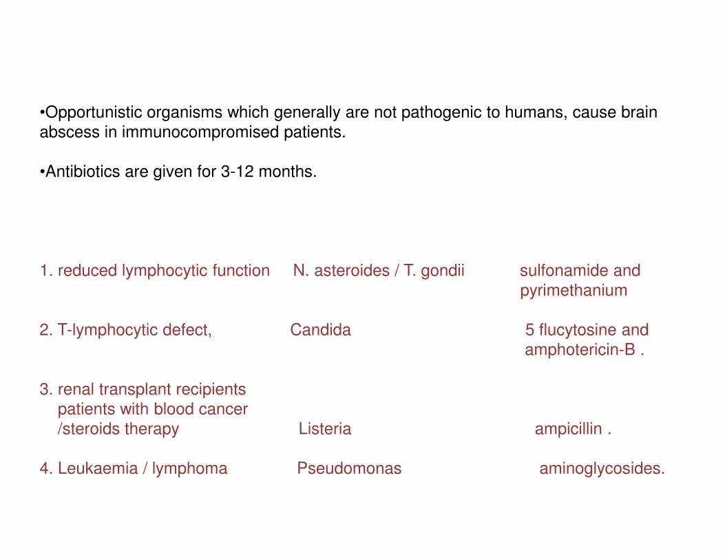
•Opportunistic organisms which generally are not pathogenic to humans, cause brain
abscess in immunocompromised patients.
•Antibiotics are given for 3-12 months.
1. reduced lymphocytic function N. asteroides / T. gondii sulfonamide and
pyrimethanium
2. T-lymphocytic defect, Candida 5 flucytosine and
amphotericin-B .
3. renal transplant recipients
patients with blood cancer
/steroids therapy Listeria ampicillin .
4. Leukaemia / lymphoma Pseudomonas aminoglycosides.

ANTICONVULSANT THERAPY
• Legg* advocated anticonvulsant therapy to all
pts of cerebral abscess for 5 yrs.
• To be discontinued only when pt is seizure
free for 2 yrs or EEG shows on epileptic free for 2 yrs or EEG shows on epileptic
activity
*Legg NJ, Gupta PC,ScottDF: Epilepsy following cerebral abscess: A clinical and EEG
study of 70 pts. Brain1973;96:259-68

• Surgical drainage provides most optimal therapy
• Aspiration is most common procedure and is often performed using a stereotactic procedure
• CAN BE USED IN ALL STAGES OF ABSCESS
SURGICAL MANAGEMENT
• CAN BE USED IN ALL STAGES OF ABSCESS
• In case of multiple abscesses or in abscesses in essential brain areas, repeated aspirations preferred to complete excision
• Aspiration can be guided by real time usg and stereotaxy.


- Recently, encouraging results with endoscopic
stereotactic evacuation of brain abscess.
- Neuroendoscopic treatment, when compared to
stereotactic aspiration, has additional advantage of more
complete drainage and lavage.*complete drainage and lavage.*
* Hellwig D, Benes L, Bertalanffy H et al: Endoscopic stereotaxy - an [eight] years experience.
Stereotact Funct Neurosurg 1997; 68: 90-97.
Fritsch M, Manwaring KH: Endoscopic treatment of brain abscess in children. Minim Invasive
Neurosurg 1997; 40: 103-106


PROGNOSIS
Improved microbial isolation technique
particularly for anaerobes
availability of newer effective antibiotics with
better blood brain barrier (BBB) penetration
for gram negative and anaerobes
improvements in surgical techniques of aspiration with stereotactic or real
time ultrasound localization
reduction in mortality rate
from 40-60% in pre CT era to
current rate of 0-10%.*
anaerobeslocalization
* Yang SY: Brain abscess: A review of 400 cases. J Neurosurg 1981; 55: 794-799.

SEQUELAE
• 30-50% of survivors are found to have neurological sequelae.
• The incidence of residual neural deficits - hemiparesis, cognitive and learning deficits in children, is less with aspiration than excision.
• About 72% of patients can have epileptic seizures upto five years of diagnosis. This incidence is less with aspiration than excision.
• 5 to 10% abscesses recur due to inadequate or inappropriate • 5 to 10% abscesses recur due to inadequate or inappropriate antibiotics, failure of removal of foreign body, dural fistula or failure of eradication of primary source.
• Hydrocephalus may also develop.
Koszewskiw M: Epilepsy following brain abscess. The evaluation of possible risk factors with emphasis on new concepts of epileptic focus formation. Acta Neurochir (Wien) 1991; 113: 117-120.

SPECIAL TYPES
• MULTIPLE ABSCESS
• 5% to 50% of all brain abscess patients harbor multiple abscesses
• detection rate has increased since the advent of CT.
• Result from Hematogenous spread from systemic infections and the organisms • Result from Hematogenous spread from systemic infections and the organisms may be found in peripheral source.
• Streptococcus and Staphylococcus are common organisms.
• More often in immunocompromised patients .
• The frequency of intraventricular rupture is also more in these cases.
Sharma BS, Khosla VK, Kak VK et al: Multiple pyogenic brain abscesses. Acta Neurochir (Wein) 1995; 133: 36-43.

SPECIAL TYPES
• CEREBELLAR ABSCESS:
• Cerebellar abscesses comprise 6-35% of all brain abscesses.
• They are often ominously silent and carry significant mortality.
• can cause sudden total occlusion of CSF pathways early in the course of disease.
• Presence of periventricular lucency is an absolute indication of immediate • Presence of periventricular lucency is an absolute indication of immediate ventricular drainage regardless of level of consciousness i.e. even if patient is fully conscious.
• burr hole aspiration has emerged as a satisfactory method.
Brydon HL, Hardwidge C: The management of cerebellar abscess since the introduction of CT scanning. Br J Neurosurg 1994; 8: 447-455.

SPECIAL TYPES
• FUNGAL ABSCESS:
• Affects Immunocompromised hosts
• Aspergillus/ candida more common
• Infection either by direct extension or by hematogenous spread
• Definitive diagnosis only by biopsy but prompt exam. possible by KOH wet • Definitive diagnosis only by biopsy but prompt exam. possible by KOH wet mount prep.
• Immunodiagnostic assays helpful in diagnosis
• Definitive therapy is antifungal chemotherapy + surgical excision
* infection of central nervous system : By W.M. Michael scheld, Richard . J.
whiley

SPECIAL TYPES
• Fungal abscess:• i.v. AMB is the mainstay of therapy
• Lipid formulation is preferred.
• Can cause nephrotoxicity, hypokalemia, hypomagnesaemia, hepatotoxicity, leucopenia, thrombopenia, cardiac arrhythmias , cardiac failure.failure.
• 5-10mg given initially as a test dose.
• Started initially at 0.3mg/kg at a rate of o.1mg /ml . Dose escalated gradually to 0.7mg/kg
• Candida infection requires :
AMB 0.7mg/kg/day+5-FU at 100mg/kg/day for 2wks f/b
Fluconazole 400-800mg/day for 6 wks
• Aspergillosis : voriconazole 300mg/day for 6mths -1yr
• Therapy is continued till lesions resolve or sustained stabilizations occur.

SPECIAL TYPESNOCARDIAL ABSCESSES
Nocardia are aerobic, gram-
positive, partially acid-fast,
filamentous bacteria.
Nocardia asteroids -most
common pathogen
Affects immunocompromised
hostshosts
Pulmonary infection is the most
common initial infection, with
hematogenous spread to other
organs Coronal T1-weighted spin-echo
gadolinium-enhanced MRI
demonstrates zone of enhancement
, with a zone of decreased
brightness (edema, white arrow).
Nocardia organisms were cultured
from within the abscess cavity.

SPECIAL TYPES
• Tubercular abscess
• Very rare
• Caseation →→ liquefaction→→ necrosis
• Granulomatous changes absent• Granulomatous changes absent
• Pus contains viable AFB bacilli
• Differentiated from tubercular cyst which contains clear
yellowish fluid + granulomatous changes in the wall
• ATT mainstay of therapy
• Surgical drainage of the abscess required for diagnosis
& Improving the neurological condition