Embed Size (px)
Citation preview

Brain Engineering Modalities Review MRI X-ray CT Ultrasound PET PET-CT PET-MRI

Lecture Outline
• Overview of different imaging systems • Review of basic signals and systems • Image quality assessment
Medical Imaging Engineering Hyoungsuk Yoo, University of Ulsan 2

What is Medical Imaging? – Quiz 1
• Using an instrument to see the inside of a human body • The properties imaged vary depending on the imaging modality
- Non-invasive
- Some with exposure to small amount of radiation
(X-ray, CT and nuclear medicine)
- Some w/o (MRI and ultrasound)
- X-ray (projection or CT): attenuation coefficient to X-ray
- Ultrasound: sound reflectivity
- MRI: hydrogen proton density, spin relaxation.
3 Medical Imaging Engineering Hyoungsuk Yoo, University of Ulsan

Projection vs. Tomography • Projection:
- A single image is created for a 3D body,
which is a “shadow” of the body in a
particular direction (integration through
the body)
4 Medical Imaging Engineering Hyoungsuk Yoo, University of Ulsan

Projection vs. Tomography – Quiz 1 • Tomography
- A series of images are generated, one from each slice of a 3D object in a particular direction (axial, coronal, sagital)
- To form image of each slice, projections along different directions are first obtained, images are then reconstructed from projections (back- projection, Radon transform)
5 Medical Imaging Engineering Hyoungsuk Yoo, University of Ulsan

Anatomical vs. Functional Imaging • Some modalities are very good at depicting anatomical (bone)
structure - X-ray, X-ray CT - MRI
• Some modalities do not depict anatomical structures well, but reflect the functional status (blood flow, oxygenation, etc.) - Ultrasound
Functional - PET, functional MRI
MRI PET CT
6 Medical Imaging Engineering Hyoungsuk Yoo, University of Ulsan

Common Imaging Modalities
• Projection radiography (X-ray)
• Computed Tomography (CT scan or CAT Scan)
• Nuclear Medicine (SPECT, PET)
• Ultrasound imaging
• MRI
7 Medical Imaging Engineering Hyoungsuk Yoo, University of Ulsan

Projection Radiography
8 Medical Imaging Engineering Hyoungsuk Yoo, University of Ulsan

Projection Radiography
Medical Imaging Engineering Hyoungsuk Yoo, University of Ulsan

• Year discovered: 1895 (Röntgen, NP 1905) • Form of radiation: X-rays = electromagnetic radiation (photons)
• Energy / wavelength of
radiation: 0.1 - 100 keV / 10 - 0.01 nm (ionizing)
• Imaging principle: X-rays penetrate tissue and create “ shadowgram" of differences in density.
• Imaging volume: Whole body
• Resolution: Very high (sub-mm)
• Applications: Mammography, lung diseases, orthopedics, dentistry, cardiovascular.
10
Projection Radiography
Medical Imaging Engineering Hyoungsuk Yoo, University of Ulsan
Dahyun! Read five dots in page 8 and translate it!
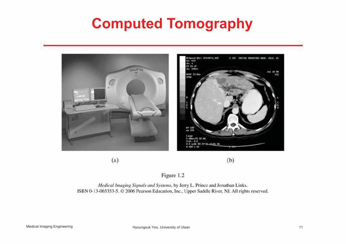
Computed Tomography
11 Medical Imaging Engineering Hyoungsuk Yoo, University of Ulsan

Computed Tomography
Medical Imaging Engineering Hyoungsuk Yoo, University of Ulsan

• Year discovered: 1972 (Hounsfield, NP 1979) • Form of radiation: X-rays
• Energy / wavelength of radiation:
10 - 100 keV / 0.1 - 0.01 nm (ionizing)
• Imaging principle: X-ray images are taken under many angles from which views are tomographic ("sliced") computed
• Imaging volume: Whole body
• Resolution: High (mm)
• Applications: Soft tissue imaging (brain, cardiovascular)
13
Computed Tomography
Medical Imaging Engineering Hyoungsuk Yoo, University of Ulsan

Nuclear Medicine • Images can only be made when appropriate radioactive
substances (called radiotracer) are introduced into the body that emit gamma rays.
• A nuclear medicine image reflects the local concentration of a radiotracer within the body • Three types
-Conventional radionuclide imaging or scintigraphy -Single photon emission computed tomography (SPECT) - Positron emission tomography (PET)
14 Medical Imaging Engineering Hyoungsuk Yoo, University of Ulsan

SPECT
15 Medical Imaging Engineering Hyoungsuk Yoo, University of Ulsan
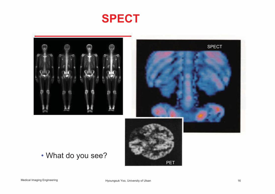
SPECT
• What do you see? PET
16
SPECT
Medical Imaging Engineering Hyoungsuk Yoo, University of Ulsan

• Year discovered: 1953 (PET), 1963 (SPECT) • Form of radiation: Gamma rays • Energy / wavelength of radiation:
> 100 keV / < 0.01 nm (ionizing)
• Imaging principle: Accumulation or "washout" of radioactive isotopes in the body cameras are imaged with x-ray.
• Imaging volume: Whole body
• Resolution: Medium - Low (mm - cm)
• Applications: Functional imaging (cancer detection, metabolic processes, myocardial infarction)
SPECT
Medical Imaging Engineering Hyoungsuk Yoo, University of Ulsan

• High frequency sound are emitted into the imaged body, time of return of these sound pulses are measured • Comparatively inexpensive and completely non-invasive • Image quality is relatively poor
18
Ultrasound Imaging
Medical Imaging Engineering
Hyoungsuk Yoo, University of Ulsan

• What do you see?
19
Ultrasound Imaging
Medical Imaging Engineering Hyoungsuk Yoo, University of Ulsan

• Year discovered: 1952 (clinical: 1962) • Form of radiation: Sound waves (non-ionizing)
NOT EM radiation! • Frequency / wavelength of radiation:
1 - 10 MHz / 1 - 0.1 mm
• Imaging principle: Echoes from discontinuities in tissue density/speed of sound are registered.
• Imaging volume: < 20 cm • Resolution: High (mm) • Applications: Soft tissue, blood flow
20
Ultrasound Imaging
Medical Imaging Engineering Hyoungsuk Yoo, University of Ulsan
Jiyoung! Read four dots in page 11-12 and translate it!

![Ln[DO3A-N -(pyrenebutanamido)propionate] complexes ...€¦ · Magnetic Resonance Imaging (MRI), Ultra Sound (US), and X-ray Computerized Axial Tomography (CAT), are imaging modalities](https://img.pdfslide.net/doc/110x75/608645510e0cde1f8e352066/lndo3a-n-pyrenebutanamidopropionate-complexes-magnetic-resonance-imaging.jpg)