Embed Size (px)
Citation preview
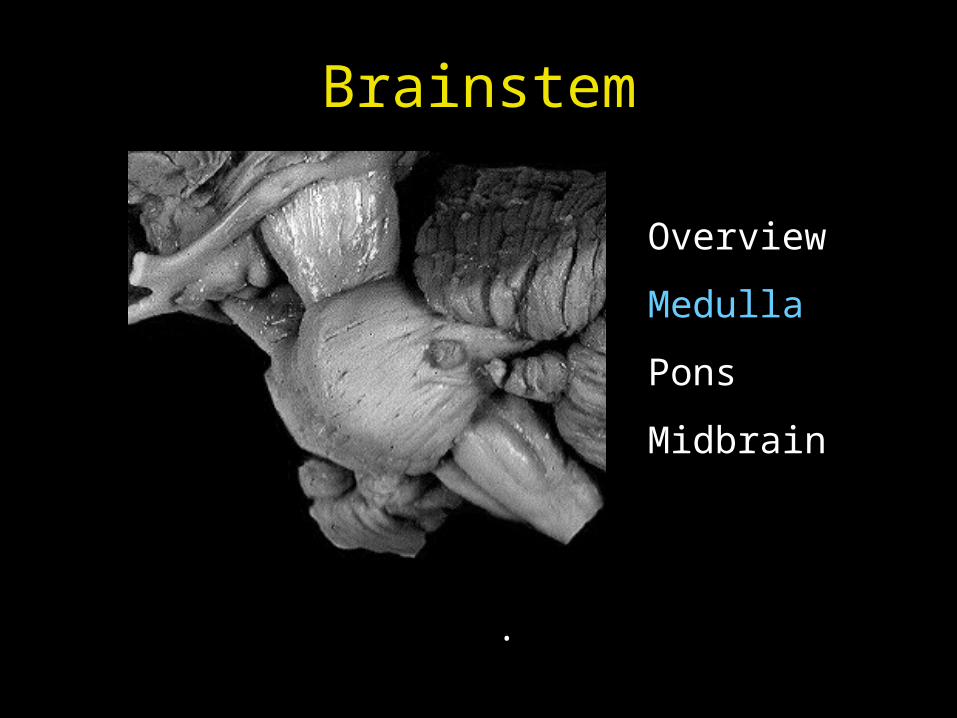
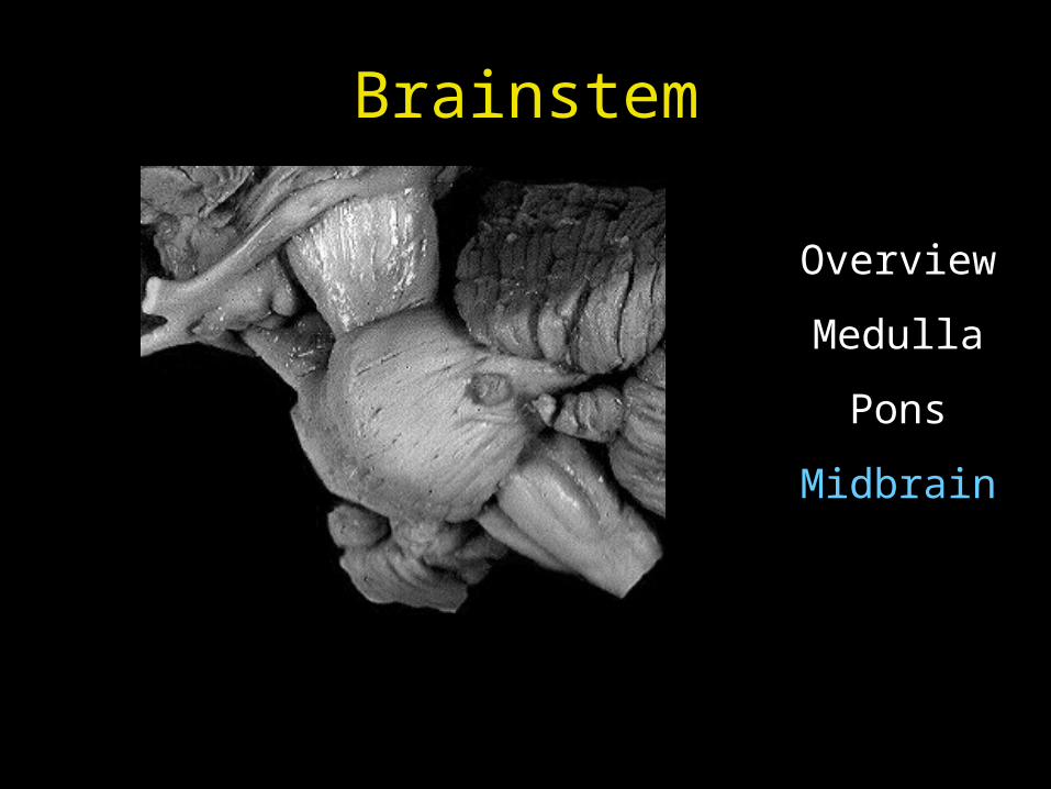
Brainstem
Overview
Medulla
Pons
Midbrain
January 12, 2009
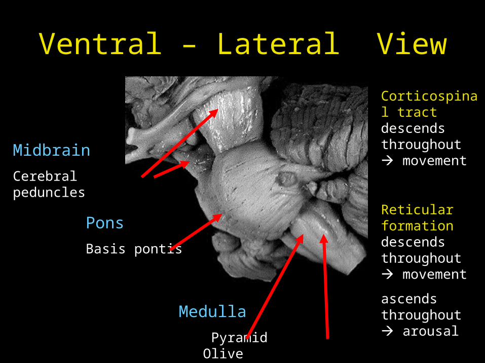
Ventral – Lateral View
Midbrain
Cerebral peduncles
Pons
Basis pontis
Medulla
Pyramid Olive
Corticospinal tract descends throughout movement
Reticular formation descends throughout movement
ascends throughout arousal
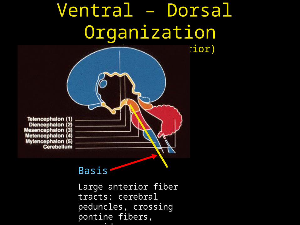
Ventral – Dorsal Organization
(anterior - posterior)
Basis
Large anterior fiber tracts: cerebral peduncles, crossing pontine fibers, pyramids
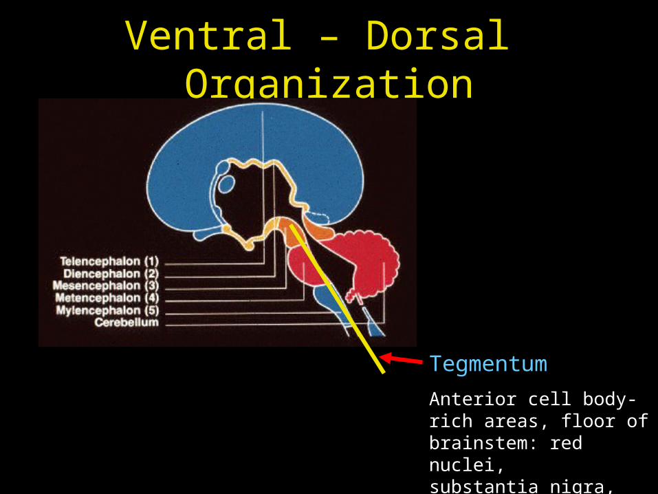
Ventral – Dorsal Organization
Tegmentum
Anterior cell body-rich areas, floor of brainstem: red nuclei, substantia nigra, reticular formation
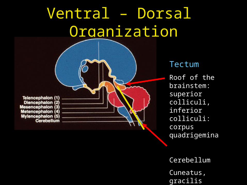
Ventral – Dorsal Organization
Tectum
Roof of the brainstem: superior colliculi, inferior colliculi: corpus quadrigemina
Cerebellum
Cuneatus, gracilis (medulla)
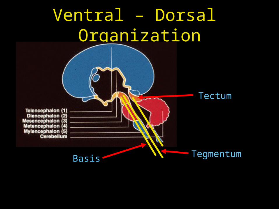
Ventral – Dorsal Organization
Basis Tegmentum
Tectum
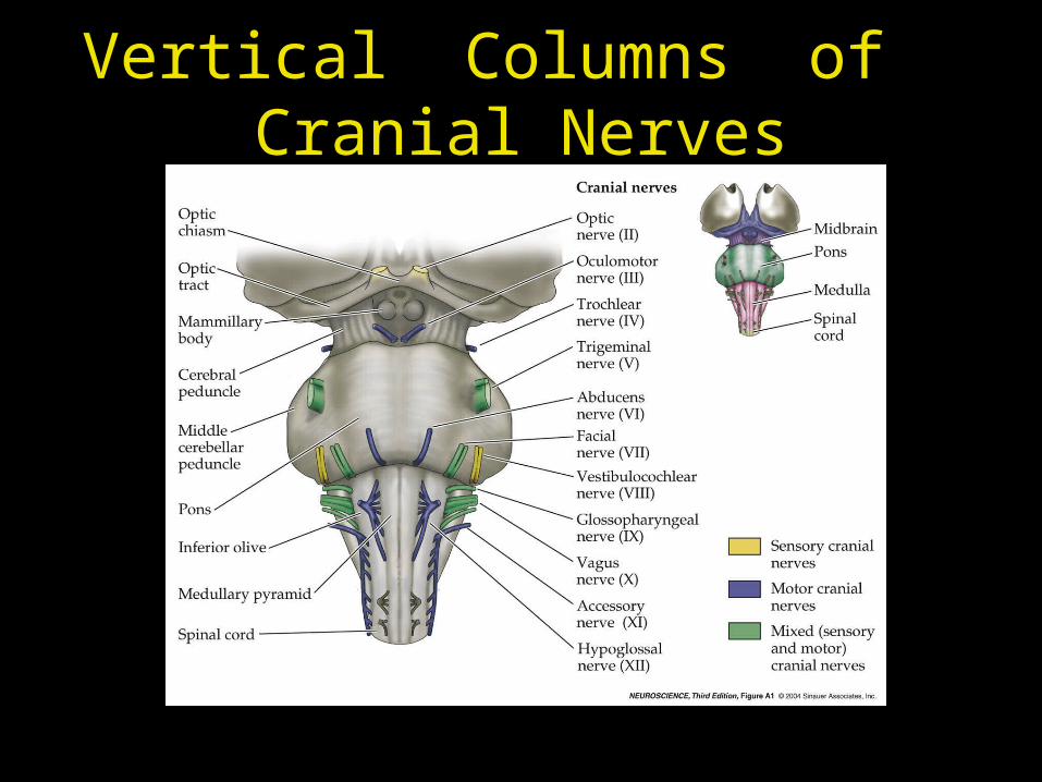
Vertical Columns of Cranial Nerves
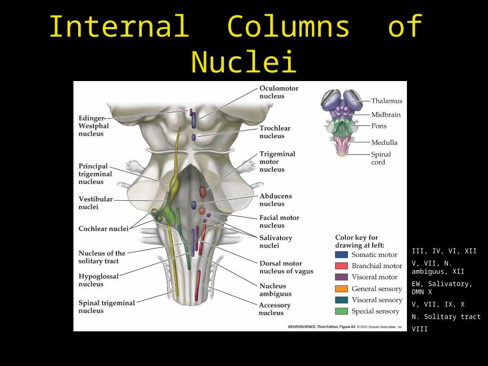
Internal Columns of Nuclei
III, IV, VI, XII
V, VII, N. ambiguus, XII
EW, Salivatory, DMN X
V, VII, IX, X
N. Solitary tract
VIII
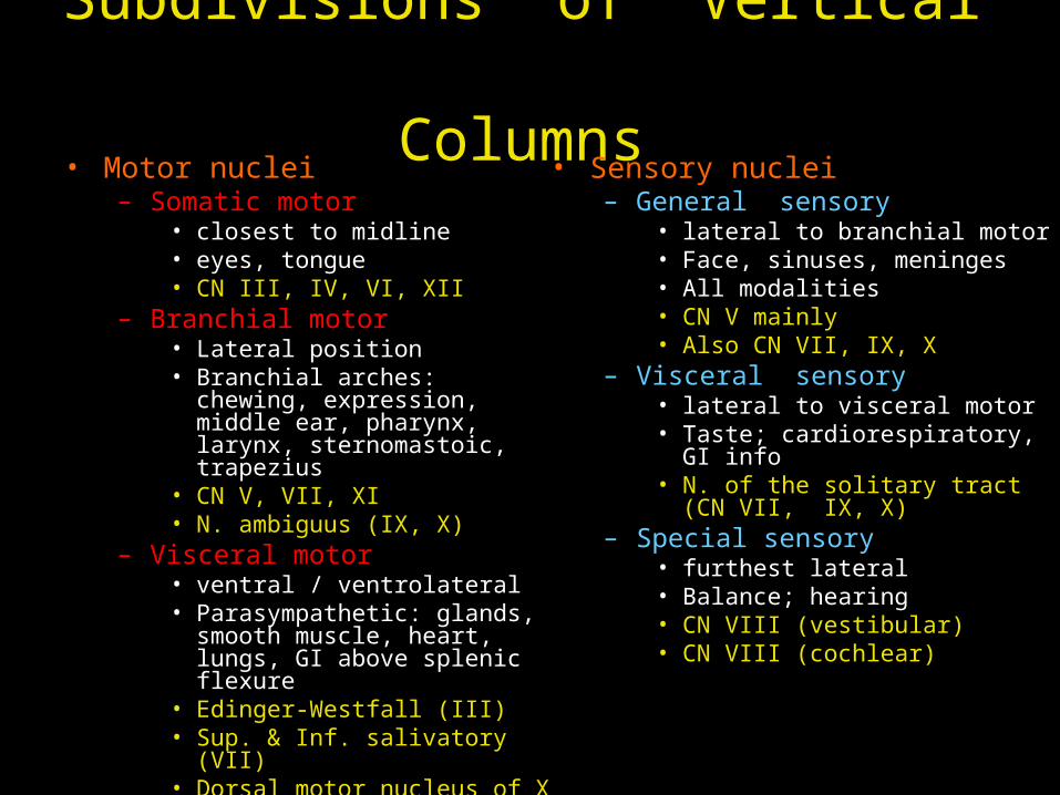
Subdivisions of Vertical Columns
• Motor nuclei– Somatic motor
• closest to midline• eyes, tongue• CN III, IV, VI, XII
– Branchial motor• Lateral position• Branchial arches: chewing,
expression, middle ear, pharynx, larynx, sternomastoic, trapezius
• CN V, VII, XI• N. ambiguus (IX, X)
– Visceral motor• ventral / ventrolateral• Parasympathetic: glands, smooth
muscle, heart, lungs, GI above splenic flexure
• Edinger-Westfall (III)• Sup. & Inf. salivatory (VII)• Dorsal motor nucleus of X
• Sensory nuclei– General sensory
• lateral to branchial motor• Face, sinuses, meninges• All modalities• CN V mainly• Also CN VII, IX, X
– Visceral sensory• lateral to visceral motor• Taste; cardiorespiratory, GI info• N. of the solitary tract (CN VII, IX,
X)– Special sensory
• furthest lateral• Balance; hearing• CN VIII (vestibular)• CN VIII (cochlear)
Brainstem
Overview
Medulla
Pons
Midbrain
.

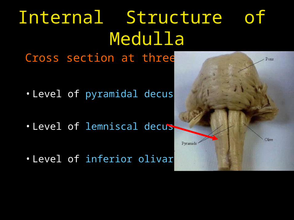
Internal Structure of Medulla
Cross section at three levels
• Level of pyramidal decussation
• Level of lemniscal decussation
• Level of inferior olivary nuclei
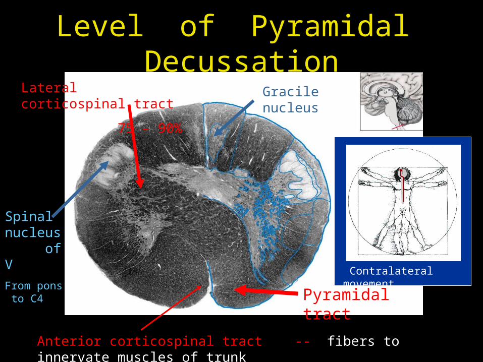
Level of Pyramidal Decussation
Pyramidal tract
Lateral corticospinal tract
75 – 90%
Anterior corticospinal tract -- fibers to innervate muscles of trunk
Spinal nucleus of V
From pons to C4
Gracile nucleus
Contralateral movement
Internal Structure of Medulla
Cross section at three levels
• Level of pyramidal decussation
• Level of lemniscal decussation
• Level of inferior olivary nuclei
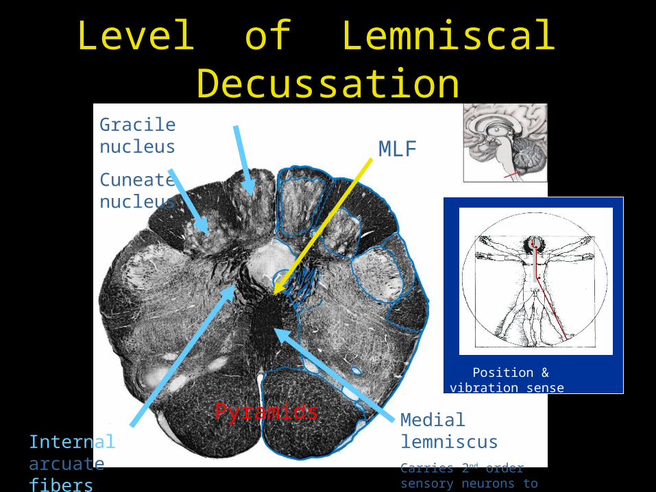
Level of Lemniscal Decussation
Medial lemniscusCarries 2nd order sensory neurons to VPL thalamus
Gracile nucleus
Cuneate nucleus
Internal arcuate fibers
MLF
Pyramids
Position & vibration sense

Internal Structure of Medulla
Cross section at three levels
• Level of pyramidal decussation
• Level of lemniscal decussation
• Level of inferior olivary nuclei
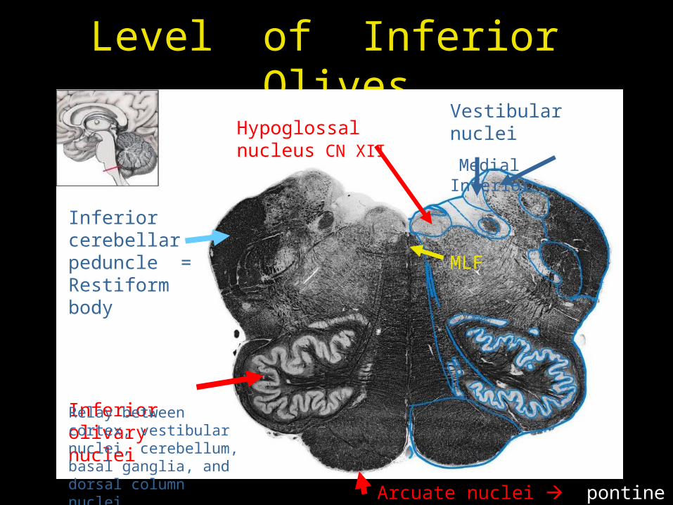
Level of Inferior Olives
Inferior cerebellar peduncle = Restiform body
Inferior olivary nucleiRelay between cortex, vestibular nuclei, cerebellum, basal ganglia, and dorsal column nuclei
MLF
Vestibular nuclei
Medial InferiorHypoglossal nucleus CN XII
Arcuate nuclei pontine nuclei
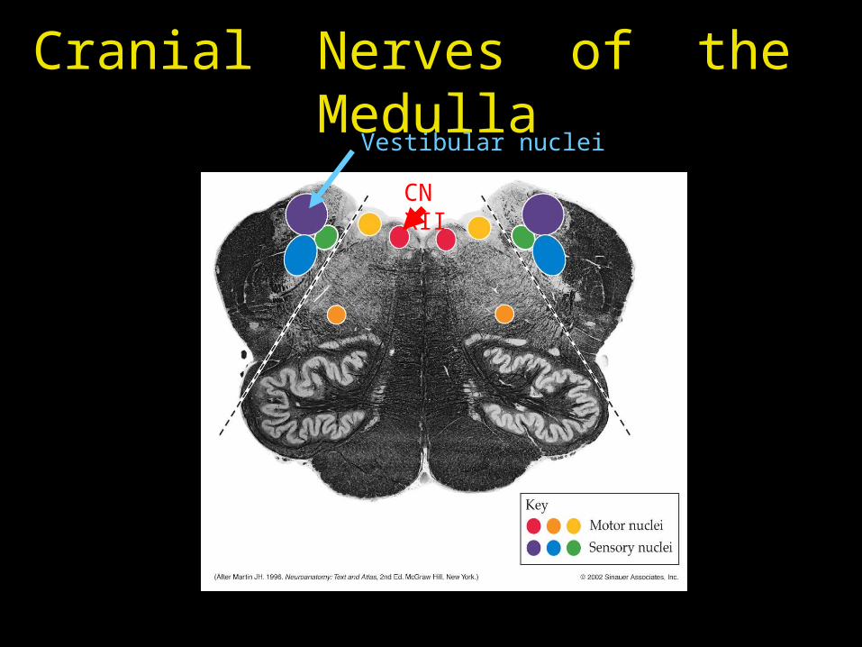
Cranial Nerves of the Medulla
Vestibular nuclei
CN XII
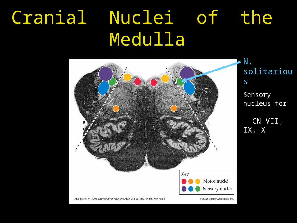
Cranial Nuclei of the Medulla
N. solitarious
Sensory nucleus for CN VII, IX, X
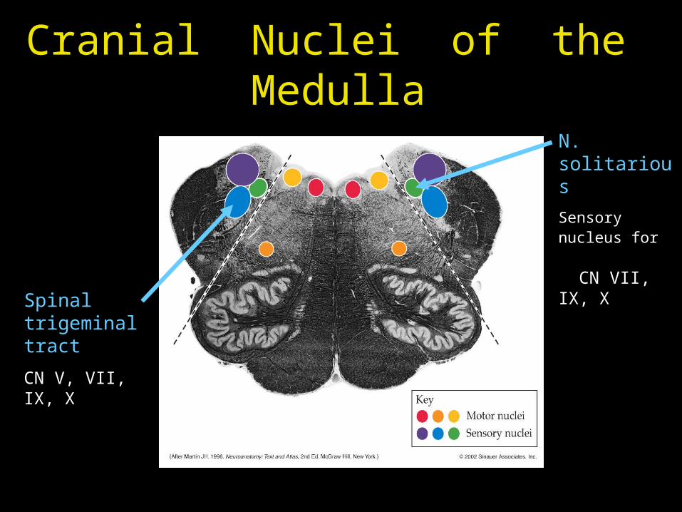
Cranial Nuclei of the Medulla
N. solitarious
Sensory nucleus for CN VII, IX, X
Spinal trigeminal tract
CN V, VII, IX, X
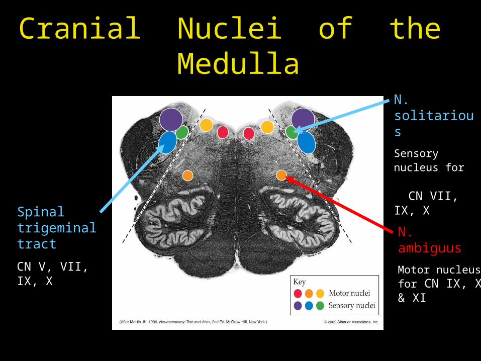
Cranial Nuclei of the Medulla
N. ambiguus
Motor nucleus for CN IX, X & XI
N. solitarious
Sensory nucleus for CN VII, IX, X
Spinal trigeminal tract
CN V, VII, IX, X
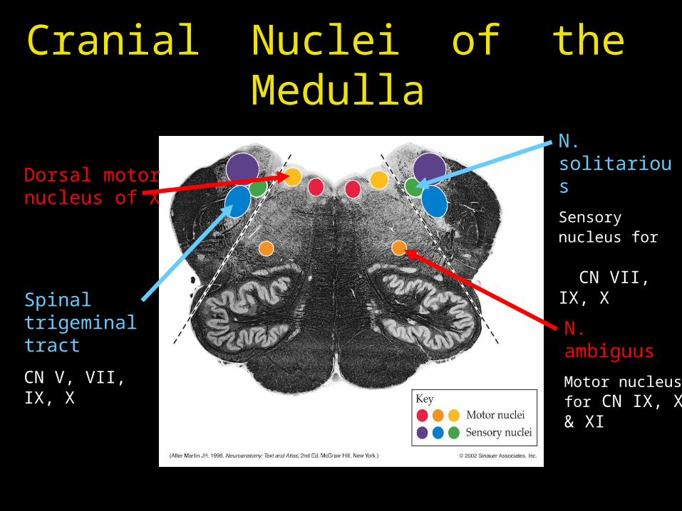
Cranial Nuclei of the Medulla
N. ambiguus
Motor nucleus for CN IX, X & XI
N. solitarious
Sensory nucleus for CN VII, IX, X
Spinal trigeminal tract
CN V, VII, IX, X
Dorsal motor nucleus of X
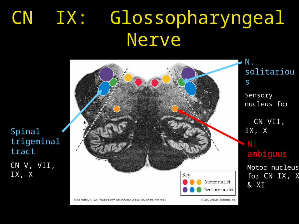
CN IX: Glossopharyngeal Nerve
N. ambiguus
Motor nucleus for CN IX, X & XI
N. solitarious
Sensory nucleus for CN VII, IX, X
Spinal trigeminal tract
CN V, VII, IX, X
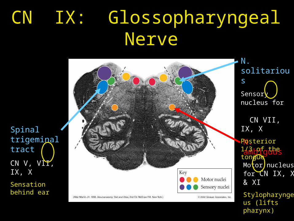
CN IX: Glossopharyngeal Nerve
N. ambiguus
Motor nucleus for CN IX, X & XI
N. solitarious
Sensory nucleus for CN VII, IX, X
Posterior 1/3 of the tongue
Spinal trigeminal tract
CN V, VII, IX, X

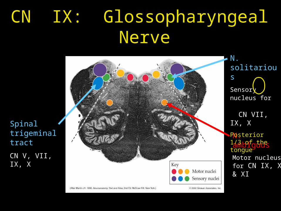
CN IX: Glossopharyngeal Nerve
N. ambiguus
Motor nucleus for CN IX, X & XI
N. solitarious
Sensory nucleus for CN VII, IX, X
Posterior 1/3 of the tongue
Spinal trigeminal tract
CN V, VII, IX, X
Sensation behind ear
CN IX: Glossopharyngeal Nerve
N. ambiguus
Motor nucleus for CN IX, X & XI
Stylopharyngeus (lifts pharynx)
N. solitarious
Sensory nucleus for CN VII, IX, X
Posterior 1/3 of the tongue
Spinal trigeminal tract
CN V, VII, IX, X
Sensation behind ear
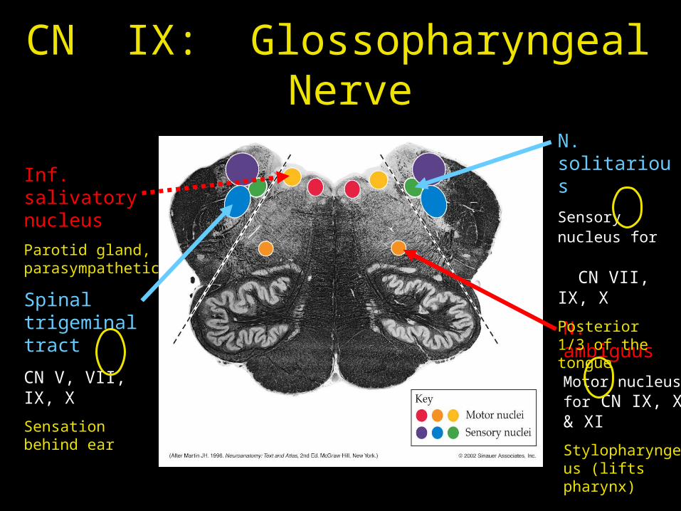
CN IX: Glossopharyngeal Nerve
N. ambiguus
Motor nucleus for CN IX, X & XI
Stylopharyngeus (lifts pharynx)
N. solitarious
Sensory nucleus for CN VII, IX, X
Posterior 1/3 of the tongue
Inf. salivatory nucleus
Parotid gland, parasympathetic
Spinal trigeminal tract
CN V, VII, IX, X
Sensation behind ear
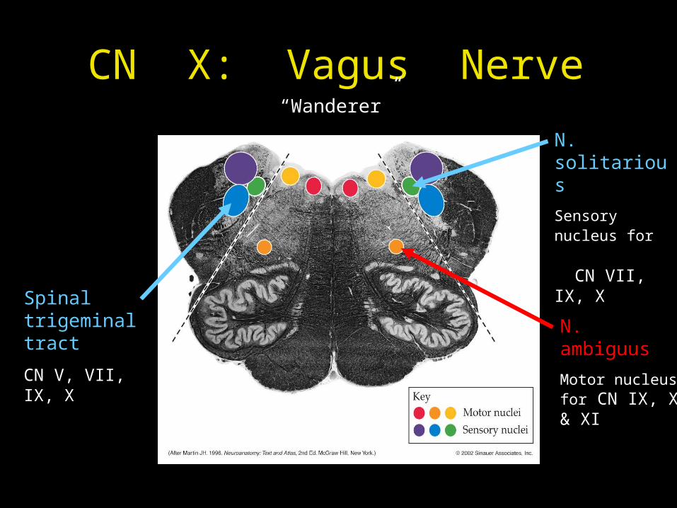
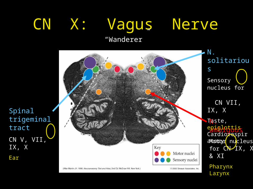
CN X: Vagus Nerve
N. ambiguus
Motor nucleus for CN IX, X & XI
N. solitarious
Sensory nucleus for CN VII, IX, X
Spinal trigeminal tract
CN V, VII, IX, X
“Wanderer”
CN X: Vagus Nerve
N. ambiguus
Motor nucleus for CN IX, X & XI
N. solitarious
Sensory nucleus for CN VII, IX, X
Taste, epiglottis Cardiorespiratory
Spinal trigeminal tract
CN V, VII, IX, X
“Wanderer”

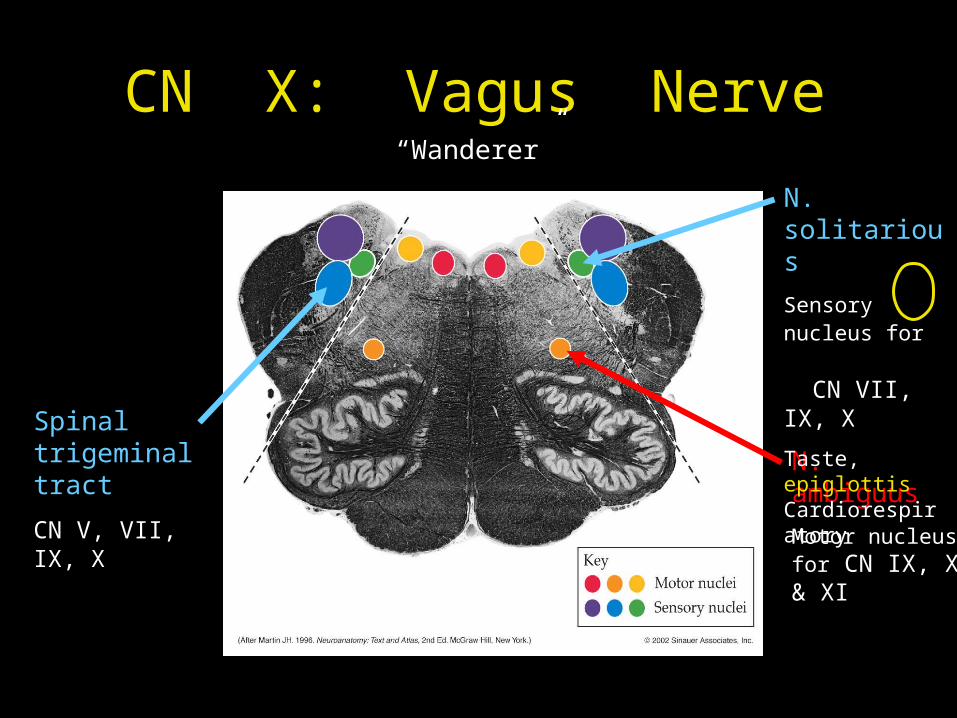
CN X: Vagus Nerve
N. ambiguus
Motor nucleus for CN IX, X & XI
N. solitarious
Sensory nucleus for CN VII, IX, X
Taste, epiglottis Cardiorespiratory
Spinal trigeminal tract
CN V, VII, IX, X
Ear
“Wanderer”
CN X: Vagus Nerve
N. ambiguus
Motor nucleus for CN IX, X & XI
Pharynx Larynx
N. solitarious
Sensory nucleus for CN VII, IX, X
Taste, epiglottis Cardiorespiratory
Spinal trigeminal tract
CN V, VII, IX, X
Ear
“Wanderer”
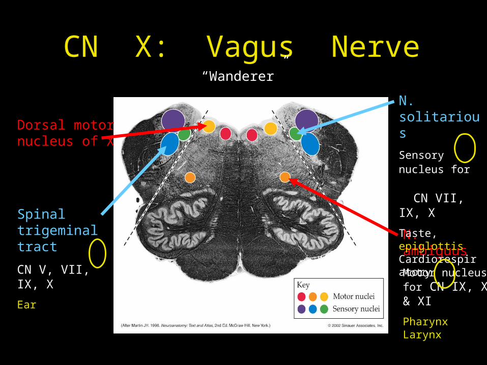
CN X: Vagus Nerve
N. ambiguus
Motor nucleus for CN IX, X & XI
Pharynx Larynx
N. solitarious
Sensory nucleus for CN VII, IX, X
Taste, epiglottis Cardiorespiratory
Dorsal motor nucleus of X
Spinal trigeminal tract
CN V, VII, IX, X
Ear
“Wanderer”
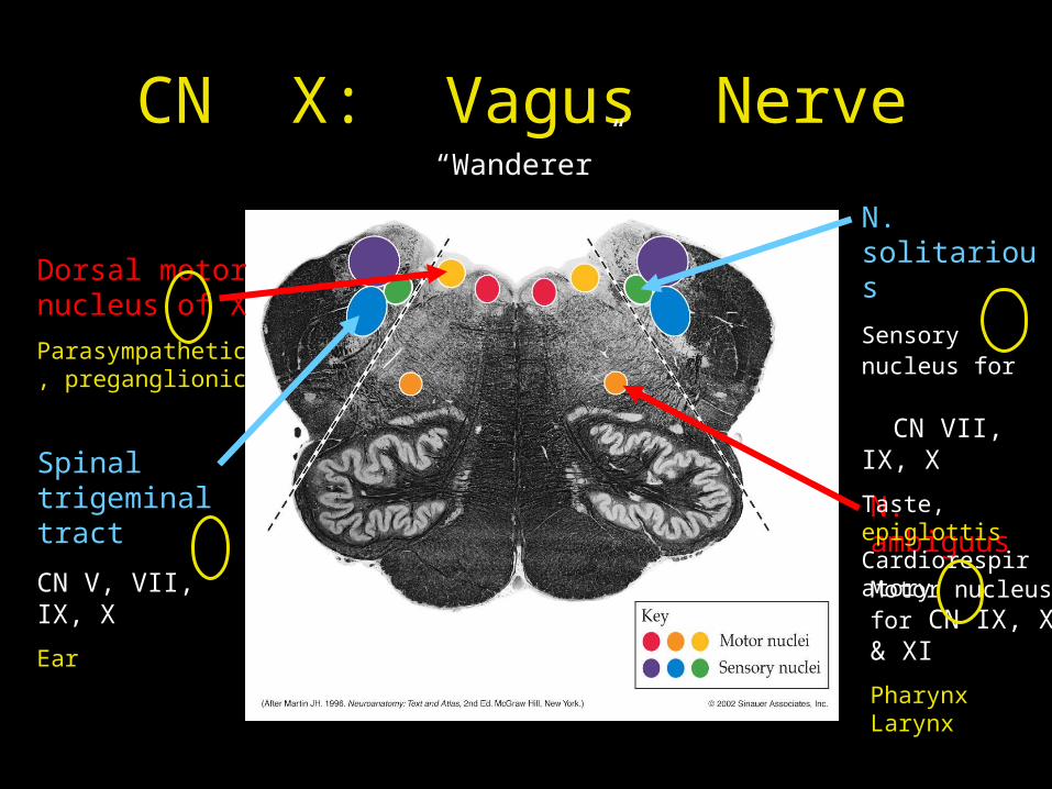
CN X: Vagus Nerve
N. ambiguus
Motor nucleus for CN IX, X & XI
Pharynx Larynx
N. solitarious
Sensory nucleus for CN VII, IX, X
Taste, epiglottis Cardiorespiratory
Dorsal motor nucleus of X
Parasympathetic, preganglionic
Spinal trigeminal tract
CN V, VII, IX, X
Ear
“Wanderer”
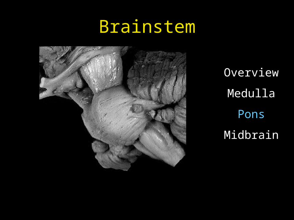
Brainstem
Overview
Medulla
Pons
Midbrain
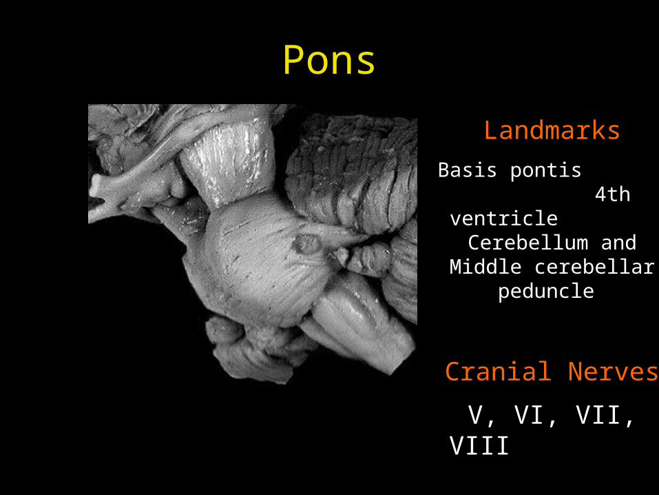
Pons
Landmarks
Basis pontis 4th ventricle
Cerebellum and Middle cerebellar peduncle
Cranial Nerves
V, VI, VII, VIII
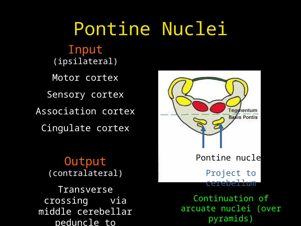
Pontine Nuclei
Pontine nuclei
Project to cerebellum
Continuation of arcuate nuclei (over pyramids)
Input (ipsilateral)
Motor cortex
Sensory cortex
Association cortex
Cingulate cortex
Output (contralateral)
Transverse crossing via middle cerebellar
peduncle to cerebellum
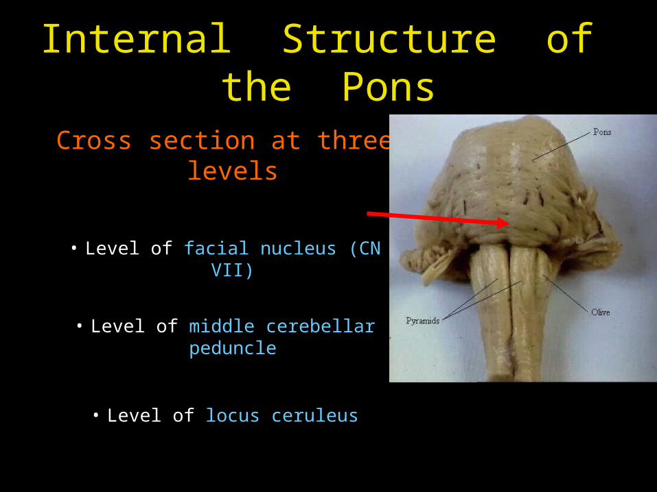
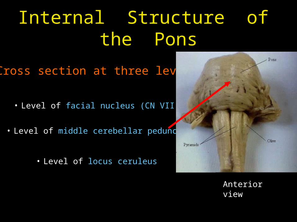
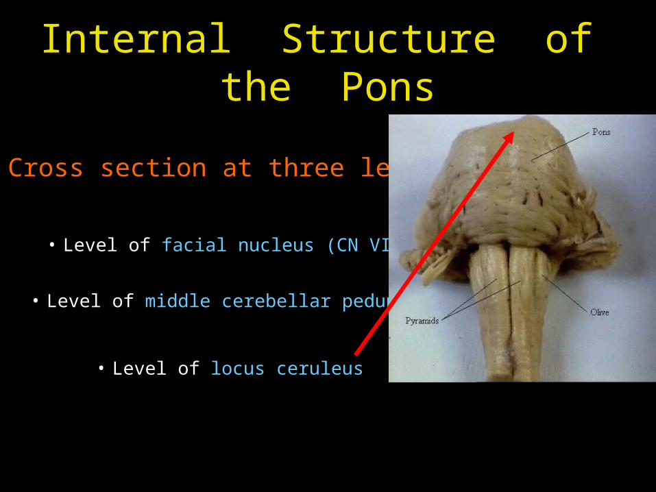
Internal Structure of the Pons
Cross section at three levels
• Level of facial nucleus (CN VII)
• Level of middle cerebellar peduncle
• Level of locus ceruleus
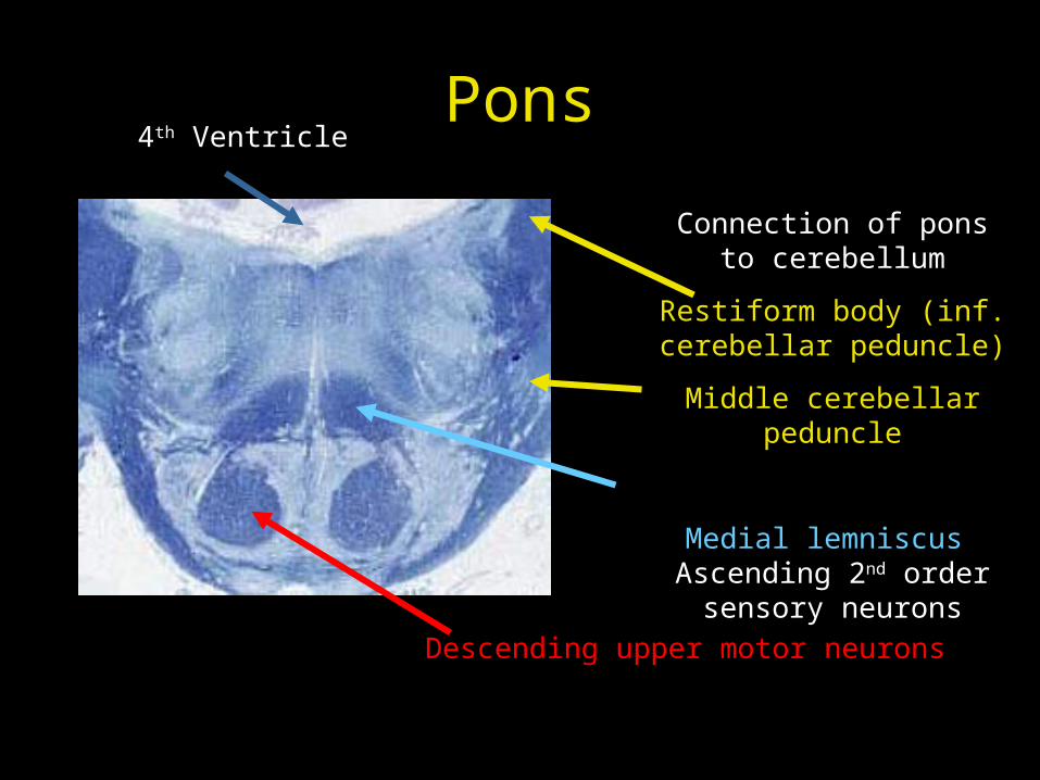
Pons
Descending upper motor neurons
Connection of pons to cerebellum
Restiform body (inf. cerebellar peduncle)
Middle cerebellar peduncle
Medial lemniscus Ascending 2nd order
sensory neurons
4th Ventricle
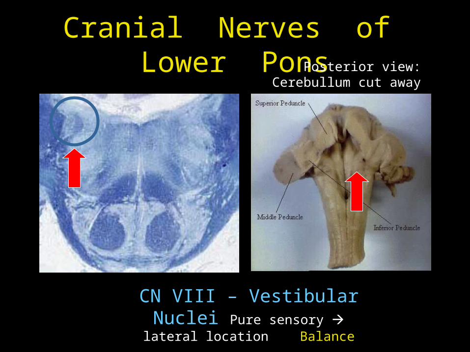
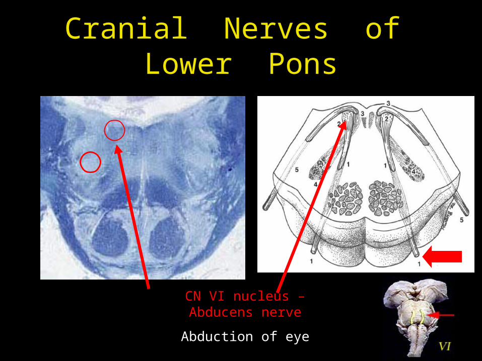
Cranial Nerves of Lower Pons
CN VIII – Vestibular Nuclei Pure sensory lateral location
Balance
Posterior view: Cerebullum cut away
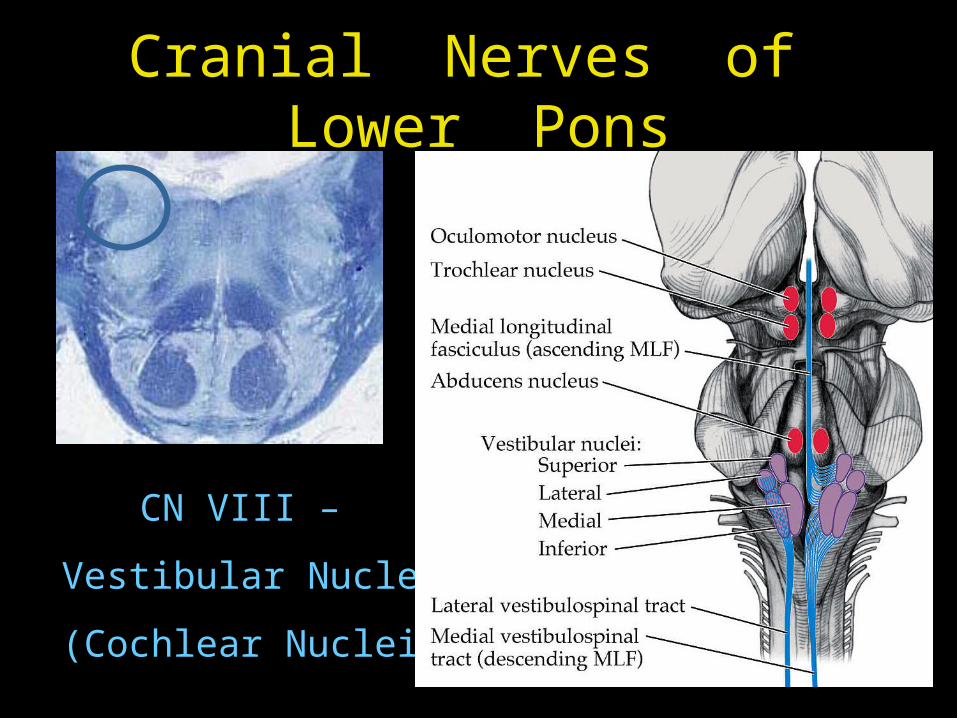
Cranial Nerves of Lower Pons
CN VIII –
Vestibular Nuclei
(Cochlear Nuclei)
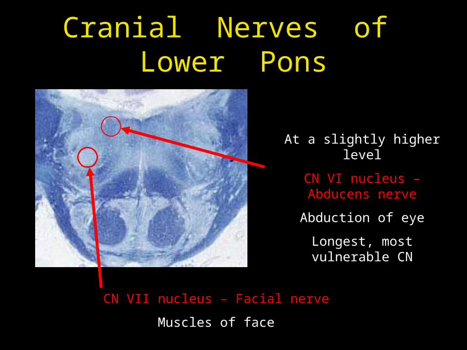
Cranial Nerves of Lower Pons
CN VII nucleus – Facial nerve
Muscles of face
At a slightly higher level
CN VI nucleus – Abducens nerve
Abduction of eye
Longest, most vulnerable CN
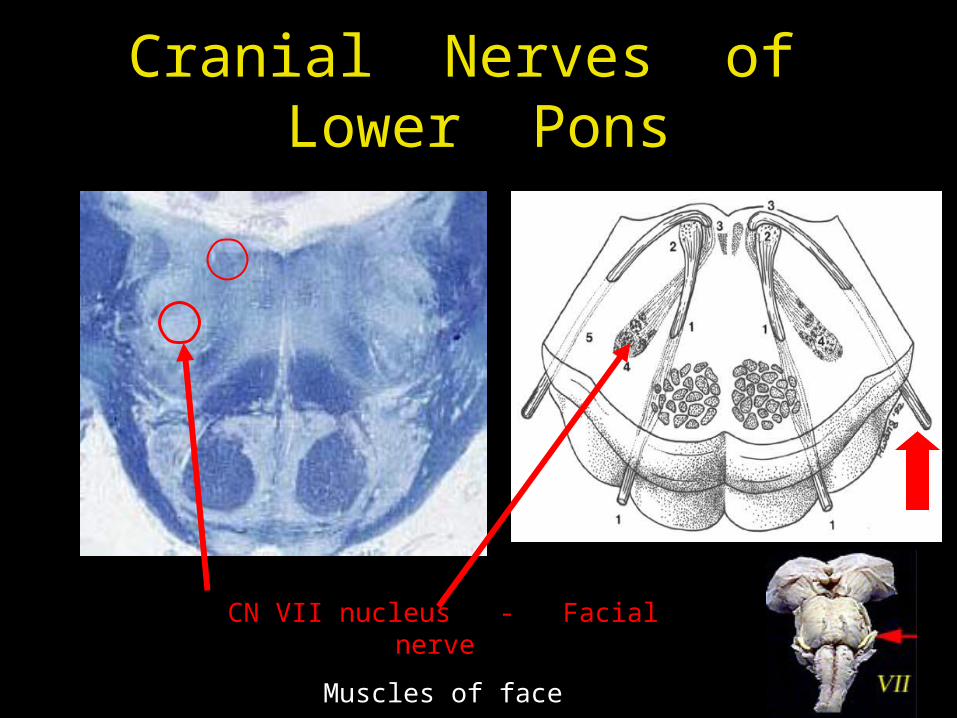
Cranial Nerves of Lower Pons
CN VII nucleus - Facial nerve
Muscles of face
Cranial Nerves of Lower Pons
CN VI nucleus – Abducens nerve
Abduction of eye
Internal Structure of the Pons
Cross section at three levels
• Level of facial nucleus (CN VII)
• Level of middle cerebellar peduncle
• Level of locus ceruleus
Anterior view
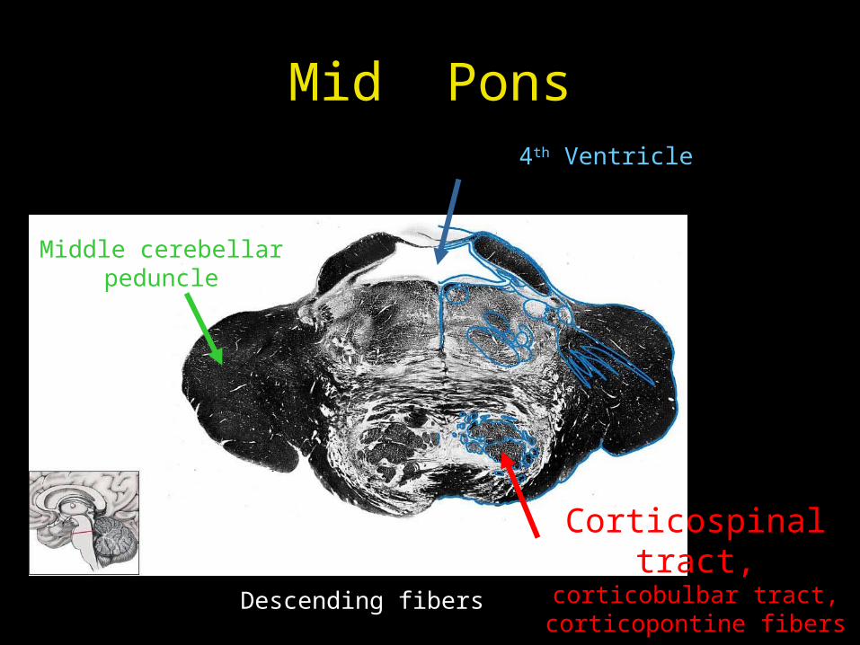
Mid Pons4th Ventricle
Middle cerebellar peduncle
Corticospinal tract, corticobulbar tract,
corticopontine fibersDescending fibers
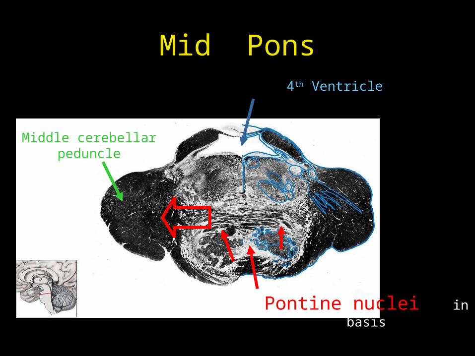
Mid Pons4th Ventricle
Middle cerebellar peduncle
Pontine nuclei in basis
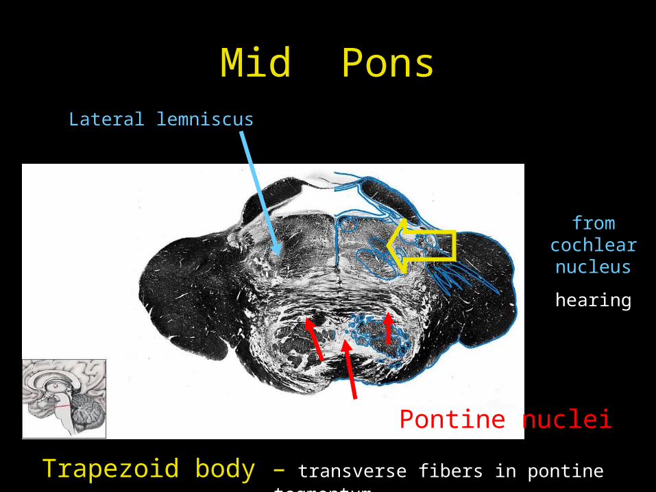
Mid Pons
Pontine nuclei
Trapezoid body – transverse fibers in pontine tegmentum
from cochlear nucleus
hearing
Lateral lemniscus
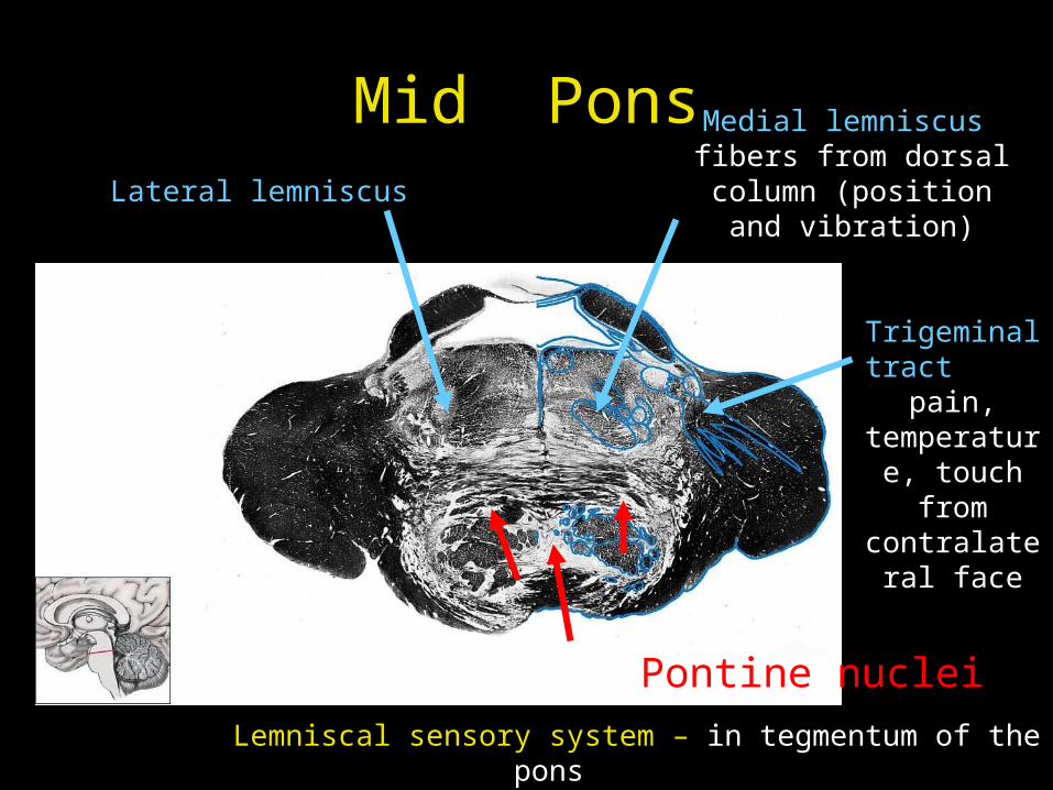
Mid Pons
Pontine nuclei
Lateral lemniscus
Medial lemniscus fibers from dorsal column
(position and vibration)
Trigeminal tract pain, temperature, touch from
contralateral face
Lemniscal sensory system – in tegmentum of the pons
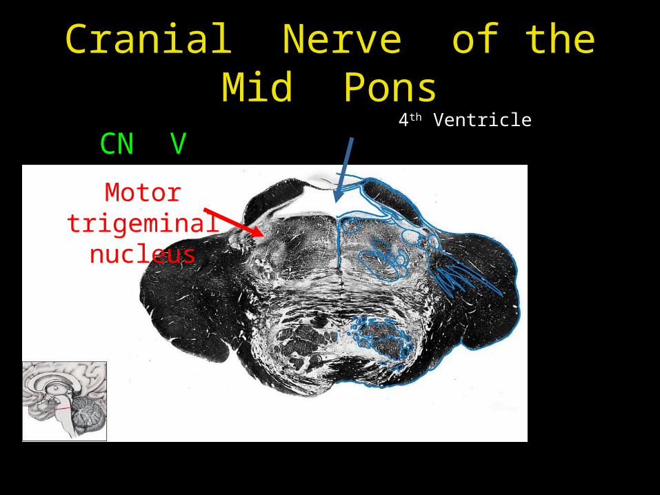
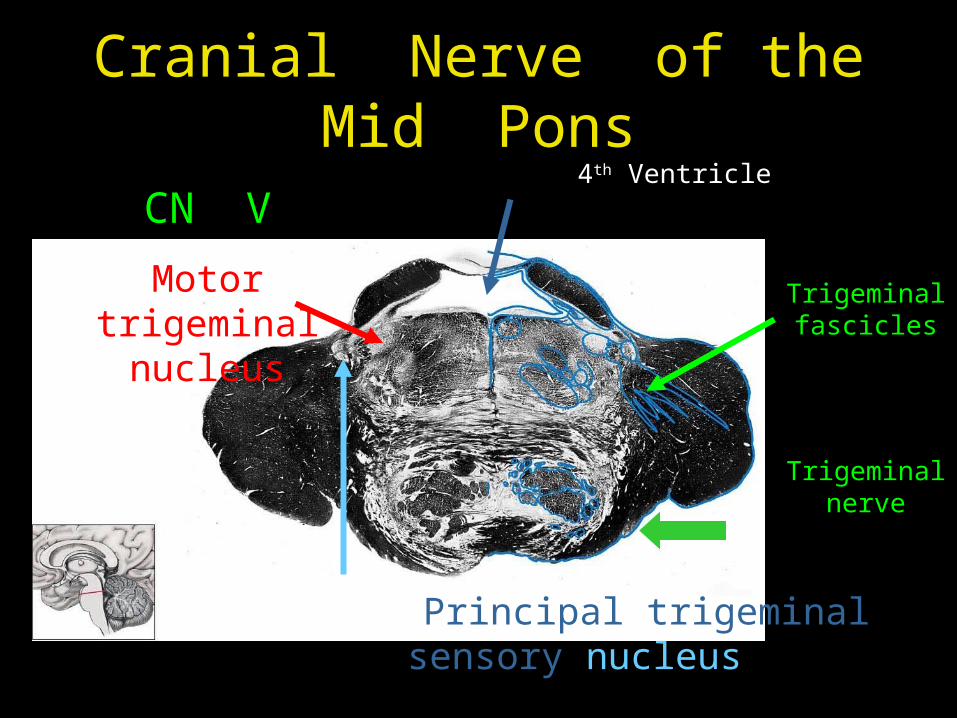
Cranial Nerve of the Mid Pons
4th Ventricle
CN V
Motor trigeminal nucleus
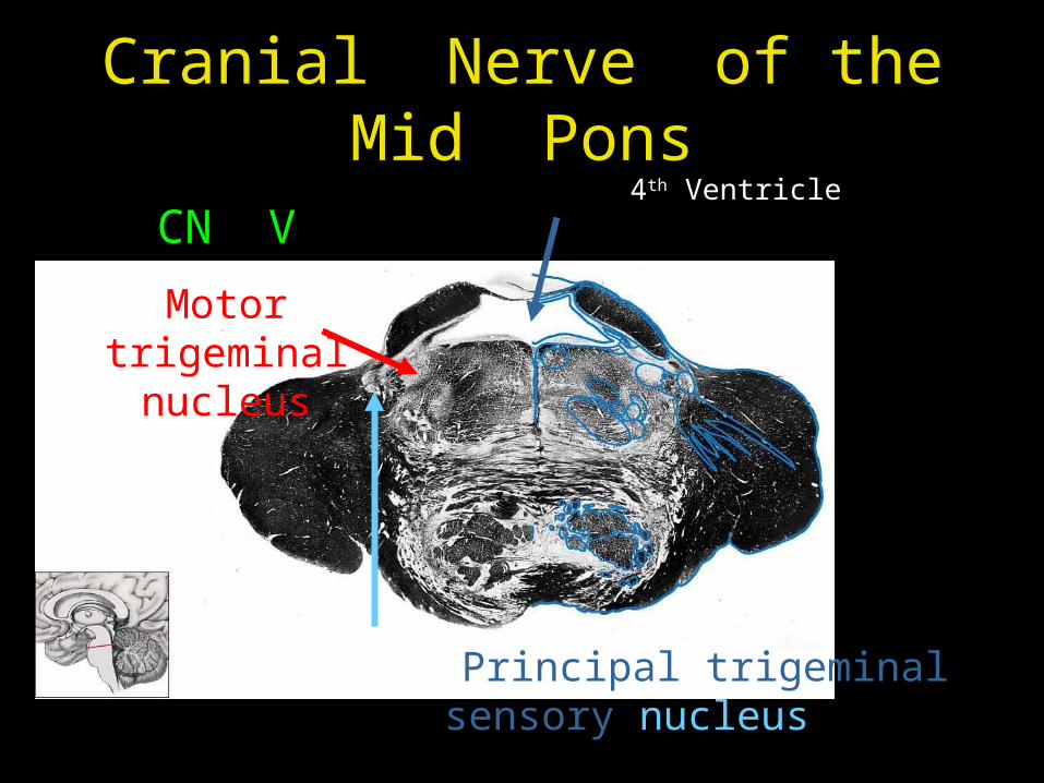
Cranial Nerve of the Mid Pons
4th Ventricle
CN V
Motor trigeminal nucleus
Principal trigeminal sensory nucleus
Cranial Nerve of the Mid Pons
4th Ventricle
CN V
Motor trigeminal nucleus
Principal trigeminal sensory nucleus
Trigeminal fascicles
Trigeminal nerve
Internal Structure of the Pons
Cross section at three levels
• Level of facial nucleus (CN VII)
• Level of middle cerebellar peduncle
• Level of locus ceruleus
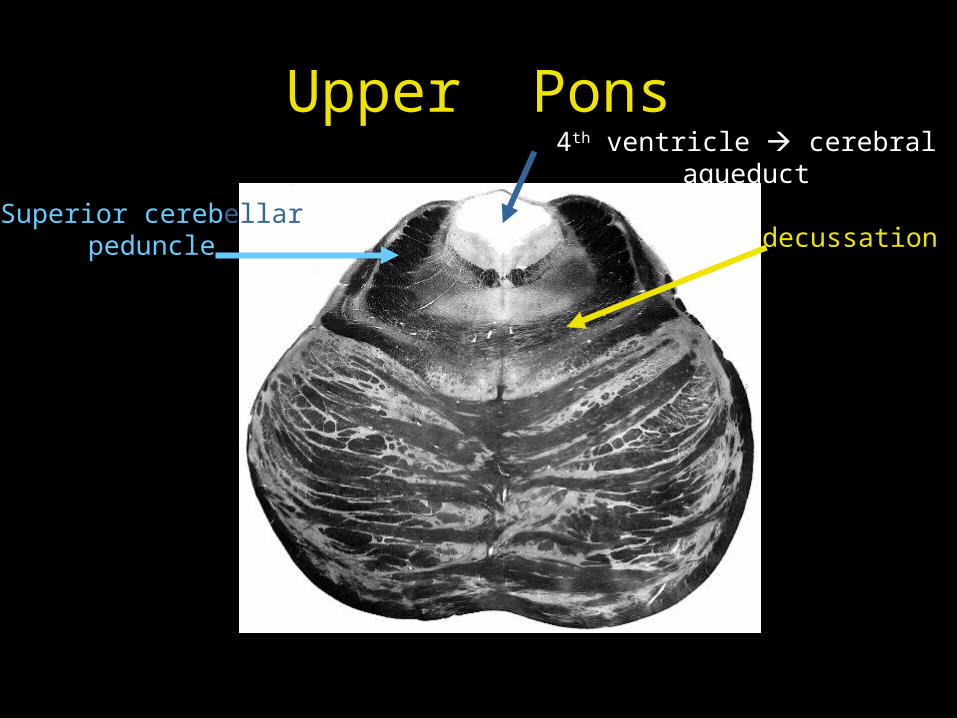
Upper Pons4th ventricle cerebral aqueduct
Superior cerebellar peduncle decussation

Upper Pons4th ventricle cerebral aqueduct
Superior cerebellar peduncle
Descending upper motor neurons
Transverse ponto-cerebellar
fibers
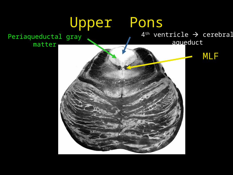
Upper Pons4th ventricle cerebral aqueduct
MLF
Periaqueductal gray matter
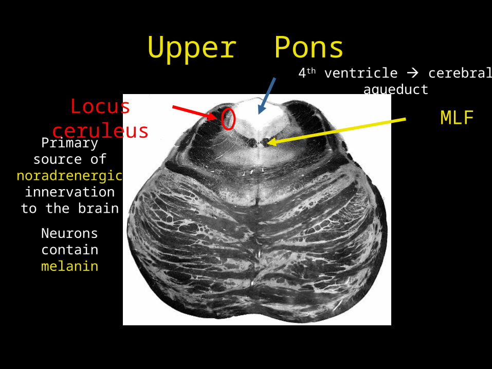
Upper Pons4th ventricle cerebral aqueduct
MLFLocus ceruleus
Primary source of noradrenergic
innervation to the brain
Neurons contain melanin
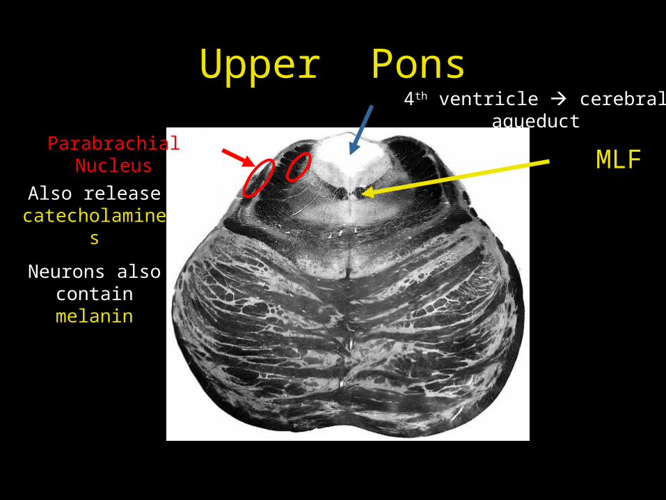
Upper Pons4th ventricle cerebral aqueduct
MLFParabrachial Nucleus
Also release catecholamines
Neurons also contain melanin

Upper Pons4th ventricle cerebral aqueduct
MLFPediculopontine Nucleus
Some neurons release
acetylcholine
Other neurons release glutamate
They assist in learning and
voluntary motor control, e.g. locomotion,
saccadic eye
Brainstem
Overview
Medulla
Pons
Midbrain
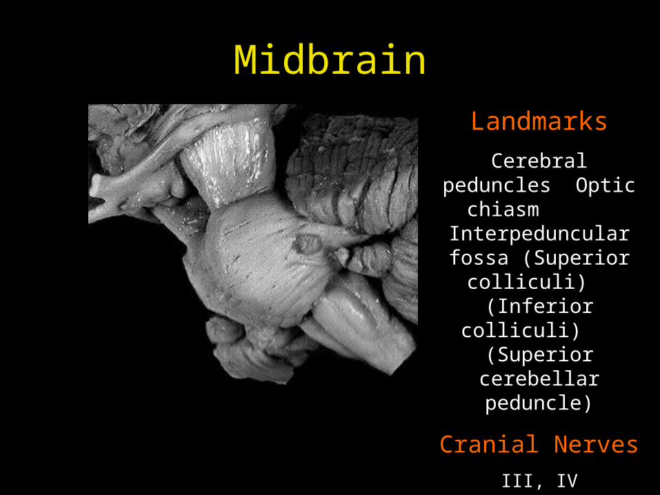
MidbrainLandmarks
Cerebral peduncles Optic chiasm
Interpeduncular fossa (Superior colliculi) (Inferior colliculi)
(Superior cerebellar peduncle)
Cranial Nerves
III, IV
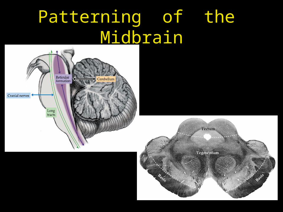
Patterning of the Midbrain
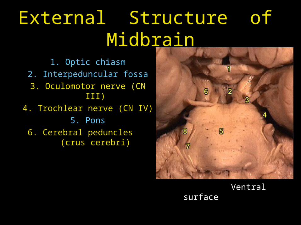
External Structure of Midbrain
1. Optic chiasm
2. Interpeduncular fossa
3. Oculomotor nerve (CN III)
4. Trochlear nerve (CN IV)
5. Pons
6. Cerebral peduncles (crus cerebri)
Ventral surface
(anterior)
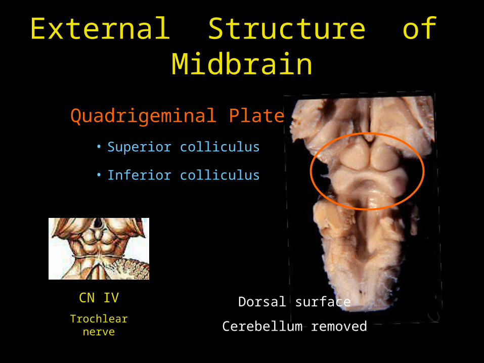
External Structure of Midbrain
Quadrigeminal Plate
• Superior colliculus
• Inferior colliculus
Dorsal surface
Cerebellum removed
CN IV
Trochlear nerve
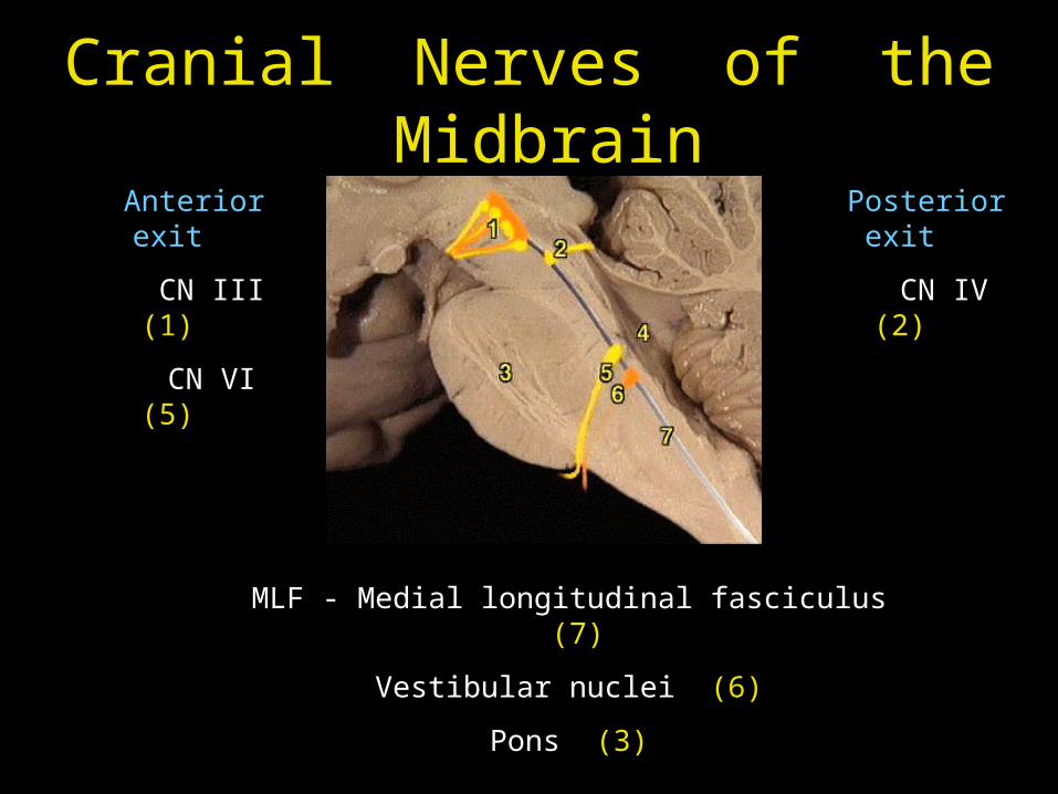
Cranial Nerves of the Midbrain
Anterior exit
CN III (1)
CN VI (5)
Posterior exit
CN IV (2)
MLF - Medial longitudinal fasciculus (7)
Vestibular nuclei (6)
Pons (3)

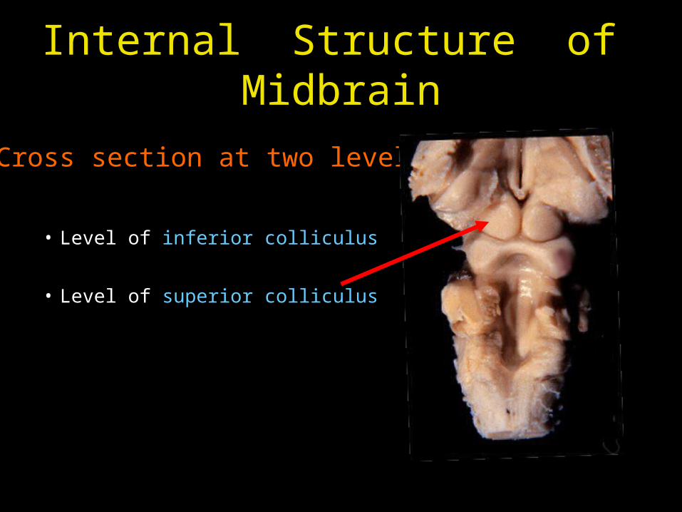
Internal Structure of Midbrain
Cross section at two levels
• Level of inferior colliculus
• Level of superior colliculus
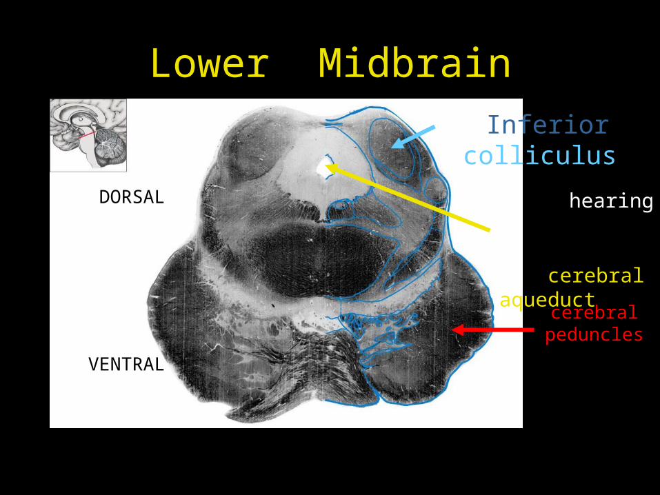
Lower MidbrainInferior colliculus
hearing
cerebral aqueduct
DORSAL
VENTRAL
cerebral peduncles
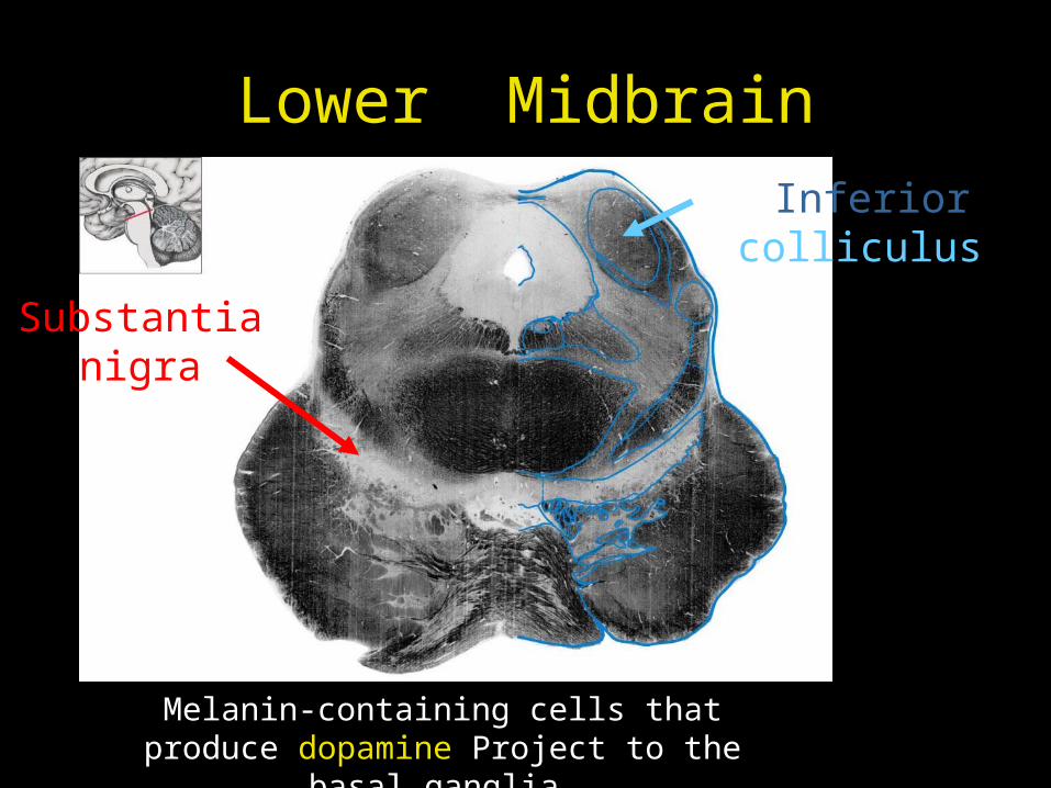
Lower MidbrainInferior colliculus
Substantia nigra
Melanin-containing cells that produce dopamine Project to the basal ganglia
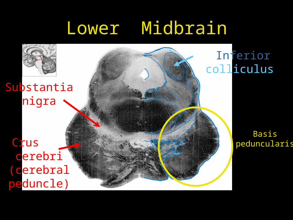
Lower MidbrainInferior colliculus
Substantia nigra
Crus cerebri
(cerebral peduncle)
Basis peduncularis
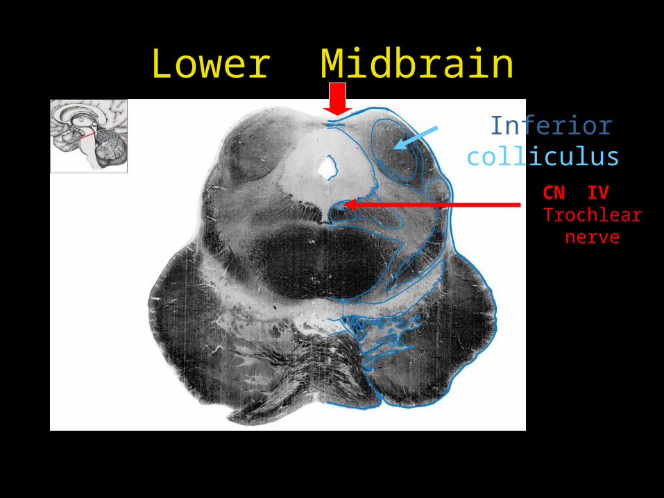
Lower MidbrainInferior colliculus
CN IV Trochlear nerve
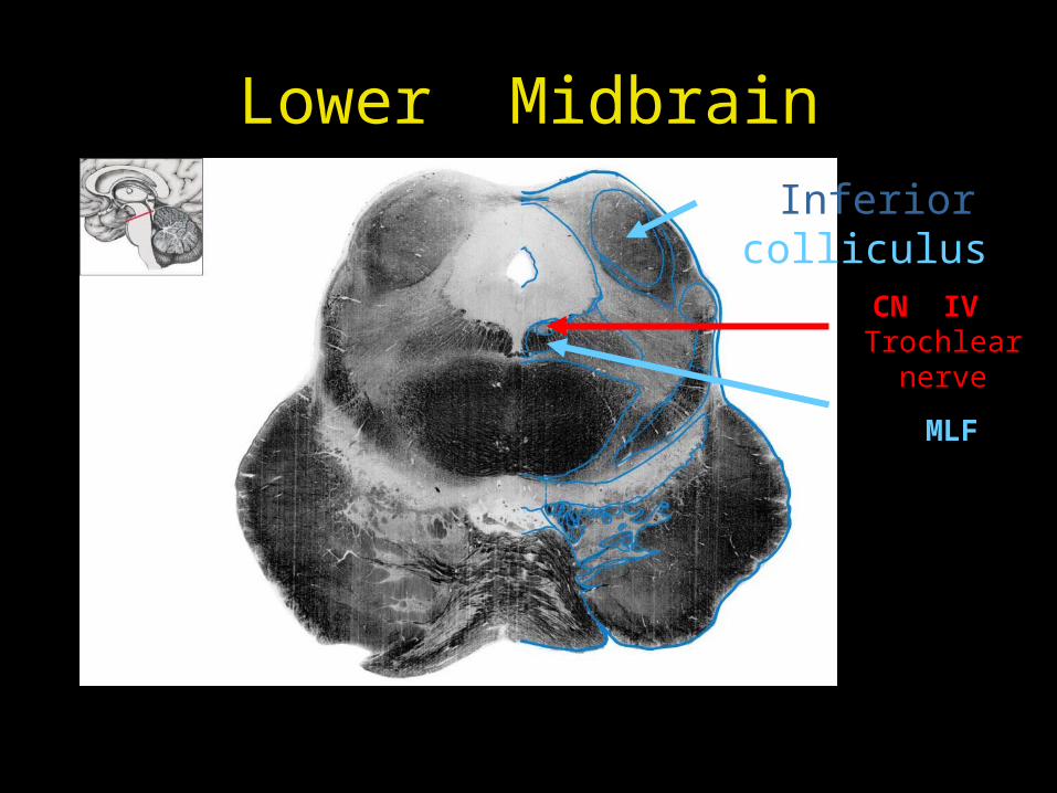
Lower MidbrainInferior colliculus
CN IV Trochlear nerve
MLF
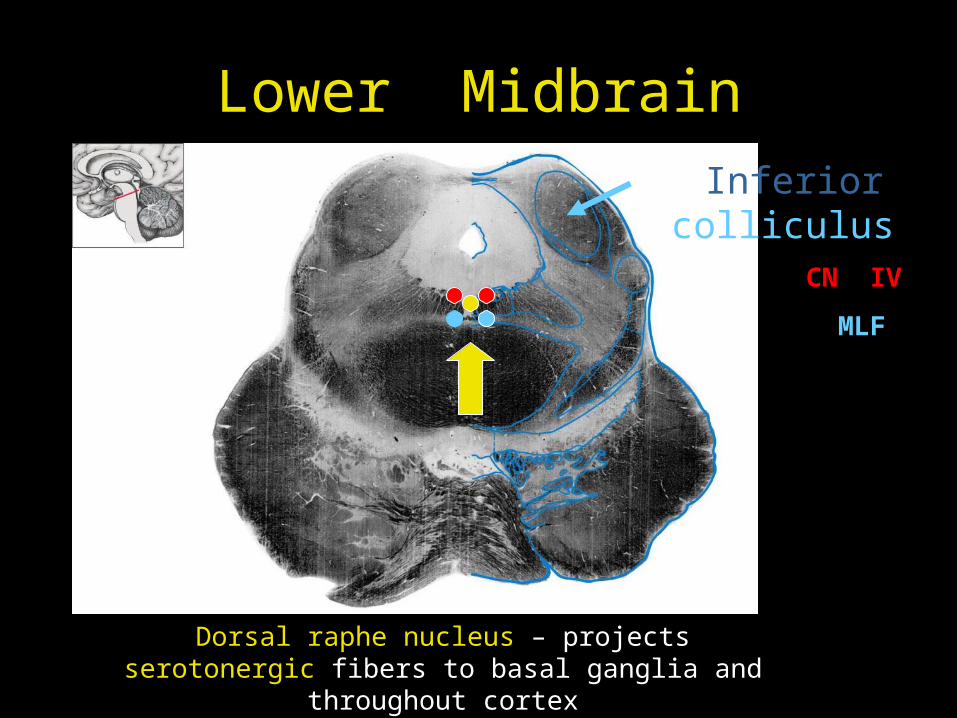
Lower MidbrainInferior colliculus
CN IV
MLF
Dorsal raphe nucleus – projects serotonergic fibers to basal ganglia and throughout cortex
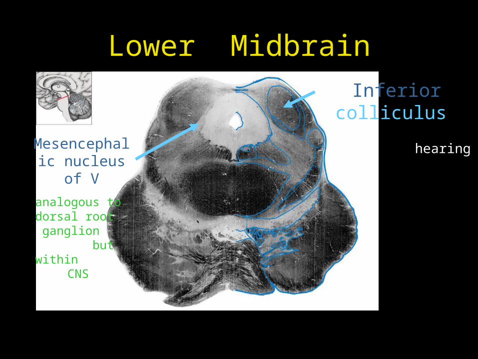
Lower MidbrainInferior colliculus
hearing Mesencephalic
nucleus of V
analogous to dorsal root
ganglion but within
CNS
Internal Structure of Midbrain
Cross section at two levels
• Level of inferior colliculus
• Level of superior colliculus
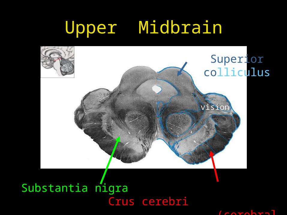
Upper Midbrain
Superior colliculus
vision
Substantia nigra Crus cerebri (cerebral
peduncle)
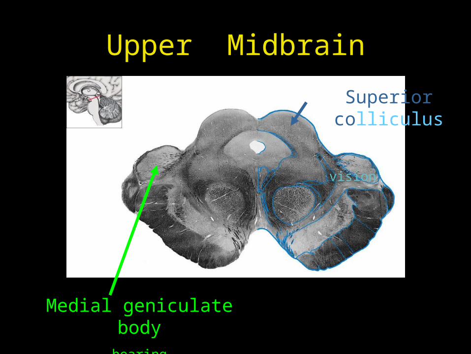
Upper Midbrain
Superior colliculus
vision
Medial geniculate bodyhearing
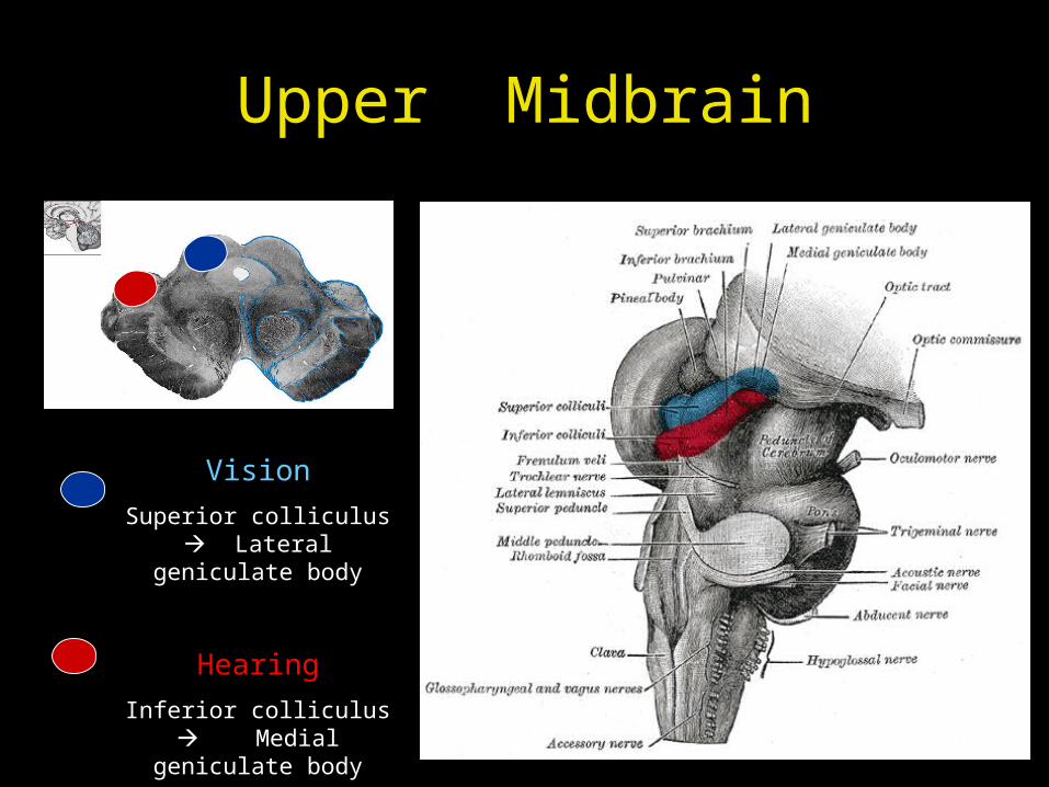
Upper Midbrain
Vision
Superior colliculus Lateral geniculate body
Hearing
Inferior colliculus Medial geniculate body
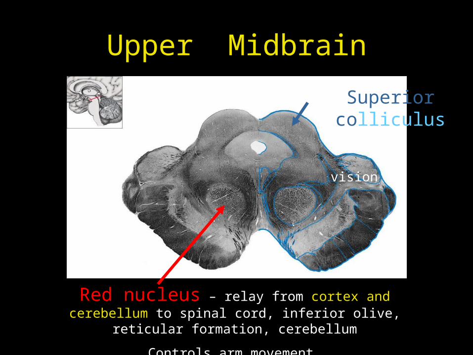
Upper Midbrain
Superior colliculus
vision
Red nucleus – relay from cortex and cerebellum to spinal cord, inferior olive, reticular formation, cerebellum
Controls arm movement
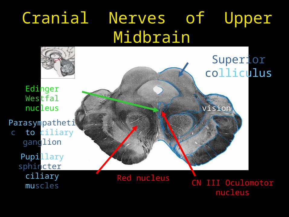
Cranial Nerves of Upper Midbrain
Superior colliculus
vision
Red nucleusCN III Oculomotor
nucleus
Edinger Westfal nucleus
Parasympathetic to ciliary ganglion
Pupillary sphincter ciliary muscles
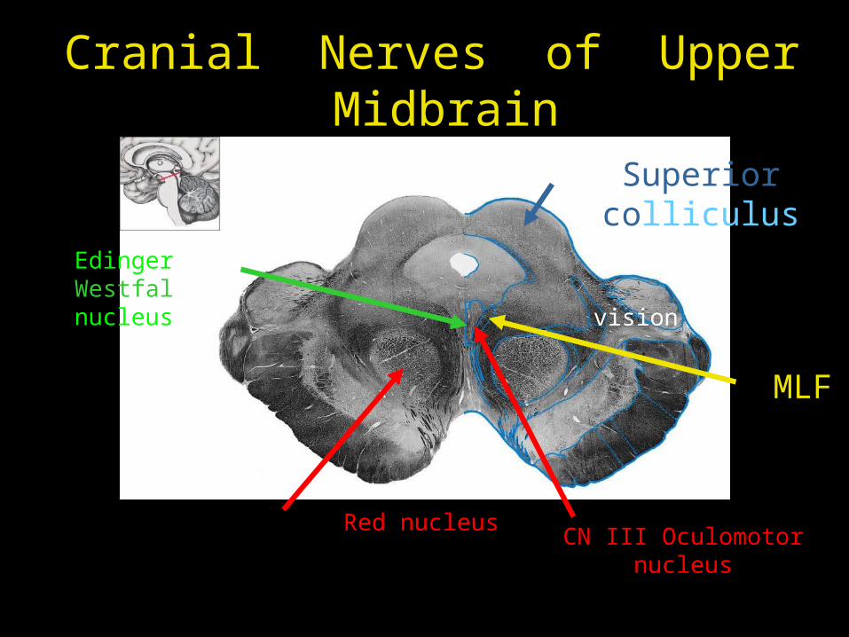
Cranial Nerves of Upper Midbrain
Superior colliculus
vision
Red nucleus
Edinger Westfal nucleus
MLF
CN III Oculomotor nucleus
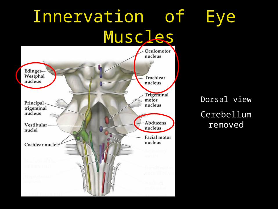
Innervation of Eye Muscles
Dorsal view
Cerebellum removed
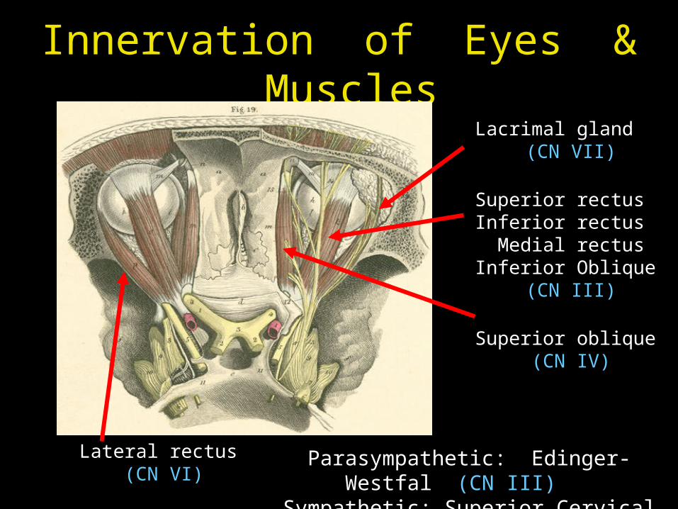
Innervation of Eyes & Muscles
Lacrimal gland (CN VII)
Superior rectus Inferior rectus Medial rectus Inferior Oblique
(CN III)
Superior oblique (CN IV)
Lateral rectus (CN VI)
Parasympathetic: Edinger-Westfal (CN III) Sympathetic: Superior Cervical Ganglion
Tuesday
Diencephalon
Thalamus
Hypothalamus