Embed Size (px)
Citation preview
Breast Cancer ScreeningWhat’s New to Know?
The Issue of Breast Density
Catherine Babcook MD
Partner, Mountain Medical Physician Specialists
Medical Director of Breast Imaging McKay Dee
Hospital Center
Disclosure
This presentation has no commercial content,
promotes no commercial vendor and is not
supported financially by any commercial
vendor. I receive no financial remuneration
from any commercial vendor related to this
presentation.
Screening Recommendations
• ACS, ACR, ACOBGYN, Intermountain
HC
– Annual mammographic screening
beginning at age 40
– Continue screening if a woman is in
good health and has a life expectancy of
5 years or more
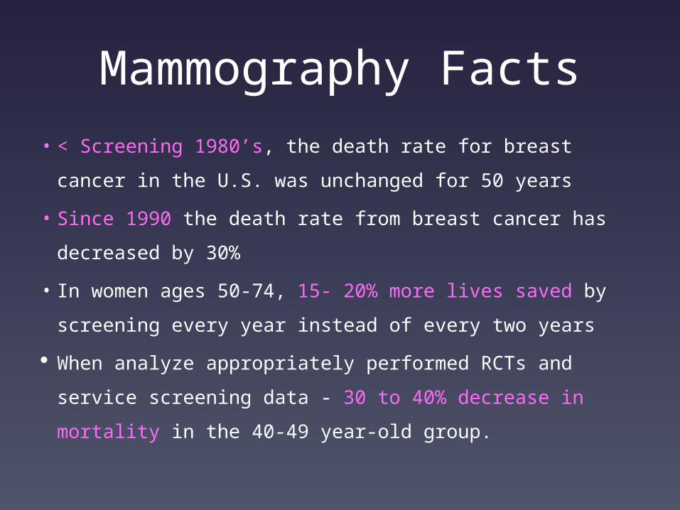
Mammography Facts
• < Screening 1980’s, the death rate for breast cancer in
the U.S. was unchanged for 50 years
• Since 1990 the death rate from breast cancer has
decreased by 30%
• In women ages 50-74, 15- 20% more lives saved by
screening every year instead of every two years
When analyze appropriately performed RCTs and
service screening data - 30 to 40% decrease in
mortality in the 40-49 year-old group.
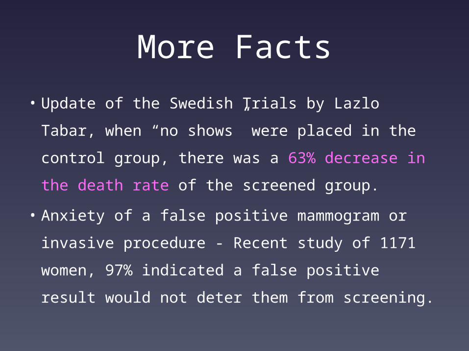
More Facts
• Update of the Swedish Trials by Lazlo Tabar, when
“no shows” were placed in the control group, there
was a 63% decrease in the death rate of the
screened group.
• Anxiety of a false positive mammogram or
invasive procedure - Recent study of 1171 women,
97% indicated a false positive result would not
deter them from screening.
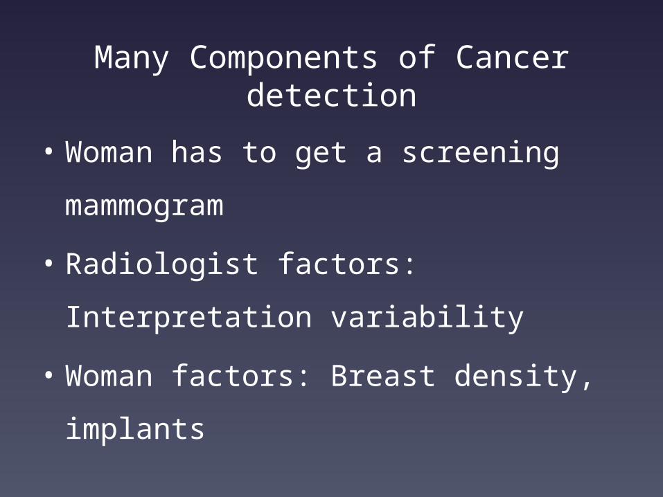
Many Components of Cancer detection
• Woman has to get a screening
mammogram
• Radiologist factors: Interpretation
variability
• Woman factors: Breast density,
implants
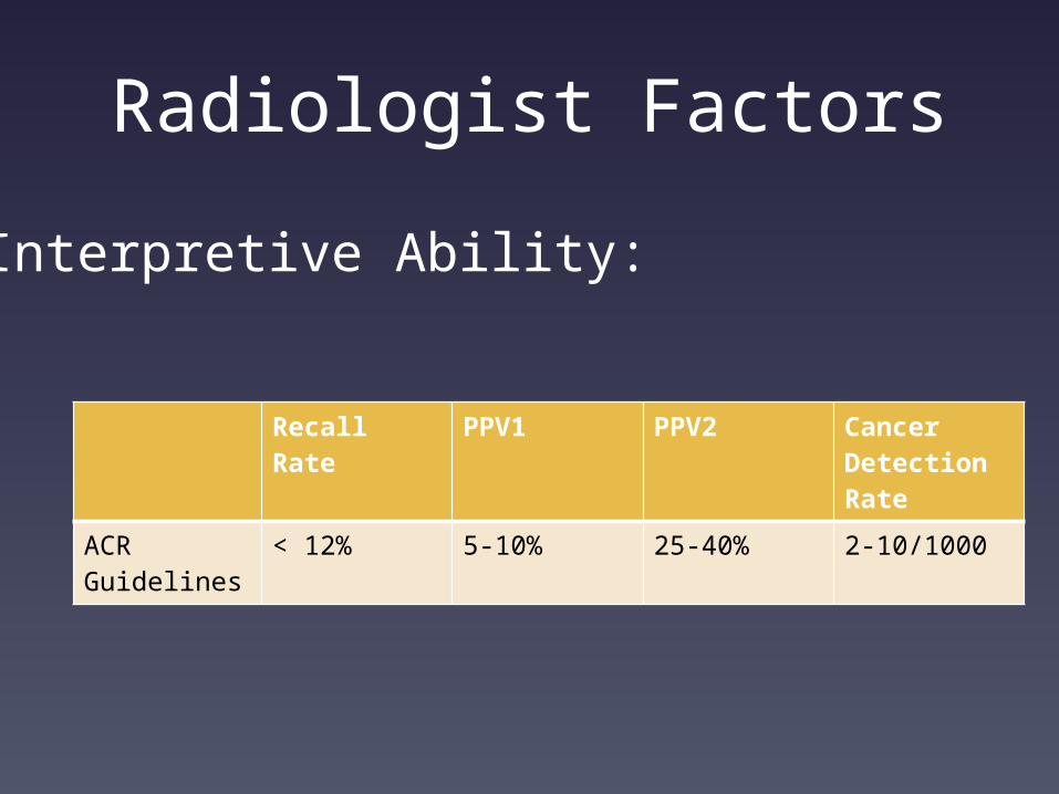
Radiologist Factors
Recall Rate PPV1 PPV2 Cancer Detection Rate
ACR Guidelines
< 12% 5-10% 25-40% 2-10/1000
Interpretive Ability:
Woman Factors
Clinically “dense” =
Mammographically Dense
Breast Tissue – Pattern and Density:
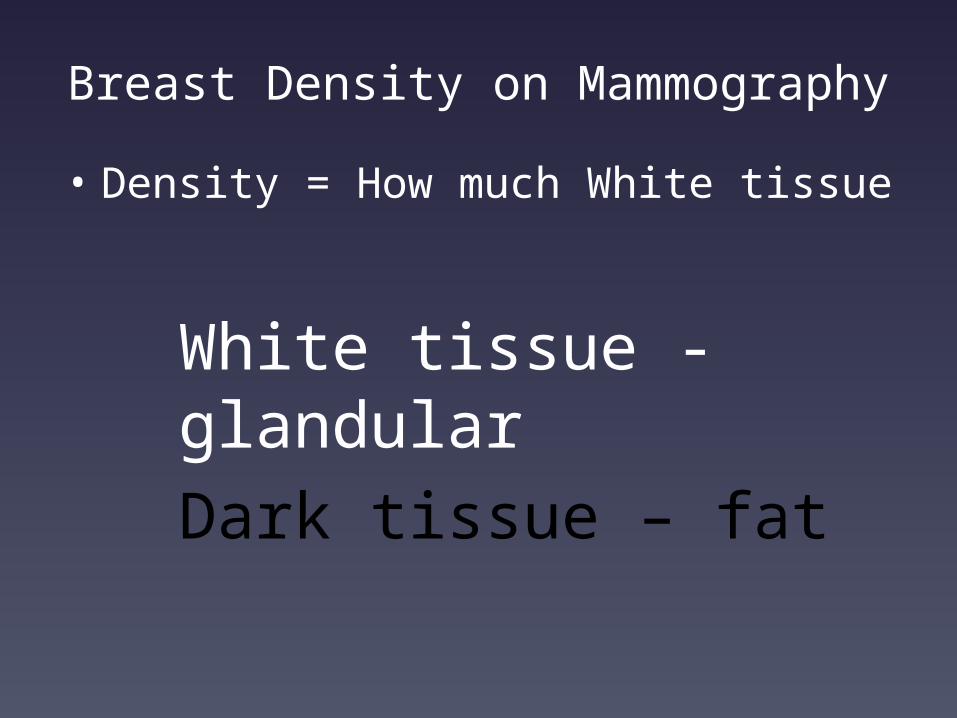
Breast Density on Mammography
• Density = How much White tissue
White tissue - glandular
Dark tissue – fat
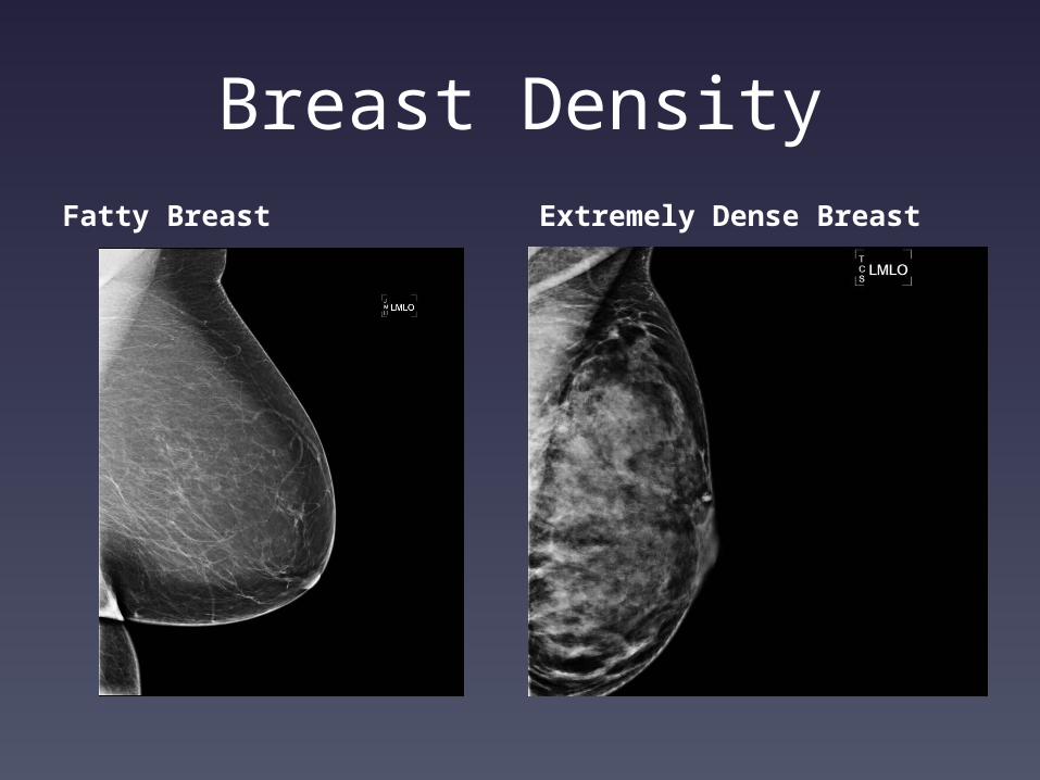
Breast DensityFatty Breast Extremely Dense Breast
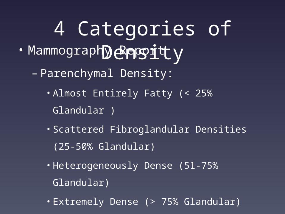
4 Categories of Density
• Mammography Report
– Parenchymal Density:
• Almost Entirely Fatty (< 25% Glandular )
• Scattered Fibroglandular Densities (25-50%
Glandular)
• Heterogeneously Dense (51-75% Glandular)
• Extremely Dense (> 75% Glandular)
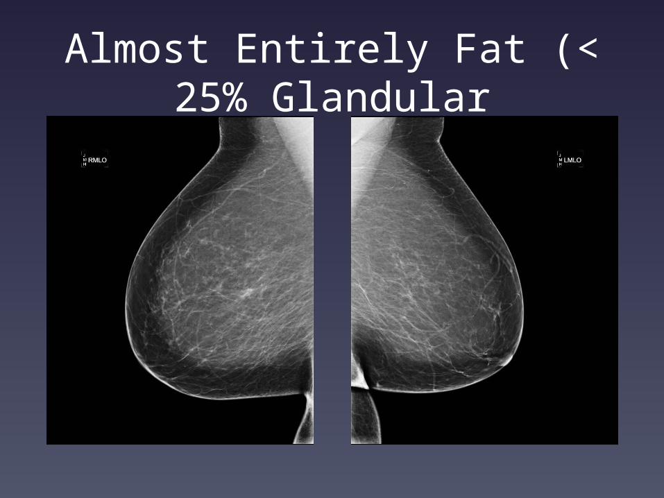
Almost Entirely Fat (< 25% Glandular

Scattered Fibroglandular Densities (25-50% Glandular)
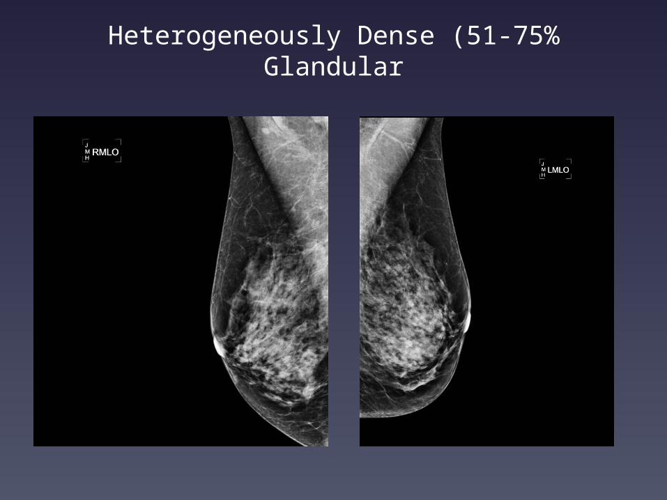
Heterogeneously Dense (51-75% Glandular
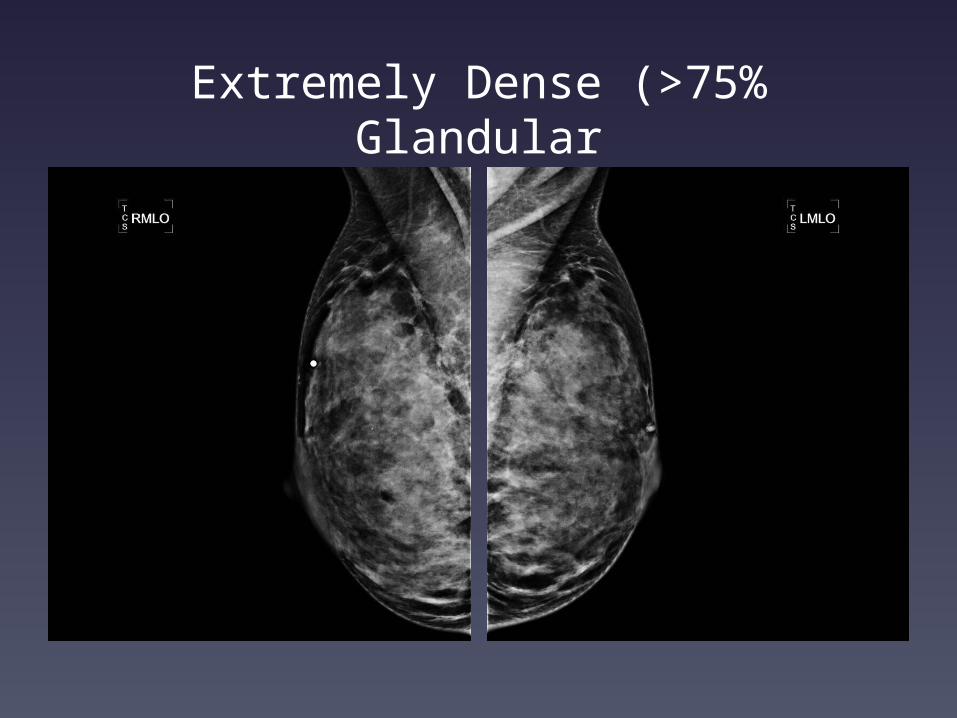
Extremely Dense (>75% Glandular
Why Does it Matter?
• Cancer is WHITE on
mammograms
• Amount of WHITE glandular
tissue impacts visibility of
WHITE cancer
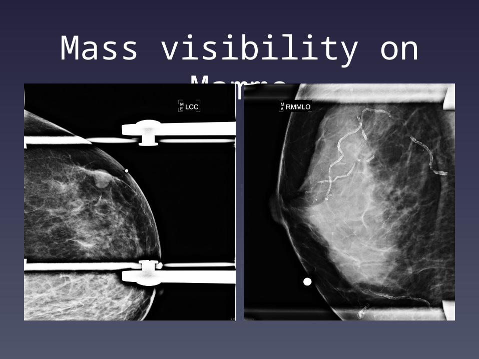
Mass visibility on Mammo
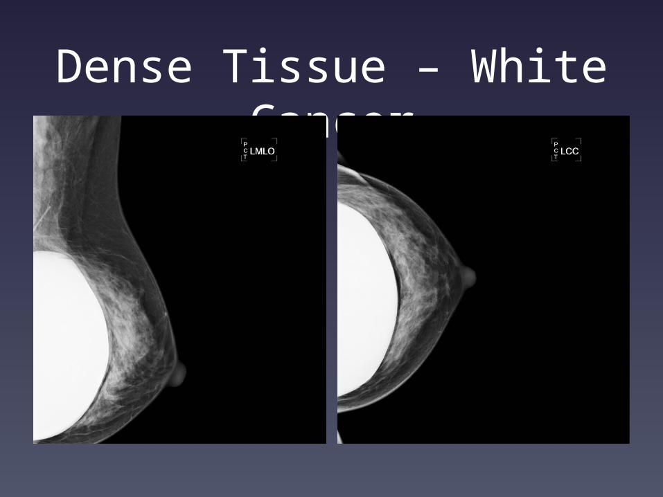
Dense Tissue – White Cancer
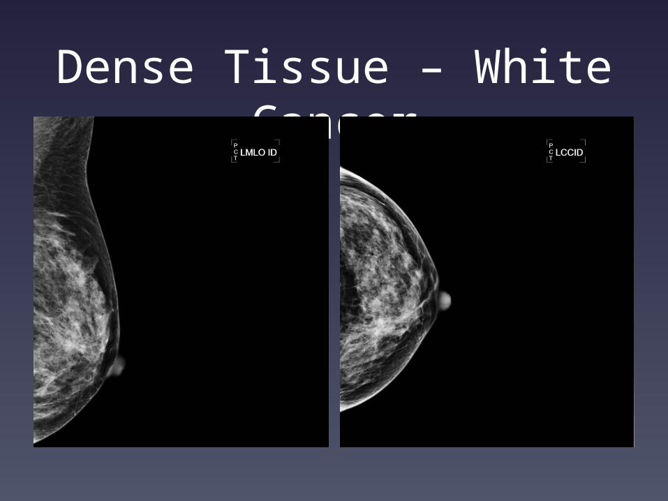
Dense Tissue – White Cancer
Cancer Can be Hidden by Glandular Tissue on Mammo
• ‘Snowflakes in a snowball’, ‘polar
bear in a snowstorm’
• What do we do:
– Wait until it’s big enough to feel
– Add a test that improves cancer
detection in white glandular tissue
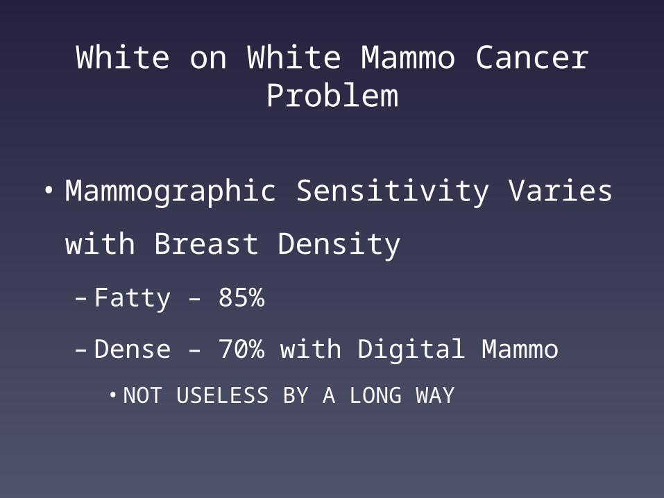
White on White Mammo Cancer Problem
• Mammographic Sensitivity Varies
with Breast Density
– Fatty – 85%
– Dense – 70% with Digital Mammo
• NOT USELESS BY A LONG WAY
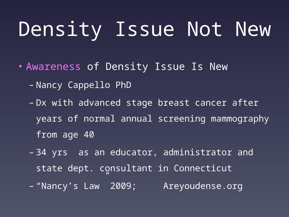
Density Issue Not New
• Awareness of Density Issue Is New
– Nancy Cappello PhD
– Dx with advanced stage breast cancer after years
of normal annual screening mammography from
age 40
– 34 yrs as an educator, administrator and state
dept. consultant in Connecticut
– “Nancy’s Law” 2009; Areyoudense.org
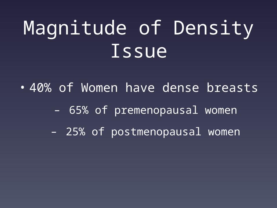
Magnitude of Density Issue
• 40% of Women have dense breasts
– 65% of premenopausal women
– 25% of postmenopausal women
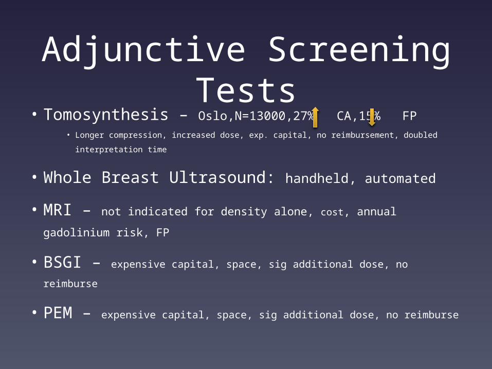
Adjunctive Screening Tests
• Tomosynthesis – Oslo,N=13000,27% CA,15% FP• Longer compression, increased dose, exp. capital, no reimbursement, doubled
interpretation time
• Whole Breast Ultrasound: handheld, automated
• MRI – not indicated for density alone, cost, annual gadolinium
risk, FP
• BSGI – expensive capital, space, sig additional dose, no reimburse
• PEM – expensive capital, space, sig additional dose, no reimburse
Breast Ultrasound
• Glandular Tissue is WHITE on
ultrasound just like mammography
• Cancer is DARK on ultrasound
Contrast advantage
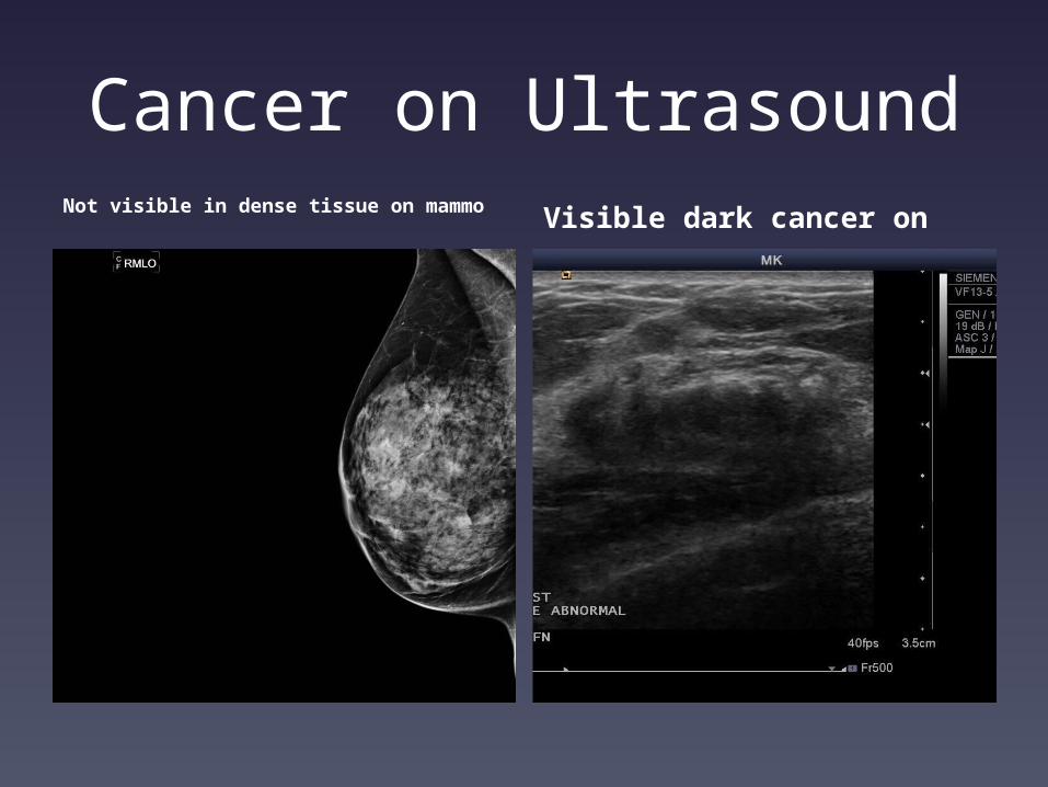
Cancer on UltrasoundNot visible in dense tissue on mammo Visible dark cancer on
ultrasound
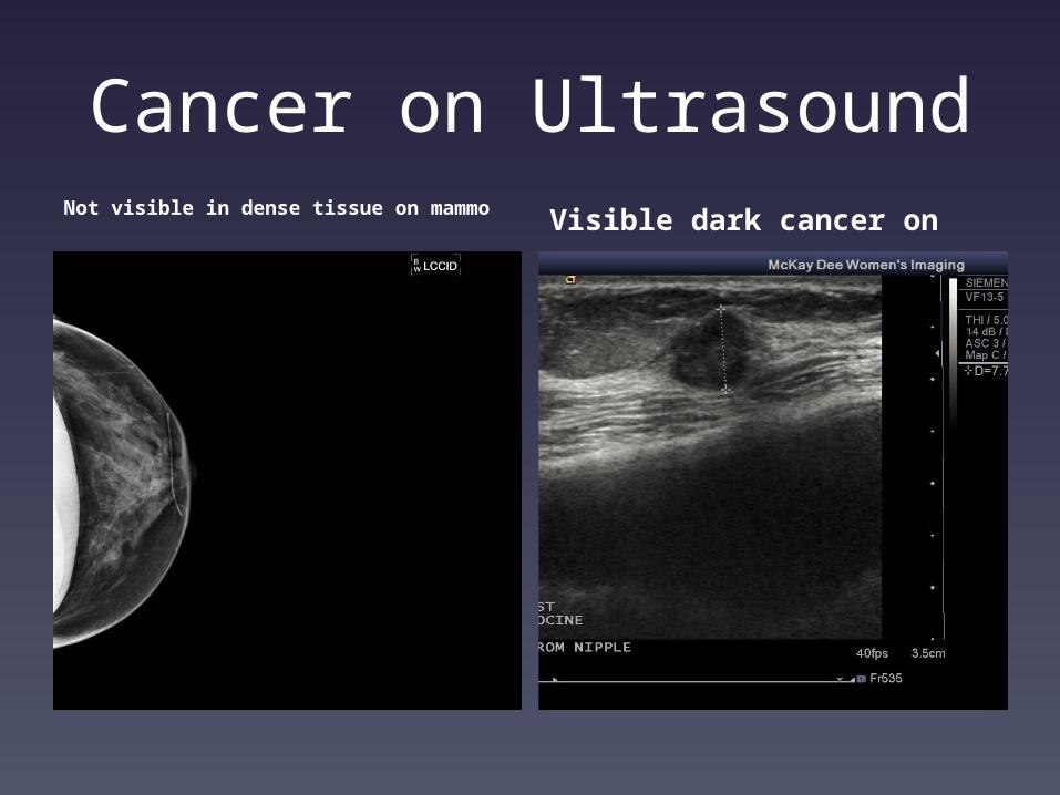
Cancer on UltrasoundNot visible in dense tissue on mammo Visible dark cancer on
ultrasound
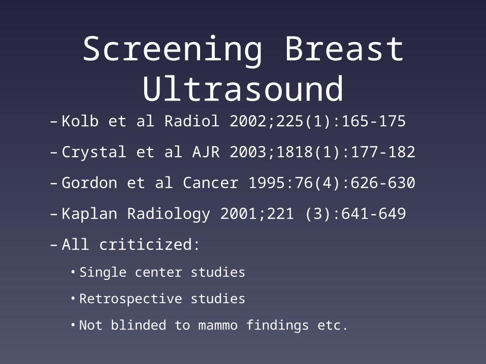
Screening Breast Ultrasound
– Kolb et al Radiol 2002;225(1):165-175
– Crystal et al AJR 2003;1818(1):177-182
– Gordon et al Cancer 1995:76(4):626-630
– Kaplan Radiology 2001;221 (3):641-649
– All criticized:
• Single center studies
• Retrospective studies
• Not blinded to mammo findings etc.
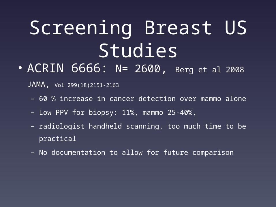
Screening Breast US Studies
• ACRIN 6666: N= 2600, Berg et al 2008 JAMA, Vol
299(18)2151-2163
– 60 % increase in cancer detection over mammo alone
– Low PPV for biopsy: 11%, mammo 25-40%,
– radiologist handheld scanning, too much time to be practical
– No documentation to allow for future comparison
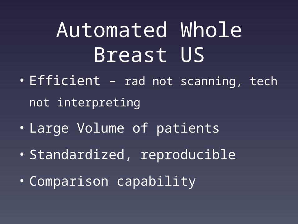
Automated Whole Breast US
• Efficient – rad not scanning, tech not
interpreting
• Large Volume of patients
• Standardized, reproducible
• Comparison capability
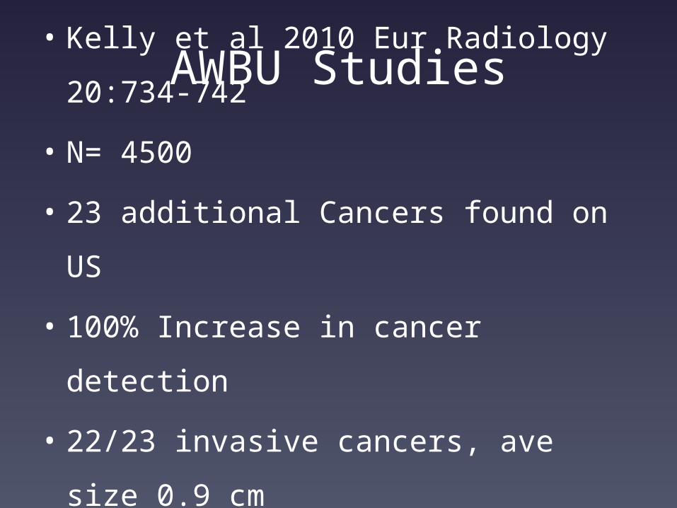
AWBU Studies• Kelly et al 2010 Eur Radiology
20:734-742
• N= 4500
• 23 additional Cancers found on US
• 100% Increase in cancer detection
• 22/23 invasive cancers, ave size 0.9
cm
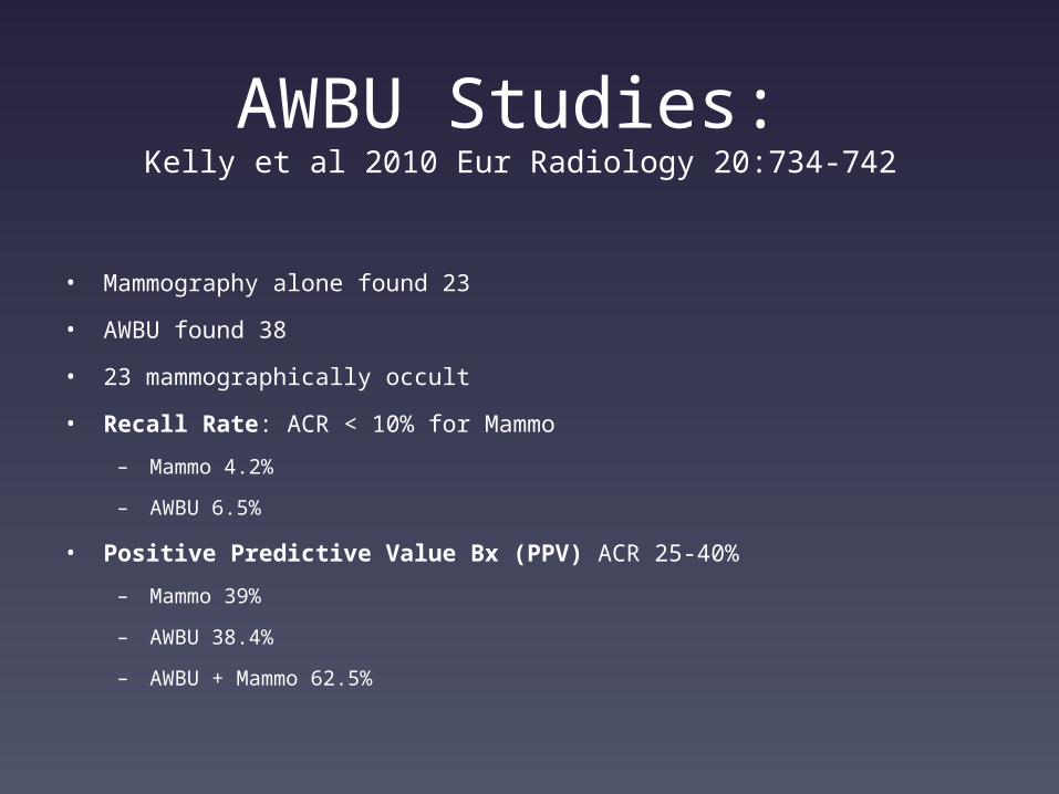
AWBU Studies: Kelly et al 2010 Eur Radiology 20:734-742
• Mammography alone found 23
• AWBU found 38
• 23 mammographically occult
• Recall Rate: ACR < 10% for Mammo
– Mammo 4.2%
– AWBU 6.5%
• Positive Predictive Value Bx (PPV) ACR 25-40%
– Mammo 39%
– AWBU 38.4%
– AWBU + Mammo 62.5%
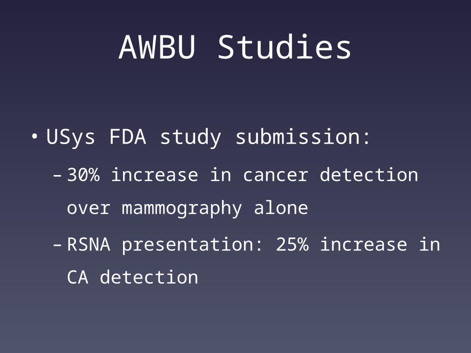
AWBU Studies
• USys FDA study submission:
– 30% increase in cancer detection over
mammography alone
– RSNA presentation: 25% increase in CA
detection
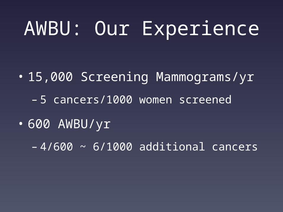
AWBU: Our Experience
• 15,000 Screening Mammograms/yr
– 5 cancers/1000 women screened
• 600 AWBU/yr
– 4/600 ~ 6/1000 additional cancers
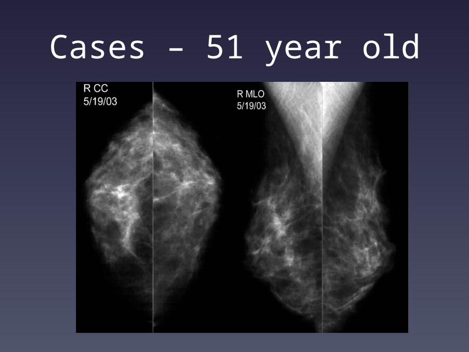
Cases – 51 year old
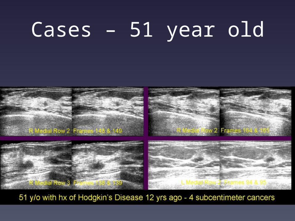
Cases – 51 year old
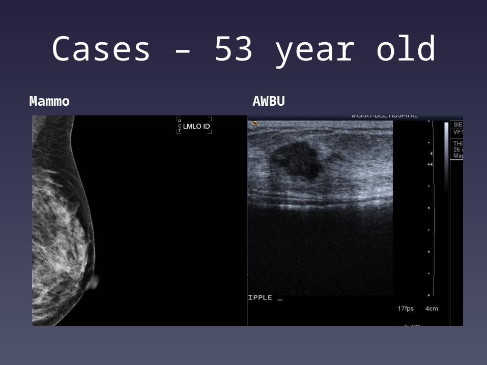
Cases – 53 year oldMammo AWBU
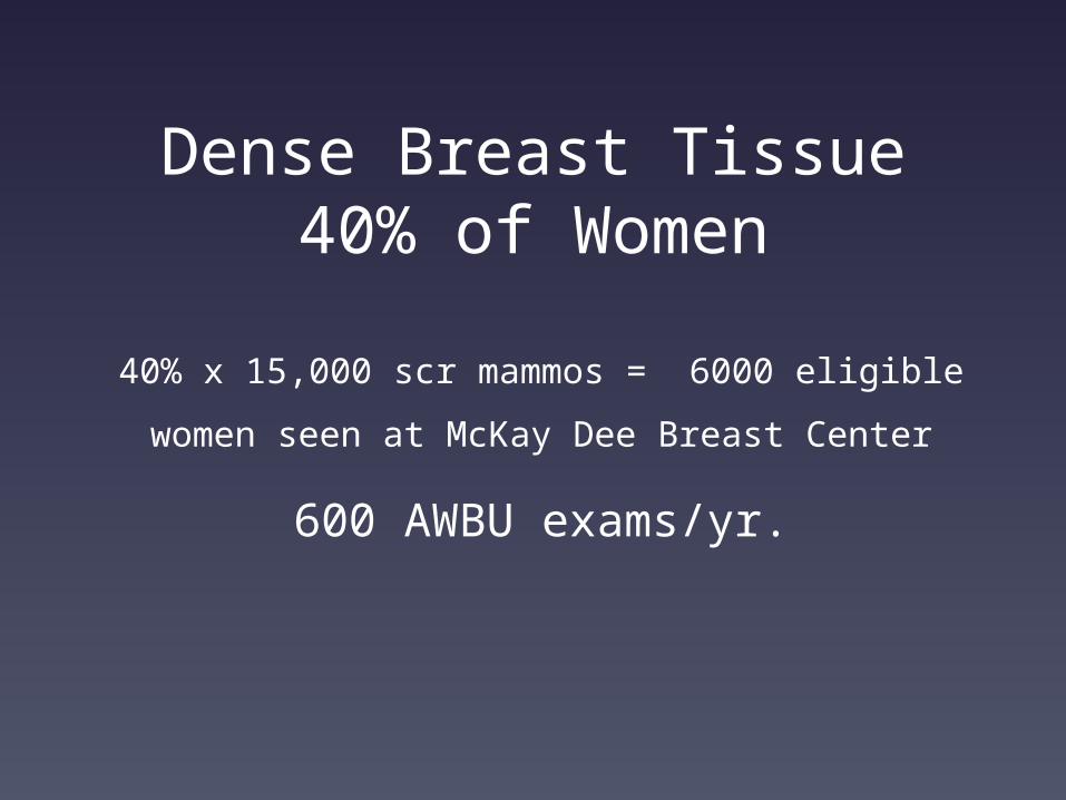
Dense Breast Tissue 40% of Women
40% x 15,000 scr mammos = 6000 eligible
women seen at McKay Dee Breast Center
600 AWBU exams/yr.
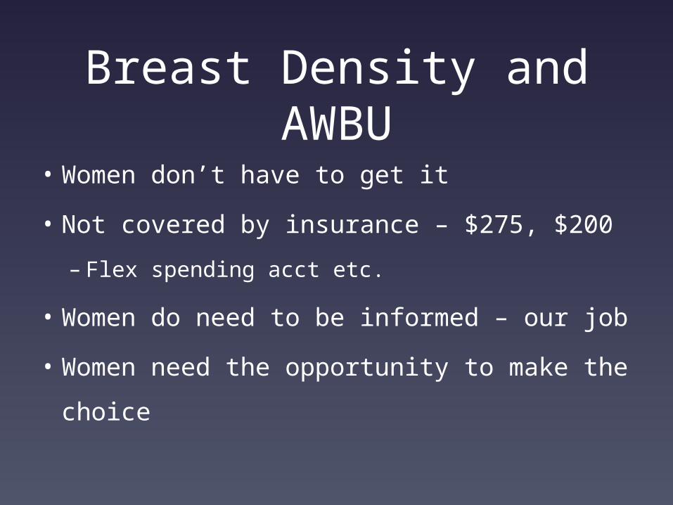
Breast Density and AWBU
• Women don’t have to get it
• Not covered by insurance – $275, $200
– Flex spending acct etc.
• Women do need to be informed – our job
• Women need the opportunity to make
the choice