Embed Size (px)
Citation preview
Breast Exam
Dr. Ruth Westra
Applied Anatomy
September 17, 2007
Goals/Objectives
Discuss Asymptomatic Female/Male Breast Assessment
Practice on Silicone Models Demonstrate Proficiency with clinical
breast exams on a professional patient model at the Duluth Family Medicine Residency
Anatomy
Milk producing glands arranged into lobules Glands connected by series of ducts to form a
common drainage path at the nipple Nipple surrounded by the areola Fibro-elastic and fatty tissue provide support Lymphatic tissue with ~90% drain into the
ipsilateral axilla and 10% drain into the Internal Thoracic nodes
EPIDEMIOLOGY
The lifetime risk for developing Breast Ca in US women is 1 in 7
Majority of women who develop breast Ca are over age 50
Majority of women with Breast Ca have no obvious risk factors
RISK FACTORS
Age Gender Race Prior History of Breast Cancer Family History of 1st Degree Relative Prolonged/Uninterrupted Exposure to
Estrogen
GAIL MODEL
Breast Cancer Risk Assessment Tool to calculate 5 year and lifetime risk for breast cancer based on several factors
Relative Risk for each factor produces a composite score
http://www.cancer.gov/bcrisktool/
SCREENING GUIDELINES
Clinical Breast Exams (CBE) part of periodic health exam every three years from women in 20s and 30s and yearly for women 40 and over.
Yearly mammogram starting at age 40 and continuing for as long as a woman is in good health.
Breast self-exam (BSE) is an option for women starting in their 20s
Women at increased risk should talk with their doctors about the benefits and limitations of screening earlier.
Taking Care of Your Breasts
Clinical Breast Exam (CBE) Mammogram Breast Self-Exam (BSE)
CLINICAL BREAST EXAM
Every 3 years for women age 20-39 Annually for women 40 and older
CLINICAL BREAST EXAM
Detection for some cancers that are missed on mammography
Follow-up on “lumps” detected by women
Screening younger women Screening women who do not follow
mammography recommendations
EXAMINATION IN DETAIL
Explain Exam Room Appropriate Draping Inspection Position Palpation
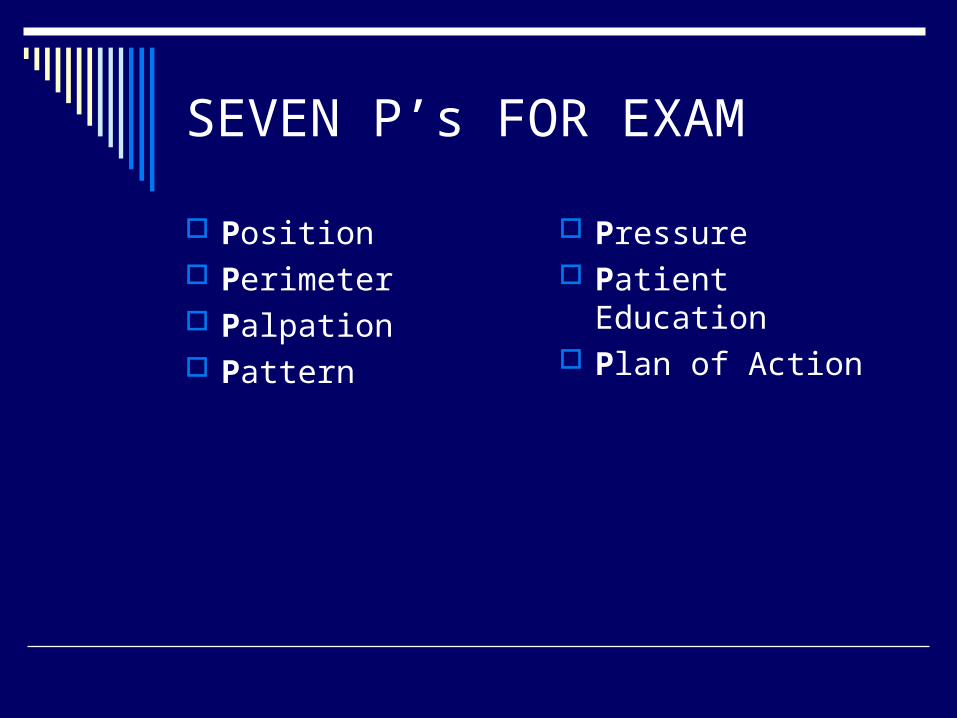
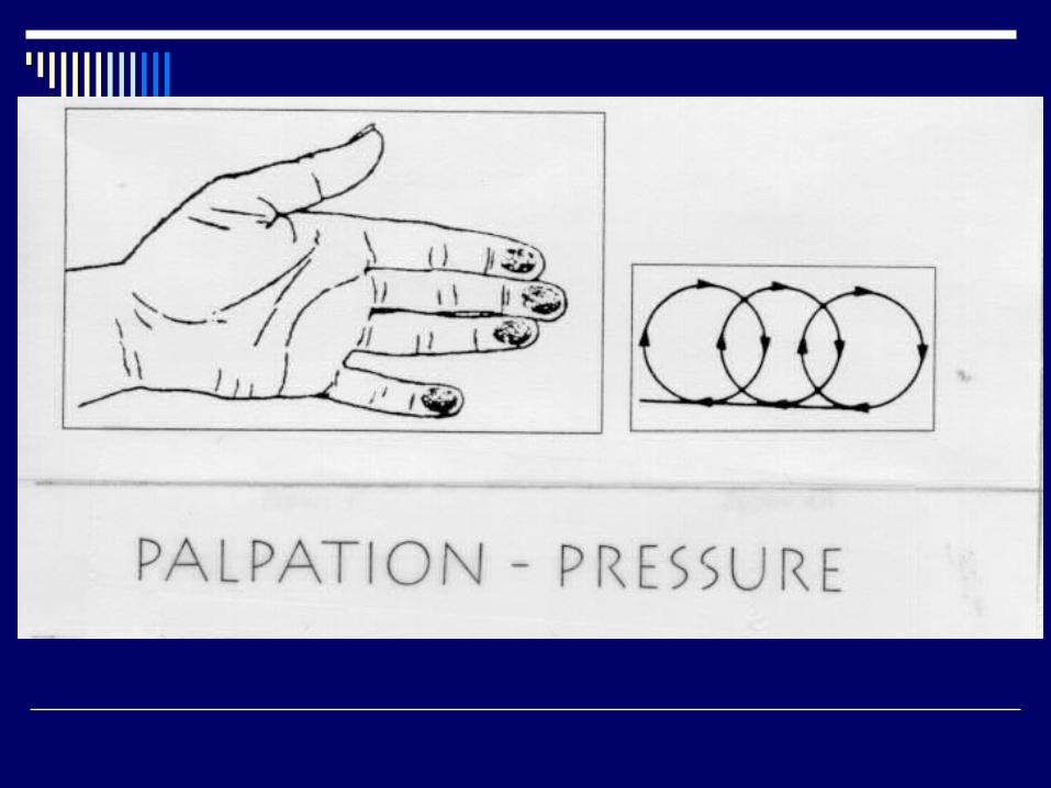
SEVEN P’s FOR EXAM
Position Perimeter Palpation Pattern
Pressure Patient Education Plan of Action


PATIENT EDUCATION AND PLAN
Discuss your Exam with the patient Check for patient understanding and
agreement Offer written material Recommend appropriate screening Document your Exam Findings Document your Patient
Recommendations
DOCUMENTATION
location size shape consistency
texture mobility tenderness
PLAN
Document your Exam Findings Document your Patient
Recommendations
COMMON ERRORS IN CBE
Missing the Auxiliary Tail Inconsistent Pressure Pattern of Search does not extend to the
Perimeter Avoiding the Nipple/Areolar Complex
MAMMOGRAM GUIDELINES
Yearly mammograms starting at age 40 and continuing for as long as a woman is in good health.
BREAST SELF-EXAM
Option starting at age 20 and older monthly
Women should report any breast change promptly
BREAST MASSES
Fibroadenoma Cysts Breast Cancer
Clinical Pearls
A clearly identifiable discrete mass requires a biopsy even if the mammogram is negative
Breast Cancer can occur in men Breast Cancer can occur in young
women If you have uncertainty, seek input
References
www.cancer.org http://medicine.ucsd.edu/clinicalmed/ Bickley, L.S. Bate’s Guide to Physical
Examination and History Taking Ninth Edition Saslow et al. “Clinical Breast Examination:
Practical Recommendations for Optimizing Performance and Reporting CA: A Cancer Journal for Clinicians Vol 54, No 6 Nov/Dec 2004
Sharon Anderson NP UMD Health Service