Embed Size (px)
Citation preview
The Breast Screening
Manual A Guide for Health Departments and Providers
Revised December 2016
Collaboration Partners: Chronic Disease and Injury Section,
Breast and Cervical Cancer Control Program
Medical Advisory Committee for the Breast and Cervical Cancer Control
Program
Women's and Children's Health Section
North Carolina Department of Health and Human Services
Division of Public Health
State of North Carolina
Department of Health and Human Services
Division of Public Health N.C. Breast and Cervical Cancer Control Program www.ncdhhs.gov www.publichealth.nc.gov
N.C. DHHS is an equal opportunity employer and provider.

MEMORANDUM
To: Local Health Directors
Nursing Directors/Supervisors
RICHARD O. BRAJER
Secretary
DANIEL STALEY Director, Division of Public Health
From: Danny Staley, MPH, Division Director
Dr. Kathleen Shapley-Quinn, Chronic Disease and Injury Medical
Consultant
Debi Nelson, MAEd, Cancer Prevention and Control Branch Head
Subject: Revised Breast Screening Manual: A Guide for Health
Departments and Providers, (December 7, 2016)
Date: December 7, 2016
Enclosed is the revision of the Breast Screening Manual, replacing “The Breast and
Cervical Screening Manual: A Guide for Health Departments” published in 2006.
The revision is an interdepartmental collaboration between the Division of Public
Health - Chronic Disease and Injury Section, North Carolina Breast and Cervical
Cancer Control Program and Woman’s and Children’s Health Section.
The current guidance from Center for Disease Control and Prevention,
National Cancer Institute, American Cancer Society, U.S. Preventive
Services Task Force, American College of Obstetricians and Gynecologists,
American Academy of Family Physicians, American Society of Breast
Surgeons and the American College of Radiology is encompassed in the
Breast Screening Manual.
The Division of Public Health document is to be used as a model and
template for writing policies and procedures to recruit, screen, diagnose
and treat women for breast cancer. In keeping with our mission to work in
partnership with local communities to improve the quality of life and save
the lives of women in North Carolina, this manual will be helpful in delivery
of health care services to the public. We thank you and appreciate the
work you do to improve the quality of life for North Carolina women.
Department of Health and Human Services | Division of Public Health
5505 Six Forks Road | 1922 Mail Service Center | Raleigh, NC 27699-1922
919 707 5300 T | 919 870 4812 F

ACKNOWLEDGEMENTS
Breast Screening Manual: A Guide for Health Departments and Providers
Revised December 2016
This Breast Screening Manual was reviewed and revised through the collaborative efforts of
representatives of the following Division of Public Health sections and programs:
Chronic Disease and Injury Section
Breast and Cervical Cancer Program
Women's and Children's Health Section
The Breast Screening Manual Committee expresses gratitude and appreciation to all
individuals who worked toward the successful completion of the Breast Screening Manual.
Breast Screening Manual Committee
Cindy Herndon, PhD, RN, WHNP, CNE Chairman
Vicki Deem, MLS, MPA, RN
Debbie Farb, MPH, BSN, RN, IBCLC
Sherry Wright, RN
The Breast Screening Manual Reviewers
Debi Nelson, MAEd
Kathleen Shapley-Quinn, MD
Belinda Pettiford, MPH Linda
Sutton, MD
Jan Wong, MD
Helene Edwards, MS BSPH
Larry Wu, MD
Kim Weaver, MSN, BSN, RN
Cherie Kuzmiak, DO
Allison Hall, MD
Barbara Toth, BSN, RN, ERRN
Molly Leatherland, WHNP, RN
Breast and Cervical Cancer Program Staff
Tammie Hobby

2 N.C. DHHS • Division of Public Health • The Breast Screening Manual: A Guide for Health Departments and Providers / December 2016
THE BREAST SCREENING MANUAL A Guide for Health Departments and Providers
Table of Contents
Breast Health .................................................................................................................................. 4
Breast Cancer Screening and Breast Cancer Risk Factors ......................................................... 5
Breast Cancer Risk Factors ........................................................................................................ 5
Components of Breast Cancer Screening in North Carolina .................................................... 7
Current Recommendations for Breast Screenings ............................................................... 7
Clinical Breast Examination (CBE) ......................................................................................... 8
Mammography Screening ...................................................................................................... 9
Breast Self-Examination (BSE) ............................................................................................. 11
Quality Assurance......................................................................................................................... 12
Quality Assurance Recommendations for Breast Screening.................................................. 12
Patient Notification Requirements .......................................................................................... 14
Management of Abnormal Clinical Findings ............................................................................... 16
I. Palpable Mass ....................................................................................................................... 16
II. Non-Palpable Masses Found on Mammography ................................................................ 17
III. Vague Thickening or Nodularity Not Suspicious for Cancer .............................................. 17
IV. Nipple Discharge or Skin Changes ..................................................................................... 18
V. Breast Pain ........................................................................................................................... 18
VI. Special Considerations ........................................................................................................ 19
Organization of the Mammography Report Reporting System (ACR BI-RADS, 2013) .............. 23
Mammography Assessment Categories ..................................................................................... 24
Appendix A: Breast Cancer Glossary ........................................................................................... 27
Appendix B: North Carolina BCCCP and Women’s Health Eligibility .......................................... 40
NC BCCCP-Eligible Population .................................................................................................. 40
Priority Populations ............................................................................................................... 40
Eligible Population ................................................................................................................ 40

3 N.C. DHHS • Division of Public Health • The Breast Screening Manual: A Guide for Health Departments and Providers / December 2016
North Carolina Women’s Health Branch Programs Eligibility ................................................. 41
Federal Poverty Guidelines ...................................................................................................... 42
Appendix C: North Carolina Division of Medical Assistance- Breast and Cervical Cancer
Medicaid ....................................................................................................................................... 43
Appendix D: NC BCCCP Policies .................................................................................................. 47
NC BCCCP & WISEWOMAN Policy for Patients Insured Under the Patient Protection and
Accordable Care Act (ACA) ....................................................................................................... 48
NBCCEDP Program Policy on Patient Navigation .................................................................... 49
NC BCCCP Eligibility for Family Planning Patients .................................................................. 51
Appendix E: Other Resources for Information and Treatment ................................................... 53
CancerCare Financial Assistance ............................................................................................ 54
CancerCare Co-Payment Assistance Foundation ................................................................... 55
Patient Advocate Foundation ................................................................................................... 56
Pretty in Pink Foundation ......................................................................................................... 57
Additional Funding Sources outside of BCCCP ....................................................................... 58
Cancer Information Resources ................................................................................................ 59
Appendix F: Breast Cancer Staging ............................................................................................. 61
Appendix G: Breast Density……………………………………………………………………………………… ....... 64
Appendix H: Understanding Genetic Risk For Breast Cancer .................................................... 66
Appendix I: Staff Directories ……………………………………………………………………………………… 78
NC Breast & Cervical Cancer Program Directory .................................................................... 78
NC Women’s Health Directory ................................................................................................. 79
References.................................................................................................................................... 80

TAB 1: Overview

BREAST HEALTH
The National Cancer Institute (NCI) estimates that women living in the United States have a
12.4%, or a 1 in 8, lifetime risk of being diagnosed with breast cancer (NCI, 2016).
Estimated risk is an average risk for all women. Individual risk factors include age, family
history, reproductive history, race and ethnicity, as well as other factors. NCI’s Surveillance,
Epidemiology, and End Results [SEER] Program, is based on breast cancer statistics for the
years 2007 through 2009. If the current incidence rate remains the same, a woman born
today has an approximate lifetime risk of 1 in 8 for being diagnosed with breast cancer at
some time during her life if she lives to an average life expectancy of 85 years.
The SEER report (NCI, 2016) estimates the risk of developing breast cancer in 10-year age
intervals. Per the current report, the risk that a woman will be diagnosed with breast cancer
during the next 10 years, starting at the following ages is as follows:
Age 30 . . . . . . 0.44 percent (or 1 in 227)
Age 40 . . . . . . 1.47 percent (or 1 in 68)
Age 50 . . . . . . 2.38 percent (or 1 in 42)
Age 60 . . . . . . 3.56 percent (or 1 in 28)
Age 70 . . . . . . 3.82 percent (or 1 in 26)
Women in North Carolina have the same lifetime risk as the national average. In their annual
projections for North Carolina, the American Cancer Society (ACS) estimates that
7,830 women will be diagnosed with breast cancer in 2016, and an estimated 1,360
women will die of breast cancer. Breast cancer is the second leading cause of cancer deaths
in North Carolina women. The burden of breast cancer falls heavily on low-income and
minority women, particularly women in rural North Carolina. In 2014, North Carolina
minority females were 30% more likely to die from breast cancer than white females (ACS,
2016).
Nationally, survival rates have increased over time for both white and African American
women; however, the American Cancer Society reports the disparity in five-year survival
rates between white women (92%) and African-American women (81%) persists. Lower
survival rates in African-American women are hypothesized to be due to later stage
detection of their breast cancers and the higher rate of more aggressive breast cancers in
young African-American women (ACS, 2016).
Early detection and treatment of breast cancer is saving lives. The American Cancer Society
reported the decline of breast cancer mortality rates across the U.S. by 36% from 1989-
2012. With improvements in early detection and treatment, more cases of breast cancer
will be diagnosed and treated at earlier stages, and breast cancer mortality will continue to
decrease (ACS, 2016).

TAB 2: Breast Cancer Screening & Risk Factors

BREAST CANCER SCREENING AND BREAST CANCER RISK FACTORS
Breast Cancer Risk Factors
Scientists and physicians cannot explain why one woman gets breast cancer and another
does not. Scientists have studied patterns and have found that environmental factors and
certain personal habits can increase a person’s chances of developing cancer. Per the
National Cancer Institute (NCI), prevention means avoiding the risk factors and increasing
the protective factors that can be controlled so that the chance of developing cancer
decreases (NCI, 2016). While risk factors can be avoided, avoidance does not necessarily
guarantee a life free of breast cancer.
The National Cancer Institute Findings:
▪ Inherited changes in certain genes (including BRCA1, BRCA2, and others)
increase the risk of breast cancer.
▪ Having a mother, sister and/or daughter with breast cancer increases the risk of
developing breast cancer, especially if they were diagnosed before age 50.
▪ Women who have a high percentage of breast tissue that appears dense on a
mammogram have a higher risk of breast cancer than women of similar age who
have little or no dense breast tissue.
▪ Populations that eat a high-fat diet are more likely to die of breast cancer.
▪ Exercise, especially in young women, may decrease hormonal levels and
decrease breast cancer risk.
▪ Breast feeding reduces breast cancer risk.
▪ Alcohol consumption may be associated with a slightly increased risk of breast
cancer.
▪ Postmenopausal weight gain after natural menopause and/or after age 60 may
increase breast cancer risk.
▪ Women who use combined estrogen and progestin menopausal hormone therapy
for 5+ years have an increased chance of developing breast cancer (NCI, 2016).
The American Cancer Society Findings:
▪ Risk factors that are not easily changed:
▪ Family history of breast cancer
▪ BRCA1/BRCA2 inherited gene mutations
▪ Having first period before twelve
▪ Not having children or not having first child until after age 30
▪ Late age at menopause
▪ High breast tissue density
▪ High bone mineral density (ACS, 2016).
▪ Modifiable risk factors:
▪ Limiting the use of hormone replacement therapy (combined estrogen and
progestin)
▪ Reducing alcohol consumption

▪ Breast feeding your child/lactating
▪ Avoiding obesity
▪ Being physically active (ACS, 2016).
The link between breast cancer and other factors such as smoking, diet and vitamin intake
and night shift work remain unclear with conflicting research findings. The 2014 Surgeon
General’s report concluded that there is “suggestive but not sufficient” evidence that
smoking increases the risk of breast cancer. While diet and vitamin intake results remain
inconsistent, maintaining a healthy weight reduces risks. In 2007, the International Agency
for Research on Cancer classified shift work with circadian disruption as a probable human
carcinogen; however, the current state of scientific knowledge does not permit a firm
conclusion that shift work increases the risk of cancer (ACS, 2016).
The Best Preventive Recommendations for Breast Cancer:
▪ Achieve and maintain a healthy weight
▪ Be physically active
▪ Maintain adequate and healthy sleep habits
▪ Limit alcoholic beverages
▪ Avoid exposure to chemicals
▪ Reduce exposure to radiation
▪ Consider the risks and benefits of hormonal replacement therapy with provider or
as a provider.
▪ Screening for breast cancer. Although screening does not protect against breast
cancer, it may detect cancer earlier and allow for earlier treatment and best
prognosis (Center for Disease Control [CDC], 2016).

Components of Breast Cancer Screening in North Carolina
There are two main components of breast cancer screening:
1. Clinical Breast Examination (CBE)
2. Age-appropriate mammogram
Current Recommendations for Breast Screenings for Average-Risk Women
Aside from genetics, personal and family history, there is no consensus on age for
mammography screening, especially for women between the ages of 40 and 49.
Whether to do a clinical breast exam is currently a controversial issue. As of publication of
this manual, NC BCCCP continues to require clinical breast examination as part of the
screening process.
Listed below is a sampling of various government and health care organizations and their
guidance.
Organization Clinical Breast Exam
recommendation
Mammogram recommendation
American Cancer Society (ACS) No CBE all ages Age 40- 44 opportunity to
screen
Age 45- 54 screen annually
Ages 55 plus screen biennially
with an opportunity to screen
annually
Upper limit Life expectancy >
10 years, if the woman is in
reasonably good health
American College of Radiology
(ACR)
Annual screening beginning at
age 40
American Academy of Family
Physicians (AAFP)
Current evidence is insufficient
to assess the benefits and
harms of clinical breast
examination (CBE) for women
aged 40 and older.
Age 50-74 screen every two
years
American Society of Breast
Surgeons, 2016
No recommendation Discussion with physician to
consider screening
mammography at age 40- 44
Annual screening mammogram
for women ages 45- 54 as
indicated by the new ACS
guidelines
Annual or biennial screening
mammogram for women 55
and older based upon a shared
decision making discussion

Organization Clinical Breast Exam
recommendation
Mammogram recommendation
regarding risk and benefits of
screening timing
Biennial screening for women
over the age of 75 if an
estimated life expectancy is
greater than 10 years (ASBrS,
2016).
US Preventive Services Task
Force (USPSTF)
No recommendation Personal decision ages 40- 49
Every 2 years, ages 50- 74
Insufficient evidence of benefit
age 75 and beyond
ACOG Women aged 29- 39 CBE every
1- 3 years;
Annual CBE beginning at age
40 (ACOG, 2016)
Annual screening beginning at
age 40
Supports shared decision
making between physician and
patient (ACOG, 2016)
National Breast and Cervical
Cancer Early Detection
Program, Center for Disease
Control (NBCCEDP)
Every 1 year, ages 40- 64 Every 1- 2 years, ages 50- 64
North Carolina Breast and
Cervical Cancer Control
Program (NC BCCCP)
Every 1 year, ages 40- 64 Federal Funds: Every 1- 2
years, ages 50- 64
State Funds: Every 1- 2 years,
ages 40- 49 and 65- 75
1. American Cancer Society, 2016
2. American College of Radiology, 2016
3. American Academy of Family Physicians, 2016
4. American Society of Breast Surgeons, 2016
5. US Preventive Services Task Force, 2016
6. American College of Obstetricians and Gynecologists, 2016
7. National Breast and Cervical Cancer Early Detection Program, Center for Disease Control,
2016
8. North Carolina Breast and Cervical Cancer Control Program, Agreement Addendum,
2016- 2017
Clinical Breast Examination (CBE)
The purpose of the clinical breast examination (CBE) is to assess breast health status. A
CBE should be thorough. The examination may be done as part of a general exam or as a
separate exam for asymptomatic or symptomatic women. In 2016, NC BCCCP continues to
require clinical breast examination as part of the screening process. Please refer to the
recommendations table on pages 8 and 9.
If you plan to conduct clinical breast examinations, and need training on the vertical strip
method, please contact NC BCCCP at 919-707-5300.
Mammography Screening
Mammography is the best way to detect breast cancer in its earliest, most treatable stage—
an average of 1–3 years before a woman can feel the lump (Duffy, 2012; National Cancer
Institute, 2016). Mammography also locates cancers too small to be felt during a clinical
breast examination.
1. Screening mammogram
a. Definition: A screening mammogram is performed on asymptomatic women to detect
early, clinically unsuspected breast cancer. (American College of Radiology [ACR],
2016)
b. Purpose: The purpose of screening mammograms is to find breast cancers before they
cause symptoms. Early detection results in the diagnosis of breast cancer before there
are palpable masses and symptoms. Breast cancers found during screening
examinations are more likely to be small, confined to the breast, may not require
chemotherapy or lymph node surgery, and increase the number of treatment options.
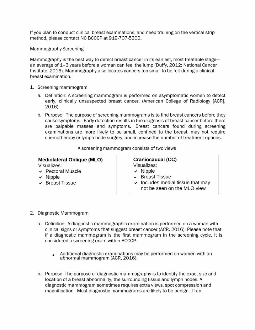
A screening mammogram consists of two views
2. Diagnostic Mammogram
a. Definition: A diagnostic mammographic examination is performed on a woman with
clinical signs or symptoms that suggest breast cancer (ACR, 2016). Please note that
if a diagnostic mammogram is the first mammogram in the screening cycle, it is
considered a screening exam within BCCCP.
• Additional diagnostic examinations may be performed on women with an
abnormal mammogram (ACR, 2016).
b. Purpose: The purpose of diagnostic mammography is to identify the exact size and
location of a breast abnormality, the surrounding tissue and lymph nodes. A
diagnostic mammogram sometimes requires extra views, spot compression and
magnification. Most diagnostic mammograms are likely to be benign. If an
Mediolateral Oblique (MLO) Visualizes: Pectoral Muscle Nipple Breast Tissue
Craniocaudal (CC) Visualizes: Nipple Breast Tissue Includes medial tissue that may
not be seen on the MLO view

10 NC DHHS • Division of Public Health • The Breast Screening Manual: A Guide for Health Departments and Providers / December 2016
abnormality is suspicious, usually an ultrasound study follows and/or a biopsy may
be ordered. If a woman has a clinically suspicious abnormality, a biopsy is the only
way to determine with certainty whether she has breast cancer (National Breast
Cancer Foundation, 2016).
Note: (1) When scheduling a mammogram, previous films should be requested and sent to
the contracted radiology facility. Films should be requested at least two weeks prior to the
woman’s appointment. (2) Results of the CBE and history of any prior breast surgery should
also be included on the referral form to the radiology facility.
NC BCCCP Guidance on Screening Mammography
The priority population for NBCCEDP (Federal Funds) screening mammography services is
the group of women between the ages of 50 and 64 who are low-income (less than 250% of
federal poverty level) and who have not been screened in the past year. At the clinician's
discretion, women age 50-64 with a history of normal screening results and no significant
risk factors may be put on an every-other-year screening cycle. The priority population for
NC BCCCP State Funds is women between the ages of 40 and 49 who are low-income (less
than 250% of federal poverty level) and who have not been screened in the past year.
(Diagnostic mammograms may be provided for symptomatic women under 40 years of age
using State Funds and under 50 years of age using Federal Funds.)
Federal BCCCP Screening Age Priorities:
Test Type National Breast and
Cervical Cancer Early
Detection Program
(NBCCEDP) Rules
Performance Indicator
Breast Cancer Screening Screening mammograms
to women 50 - 64 years of
age every 1 - 2 years
At least 75% of all initial
mammograms paid with
Federal Funds should be
within this age group
Breast Cancer Diagnostic
Imaging
Mammograms provided
for symptomatic women
under 50 years of age who
require a diagnostic work-
up or who have a family
history of breast cancer
No more than 25% of all
initial mammograms paid
with Federal Funds should
be within this age group
NBCCEDP, 2016

11 NC DHHS • Division of Public Health • The Breast Screening Manual: A Guide for Health Departments and Providers / December 2016
NC State BCCCP Screening Age Priorities:
Test Type NC Breast and Cervical
Cancer Early Detection
Program (NC BCCCP)
Rules
Performance Indicator
Breast Cancer Screening Screening mammograms
to women age 40 - 49
years and up to age 75 if
no other source of
payment is available every
1 - 2 years
NC BCCCP funds cannot
be used to screen
asymptomatic women
under the age of 40, even
if considered to be at high
risk for breast cancer.
Breast Cancer Diagnostic
Imaging
Women age 21 to 75 with
gross incomes below
250% of Federal Poverty
Level, who are uninsured
or underinsured may be
eligible subject to
limitations; Eligible women
ages 21- 39 with an
undiagnosed breast
abnormality may receive
NC BCCCP funded
diagnostic services if no
other source of healthcare
reimbursement is
available.
Eligible women ages 21-
39 May be referred from
Family Planning Services,
outside clinics or through a
self-referral for an
undiagnosed abnormality.
NC BCCCP Training Manual, 2016
Breast Self-Examination (BSE)
A woman can notice changes by being aware of how her breasts normally look and feel and
by feeling her breasts for changes, or by choosing to use a step-by-step approach and using
a specific schedule to examine her breasts. Breast self-exam is no longer recommended by
most professional organizations.
If your patients are interested in learning how to conduct breast self-exams, you may contact
the NC BCCCP to set up a train-the-trainer instructional seminar.

TAB 3: Quality Assurance

QUALITY ASSURANCE Quality Assurance Recommendations for Breast Cancer Screening
For breast cancer screening to be effective, health care providers must have systems in
place to ensure that any abnormalities detected by clinical breast exam or mammography
are followed up appropriately. Patients with abnormal tests results should be notified
promptly. Patients who need additional diagnostic tests or treatment should be tracked to
assure they receive proper follow-up care.
Five key steps are necessary for managing the results of breast cancer screening:
1. Track any imaging studies until results are obtained;
2. Follow requirements for patient notification (see page 14- 16);
3. Document that notification has occurred;
4. Refer patients with any abnormalities on clinical breast exam or imaging for
appropriate follow-up; and
5. Track referrals to make sure that patients have received follow-up.
Each clinic might have a different mechanism for ensuring that all these steps have
occurred, but all clinics should have written guidelines, standards and policies for
management of breast cancer screening programs. Written policies must be accessible to
staff. This manual contains recommendations that should be considered in the
development of local policies. Policies should be reviewed at least annually and revised as
needed.
The following integral elements are required for a follow-up system.
1. Designation of a responsible person: The person designated as having responsibility for
follow-up (or supervision of follow-up) of breast cancer screening should be a registered
nurse (RN) who has knowledge of breast cancer screening programs and familiarity with
guidelines regarding follow-up of patients with abnormal breast cancer screening results.
2. A referral plan: The referral plan will contain written procedures for referring patients
with abnormal findings, including referral resources, the process of referring and the
preparation of eligibility forms, if applicable. All education and counseling protocols
should be included, along with a list of educational materials used to assist the patient in
understanding the abnormal test result or any additional diagnostic tests that may be
done.
3. A follow-up plan: The follow-up plan will contain written procedures that ensure the
patient was referred to a provider, needed services were provided if the patient agreed
to the referral and the results of the referral were returned to the agency.
4. A tracking system: Clinical management of patients is improved with a tracking system.
Tickler files, computerized databases or written logs are common methods of tracking

patients. The system alerts staff of patients' status, especially abnormal breast
screening, and provides a simple tool for follow-up. Any tracking system must be
checked at predetermined intervals to ensure follow-up is completed. The following is a
suggested general process for breast screening tracking:
• All mammograms ordered are logged into a tracking system
• When results are received by the agency, the person responsible for follow-up
reviews the reports
• Results requiring no intervention require patient notification. The report is
initialed by the nurse or designee and filed in the medical record. The patient is
contacted and notified of the results.
• Results requiring follow-up are reviewed, the patient is notified and the plan of
care is determined based on this manual, local policy and consultation with the
medical advisor.
• The plan of care and notification of the patient are documented in the medical
record
• The nurse responsible for patient follow-up enters information in the tracking
system and monitors the progress of the patient until follow-up is complete
Tracking systems remind staff to:
• Document all patient contacts
• Obtain results of all referrals
• Contact patients with incomplete short term follow up
• Navigate patients to additional work-up/referrals as indicated
• Develop procedures to overcome patient-related barriers to follow-up, for
example, telephone reminders or mailing reminders
• Attempt to contact patients three times for results that have not been
communicated, including a certified letter as the third attempt for lost to follow-up
5. Internal quality assurance: On an annual basis, a minimum of 5 chart audits should be
performed to track the percent of women with abnormal results who receive definitive
diagnostic and therapeutic procedures. Documentation of findings and corrective action
should be on file.

Patient Notification Requirements
Mammography Quality Standards Act (MQSA)
MQSA requires the radiology facility that performed the mammogram to send the provider a
report of the examination via hard copy and or electronic copy and send the patient a lay
letter of the examination. The expectations for patient notification is as follows:
• If the mammogram is interpreted as Category 0: Incomplete, the radiologist will
obtain additional imaging view and/or compare with prior mammograms. No report
will be sent out until further imaging is complete.
• No additional follow-up is required if the mammogram is interpreted as
Category 1: Negative or
Category 2: Benign
The radiology facility is required to send a written mammography report within 30
days for categories 1 and 2. (Positive mammography reports should be available
within three business days).
• If the mammogram is interpreted as Category 3: Probably benign, the radiologist will
recommend a short-term follow up mammogram, usually in six months and send a
written report within 5 and 3 business days.
• The following are required if the mammogram is interpreted as either
Category 4 - Suspicious or
Category 5 - Highly Suggestive of Malignancy,
The facility is required to notify the patient and health care provider of positive
examinations as soon as possible (as guidance, within 5 and 3 business days
respectively). In the case of verbal communication, this may be done by documenting
such communication in the mammography report or in logs. In the case of written
communication, see two bulleted items below:
• The facility is required to send written lay summaries to the patients themselves. This
may be done by having copies of the lay summary available within five business days.
If the facility does not keep copies of the patients' lay reports, they may document
such communication in the mammography report, or in logs, or by stating in the
facility's Quality Assurance (QA) manual that the lay summary is provided within the
appropriate time frames.
• NC BCCCP strongly encourages the ordering provider to notify the patient of
mammography results, positive or negative and document appropriately.

NC Screening Provider Quality Assurance
A. Responsibilities of all Breast Screening Providers
• Notify patients who have normal (negative) mammograms of their results
(Radiologists as well as providers provide documentation.)
• Ensure follow-up of abnormal screening results with the patient
• All results from any referral will be documented in the patient's medical record
• Documentation will include all contacts with patients regarding appointments for
referral and appointments not kept
B. Additional Responsibilities of Ordering Providers (NC BCCCP and Womens Health)
• The screening provider assures follow-up on patients with abnormal screening results
is completed within 60 days of the patient's initial screening examination.
• Three attempts are required to contact patients with abnormal screening results.
The third attempt to notify a patient with abnormal screening results must be by
certified mail.
• The NC BCCCP and/or Womens Health provider will ensure clinical standards of care
will be used to manage abnormal test results. Contracts with outside medical
providers will specify program expectations.
• All NC BCCCP and/ or Womens Health providers will ensure eligible women, who have
abnormal results for any covered test are followed by the Nurse Coordinator until:
o 1. A qualified provider determines that the patient does not have cancer; or
o 2. Until the patient is under care for a diagnosed cancer; or
o 3. The Nurse Coordinator is unable to contact the patient; or
o 4. The patient declines services.
• The follow-up process includes correct entry of clinical information to support NC
BCCCP's requirements for CDC for submission and timely data reports or other
mandated reports for Womens Health programs.
• The follow-up process also includes a local protocol that recalls the NC BCCCP
patient for appropriate re-screening for breast and cervical cancer or other Womens
Health programs.

TAB 4: Management of Abnormal Clinical Findings

MANAGEMENT OF ABNORMAL CLINICAL FINDINGS If an abnormality is found on clinical breast examination or screening mammography, further
diagnostic workup is necessary to diagnose the nature of the abnormality. An algorithm that
summarizes key management decisions is provided (See pages 21 and 22).
Abnormal Clinical Findings
I. Palpable Mass
Any patient with a solid, well-defined palpable mass (or an ill-defined mass) should be
referred for breast imaging. If additional imaging does not explain the clinical finding, a
surgical referral must be made. If cyclical cysts are suspected, a repeat CBE at a different
point in a woman’s menstrual cycle (within a month) may be offered.
Women who are older than 30 years old should be referred for a diagnostic mammogram
and follow-up ultrasound at the discretion of the radiologist. Mammograms can be more
difficult to interpret after diagnostic procedures such as fine needle aspirations, so it should
be ensured that the mammogram appointment takes place prior to surgical evaluation. The
location and nature of any breast abnormality detected on examination should be noted on
the mammogram referral.
Women who are less than 30 years old should be referred for breast ultrasound. Again, the
imaging should take place prior to surgical evaluation, and abnormal findings on breast
examination should be noted on the ultrasound referral.
Referral to a surgeon should occur if breast imaging (mammogram and/or breast
ultrasound) does not explain the clinical finding, and if the finding persists greater than two
weeks. A negative mammogram in a patient with a palpable mass does not rule out breast
cancer. Any questionable pathologic findings or pathologic findings that do not correlate
with the imaging are indications for biopsy by excision to rule out the presence of occult
malignancy in the region of the mammographic abnormality (US Department of Health and
Human Services, 2016).
Mammography may miss up to 20 percent of cancers in women with dense breasts
(National Cancer Institute, 2016). When a patient has an area of palpable concern that is
limited by dense tissue, and the mammogram and spot compression magnification are
unremarkable, ultrasound is performed.
Procedures a woman might undergo when referred to a surgeon include fine needle
aspiration, core needle biopsy or surgical excisional biopsy. Fine needle aspiration (FNA) is
particularly useful for a patient in whom it is suspected that a breast mass is a simple cyst.
The procedure consists of inserting a 22- or 24-gauge needle into the mass and removing
any fluid. Fluid is sent for laboratory analysis to assess for malignancy. Core needle biopsy
consists of inserting a larger gauge needle into the mass and removing tissue for evaluation
by a pathologist. Excisional biopsy consists of surgically removing the entire mass for
evaluation by a pathologist (US Department of Health and Human Services, 2016).
.
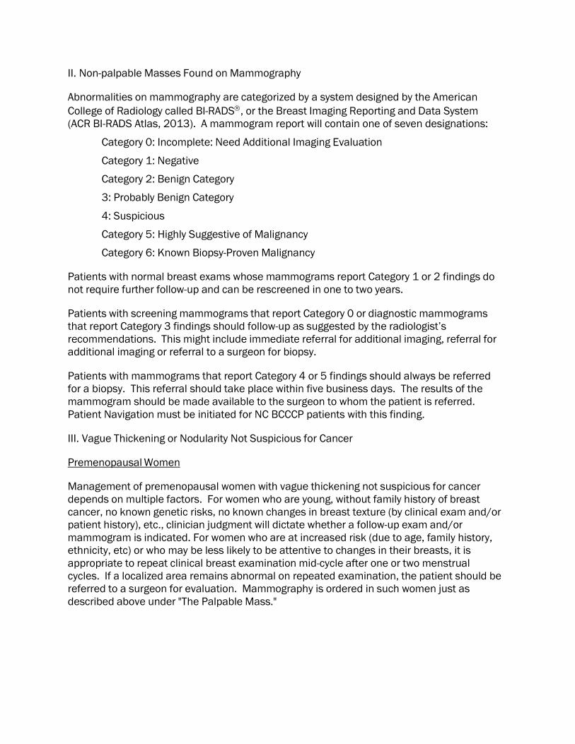
II. Non-palpable Masses Found on Mammography
Abnormalities on mammography are categorized by a system designed by the American
College of Radiology called BI-RADS, or the Breast Imaging Reporting and Data System
(ACR BI-RADS Atlas, 2013). A mammogram report will contain one of seven designations:
Category 0: Incomplete: Need Additional Imaging Evaluation
Category 1: Negative
Category 2: Benign Category
3: Probably Benign Category
4: Suspicious
Category 5: Highly Suggestive of Malignancy
Category 6: Known Biopsy-Proven Malignancy
Patients with normal breast exams whose mammograms report Category 1 or 2 findings do
not require further follow-up and can be rescreened in one to two years.
Patients with screening mammograms that report Category 0 or diagnostic mammograms
that report Category 3 findings should follow-up as suggested by the radiologist’s
recommendations. This might include immediate referral for additional imaging, referral for
additional imaging or referral to a surgeon for biopsy.
Patients with mammograms that report Category 4 or 5 findings should always be referred
for a biopsy. This referral should take place within five business days. The results of the
mammogram should be made available to the surgeon to whom the patient is referred.
Patient Navigation must be initiated for NC BCCCP patients with this finding.
III. Vague Thickening or Nodularity Not Suspicious for Cancer
Premenopausal Women
Management of premenopausal women with vague thickening not suspicious for cancer
depends on multiple factors. For women who are young, without family history of breast
cancer, no known genetic risks, no known changes in breast texture (by clinical exam and/or
patient history), etc., clinician judgment will dictate whether a follow-up exam and/or
mammogram is indicated. For women who are at increased risk (due to age, family history,
ethnicity, etc) or who may be less likely to be attentive to changes in their breasts, it is
appropriate to repeat clinical breast examination mid-cycle after one or two menstrual
cycles. If a localized area remains abnormal on repeated examination, the patient should be
referred to a surgeon for evaluation. Mammography is ordered in such women just as
described above under "The Palpable Mass."

Postmenopausal Women
Postmenopausal women with a questionable clinical breast examination should be referred
for imaging and surgical evaluation per the recommendations above under “The Palpable
Mass.”
IV. Nipple Discharge or Skin Changes
The nature of nipple discharges should be defined by a careful patient history. A patient with
a spontaneous, single duct discharge, even when non-bloody, is potentially pathologic and
should be referred to a surgeon. However, bilateral milky, green or grey nipple discharge is
typically benign. Medical work-up of galactorrhea may be appropriate for persistent milky
discharge.
Patients with any skin breakdown require treatment and follow up. A trial of topical
treatment (e.g. steroid cream) may be appropriate for a limited amount of time with re-
evaluation in 1-2 weeks. Patients without complete resolution or with recurring symptoms
require a surgery referral. Biopsy of the nipple may be necessary to differentiate eczema of
the nipple from Paget’s disease (cancer of the nipple) in certain cases.
V. Breast Pain
Breast pain includes any discomfort or pain of the breast, such as premenstrual tenderness.
Breast pain is typically benign. The question is how tolerable (or intolerable) the pain is for
the woman. There are many causes of breast pain, including hormonal fluctuations related
to menstruation or pregnancy, where some degree of pain is normal. With menopause
breast tenderness often goes away, unless a woman is taking hormone replacement therapy.
Other causes of breast pain include fibrocystic breast changes, mastitis (blocked or infected
milk duct), premenstrual syndrome (PMS), alcoholism with liver damage and injury. There
are certain medications that cause breast pain including digitalis preparations, aldomet,
aldactone and other potassium-sparing diuretics, anadrol and chlorpromazine.
If the clinical breast examination is normal, reassure the patient and explain the hormonal
causes of breast pain. Typically, the patient's mind is put at ease. The provider may
recommend a trial of non-narcotic analgesics such as acetaminophen, or topical or oral
NSAIDs. The use of a well-fitting bra that provides good support, or the use of warm
compresses may also be recommended. Although there is no clear evidence in the
literature that shows reducing dietary caffeine, salt or fat improves breast pain, some
women report anecdotal benefits from these changes. If the pain persists, a repeat breast
exam and mammogram may be provided.
If the follow-up breast examination and mammogram are normal and breast pain persists,
refer the woman to a breast specialist for further evaluation. For women with breast pain
who have a palpable mass or mammographically detected abnormality, the work-up is
identical to that of women with palpable mass. Though breast cancers are usually painless,

the presence of pain cannot reliably rule out breast cancer. There are a small percentage of
breast cancers that present as painful or uncomfortable.
VI. Special Considerations
Fibrocystic Breasts - Fibrocystic changes are the most common cause of non-cancerous
breast lumps (Mayo Clinic, 2016). They affect at least 50% of women at some point in their
lives, most commonly between the ages of 30 and 50. Fibrocystic breasts are usually not a
risk factor for breast cancer, but women with fibrocystic breasts may have diffusely lumpy
breasts, making detection of underlying breast cancer more difficult. If there is any
uncertainty about clinical breast exam in a patient with fibrocystic breasts, the patient may
be referred for mammography, ultrasound, and/or a consultation with a breast specialist.
Fibroadenoma - A noncancerous rubbery mass in the breast that is usually painless and
moves around easily when palpated. Fibroadenomas cannot be diagnosed with
mammography, sonography or histopathology. Fibroadenomas can only be diagnosed with a
biopsy.
Pregnant and Lactating Women - These women often experience breast tenderness and
engorgement, which can make detection of masses more difficult. Clogged pores and/ or
ducts may also be the culprit of breast pain or tenderness and it is recommended that if this
is thought to be a problem that adequate time be given for this to resolve prior to
intervention. Lactating women should empty their breasts prior to a mammogram.
If a palpable abnormality is found on CBE, diagnostic evaluation with ultrasound and
mammography may need to be performed. Ultrasound is the first line imaging modality to
be used in these patients since most of these findings are benign. However, if a suspicious
mass/mass highly suggestive of malignancy is detected with ultrasound then bilateral
diagnostic mammogram is recommended for further evaluation/extent of disease.
Mammography is thought to be safe to perform during pregnancy. The amount of radiation
needed for a digital breast mammogram is small and focused only on the breast. A lead
shield is placed over the lower part of the body for protection. Still, scientists cannot be
certain about the effects of radiation on an unborn baby (ACS, 2016). Mammograms should
only be used to evaluate distinct, dominant masses. The radiologist should always be
informed if the woman is pregnant. A referral to a breast surgeon should be made for a
definitive diagnosis.
Other Patients with a Difficult Breast Examination
Some women may have a difficult clinical examination that requires further evaluation. This
group may include:
• Women who have had breast reduction surgery
• Women with multiple previous biopsies and scarring
• Women with breast implants
• Women who have had a mastectomy

20 NC DHHS • Division of Public Health • The Breast Screening Manual: A Guide for Health Departments and Providers / December 2016
If a clinician is unsure of the significance of findings on clinical examination in any of the
above situations, a referral to a mammography or breast specialist should be made.
Algorithm for Management of Findings on Clinical Breast Screening (both screen and
diagnostic are on the diagram)
(See next page)

NC DHHS • Division of Public Health • The Breast Screening Manual: A Guide for Health Departments and Providers / December 2016
21
Algorithm for Management of Findings on Breast Screening
NC DHHS • Division of Public Health • The Breast Screening Manual: A Guide for Health Departments and Providers / December 2016
22

TAB 5: Interpretation of Mammography Reports

23 NC DHHS • Division of Public Health • The Breast Screening Manual: A Guide for Health Departments and Providers / December 2016
ORGANIZATION OF THE MAMMOGRAPHY REPORT REPORTING
SYSTEM (ACR BI-RADS ATLAS, 2013)
The reporting system should be concise and organized using the following structure. If a
comparison to previous studies is made, this should be indicated in the report.
A. INDICATION FOR EXAMINATION
1. A brief description of the indication for examination.
a. Screening for an asymptomatic woman
b. Recall for additional work-up of an abnormal finding
c. Evaluation of a clinical finding, specifying the finding and its location
d. Follow-up of a probably benign lesion or cancer treated with breast conservation
2. If an implant is present, both standard and implant-displaced views will be indicated.
B. SUCCINCT DESCRIPTION OF THE OVERALL BREAST COMPOSITION
1. Breast density helps indicate the relative possibility that a lesion could be obscured
by normal tissue.
2. Mammography does not identify all breast cancers. Clinical breast examination
(CBE) is an important element of screening. Abnormal CBE findings should never be
ignored, and may be especially important in dense breasts.
3. If breasts are not equally dense, the denser breast should be used to categorize
composition
4. The categories of density are:
a. Almost entirely fatty
b. Scattered areas of fibroglandular density
c. Heterogenously dense. If some areas are relatively dense but other areas are
primarily fatty, a second sentence will describe the location(s) of the denser
tissue. This category may obscure small masses.
d. Extremely dense. Mammography sensitivity is lowest in this category.
C. DESCRIPTION OF ANY IMPORTANT FINDINGS THAT MAY BE SUSPICIOUS FOR CANCER
1. Mass:
a. Size
b. Morphology (shape, margin)
c. Density
d. Associated calcifications
e. Associated features
f. Location
2. Calcifications:
a. Morphology — describe typically benign type or describe shape of particles
b. Distribution (may not be appropriate for typically benign calcifications)
c. Associated features
d. Location
24 NC DHHS • Division of Public Health • The Breast Screening Manual: A Guide for Health Departments and Providers / December 2016
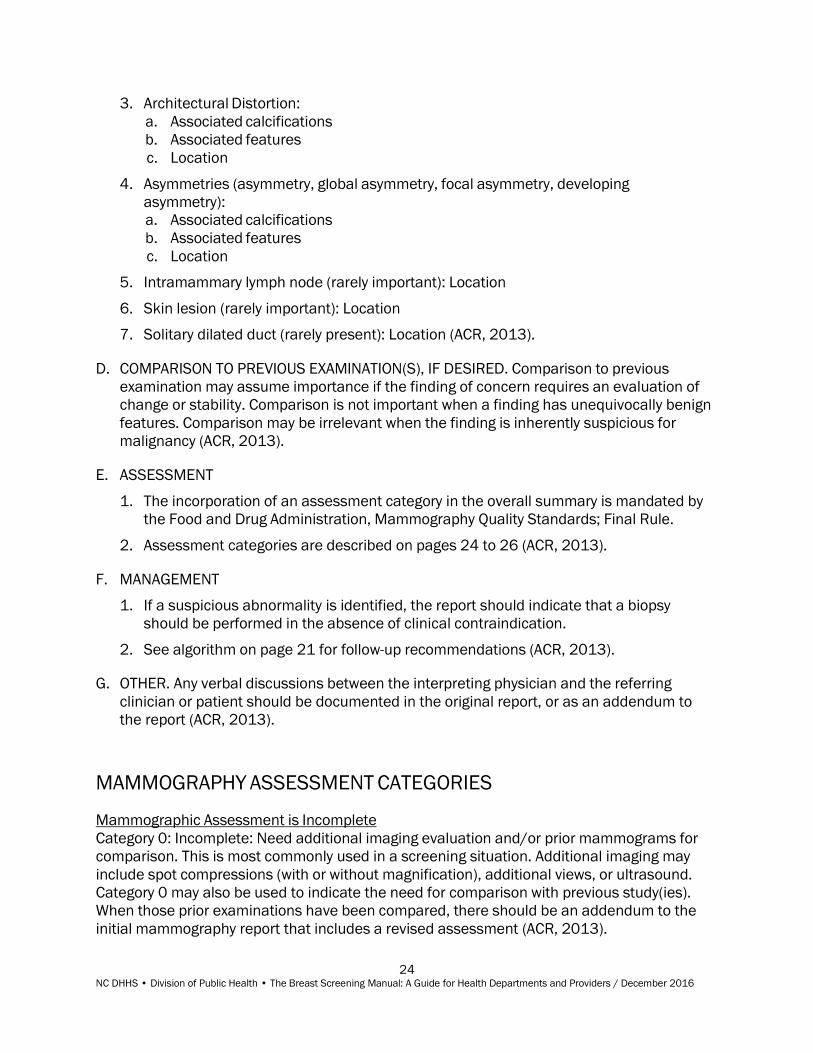
3. Architectural Distortion:
a. Associated calcifications
b. Associated features
c. Location
4. Asymmetries (asymmetry, global asymmetry, focal asymmetry, developing
asymmetry):
a. Associated calcifications
b. Associated features
c. Location
5. Intramammary lymph node (rarely important): Location
6. Skin lesion (rarely important): Location
7. Solitary dilated duct (rarely present): Location (ACR, 2013).
D. COMPARISON TO PREVIOUS EXAMINATION(S), IF DESIRED. Comparison to previous
examination may assume importance if the finding of concern requires an evaluation of
change or stability. Comparison is not important when a finding has unequivocally benign
features. Comparison may be irrelevant when the finding is inherently suspicious for
malignancy (ACR, 2013).
E. ASSESSMENT
1. The incorporation of an assessment category in the overall summary is mandated by
the Food and Drug Administration, Mammography Quality Standards; Final Rule.
2. Assessment categories are described on pages 24 to 26 (ACR, 2013).
F. MANAGEMENT
1. If a suspicious abnormality is identified, the report should indicate that a biopsy
should be performed in the absence of clinical contraindication.
2. See algorithm on page 21 for follow-up recommendations (ACR, 2013).
G. OTHER. Any verbal discussions between the interpreting physician and the referring
clinician or patient should be documented in the original report, or as an addendum to
the report (ACR, 2013).
MAMMOGRAPHY ASSESSMENT CATEGORIES
Mammographic Assessment is Incomplete
Category 0: Incomplete: Need additional imaging evaluation and/or prior mammograms for
comparison. This is most commonly used in a screening situation. Additional imaging may
include spot compressions (with or without magnification), additional views, or ultrasound.
Category 0 may also be used to indicate the need for comparison with previous study(ies).
When those prior examinations have been compared, there should be an addendum to the
initial mammography report that includes a revised assessment (ACR, 2013).

25 NC DHHS • Division of Public Health • The Breast Screening Manual: A Guide for Health Departments and Providers / December 2016
Mammography Assessment is Complete
Category 1: Negative. This is a normal exam and there is nothing to comment on (ACR,
2013).
Category 2: Benign. Like Category 1, this category indicates there is no mammographic
evidence of malignancy, and routine mammographic screening is recommended. However,
the interpreting physician has chosen to describe one or more characteristically benign
findings such as:
• Involuting calcified fibroadenomas
• Skin calcifications
• Metallic foreign bodies such as core biopsy and surgical clips
• Fat-containing lesions such as oil cysts, lipomas, galactoceles and mixed-density
hamartomas
• Intramammary lymph nodes
• Vascular calcifications
• Implants
• Architectural distortion clearly related to prior surgery (ACR, 2013).
Category 3: Probably benign. A finding in this category indicates less than 2% likelihood of
malignancy, but greater likelihood than a characteristically benign finding. It is not expected
to change over the period of surveillance, but the interpreting physician prefers to establish
stability before recommending routine screening.
Commonly reported probably benign findings include:
• Non-calcified circumscribed solid mass
• Focal asymmetry
• Solitary group of punctate calcifications
This category should never be used to assess an initial screening mammogram without
obtaining a complete diagnostic imaging evaluation. It should also not be used in the
presence of a palpable lesion; nor should it be used for a finding that is either new or
increasing in size or extent.
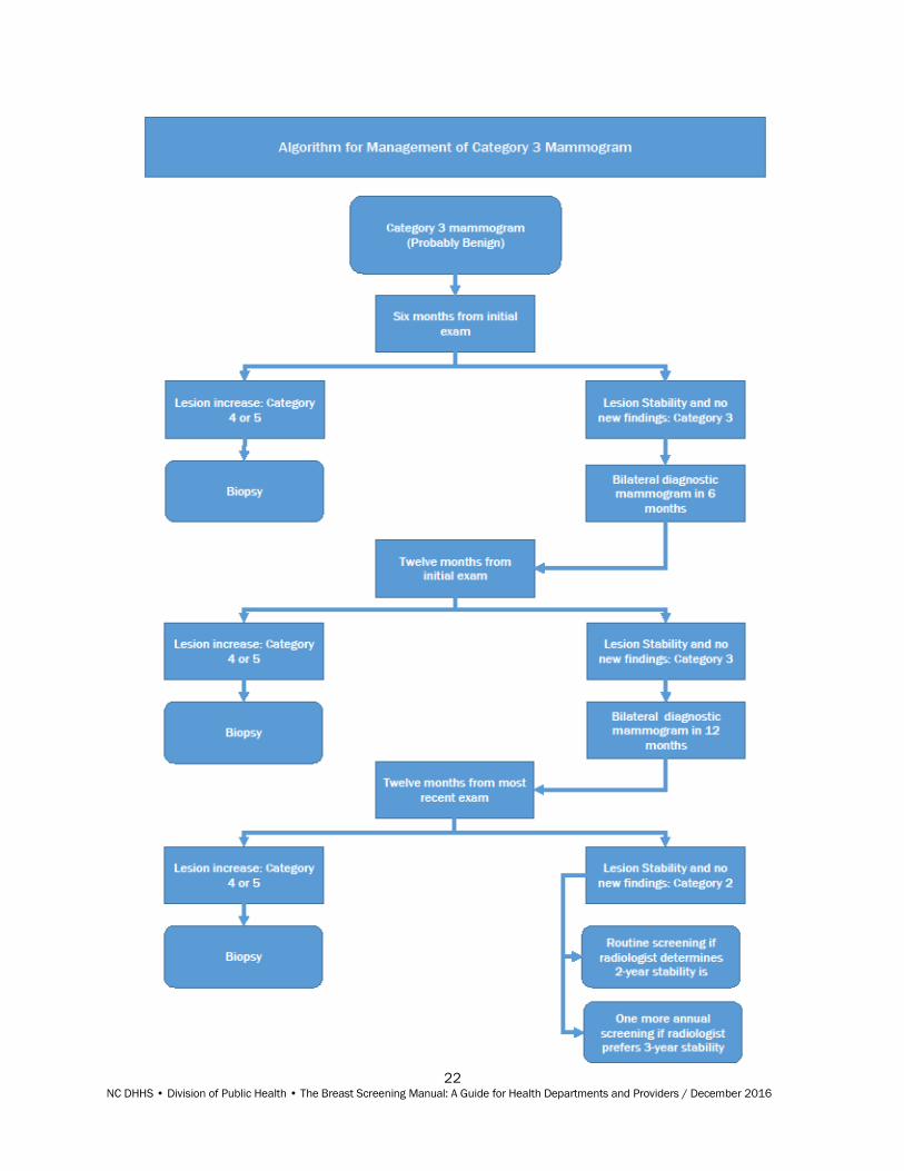
Most category 3 findings will be managed with short term (six months) mammographic
follow-up, followed by additional mammograms until long term (2-3 years) stability has been
established. Patient preference or overriding clinical concern may lead to biopsy rather than
short term follow-up (ACR, 2013).
Category 4: Suspicious. This category indicates findings that do not have the classic
appearance of malignancy, but are sufficiently suspicious to recommend biopsy. The
likelihood of a Category 4 finding being malignant ranges from greater than 2% to less than
95%. To narrow this range and better guide patients and clinicians regarding management,
Category 4 findings may be subdivided into additional categories:
• 4A: low suspicion for malignancy (>2% - 10% likelihood)
• 4B: intermediate suspicion of malignancy (>10% - 50% likelihood)
• 4C: moderate concern, but not classic for malignancy (>50% - <95% likelihood) (ACR,
2013).

26 NC DHHS • Division of Public Health • The Breast Screening Manual: A Guide for Health Departments and Providers / December 2016
Category 5: Highly Suggestive of Malignancy. Findings in this category have at least a 95%
likelihood of being malignant. This category is typically established after an inconclusive
percutaneous biopsy, and will likely result in a recommendation for repeat biopsy (usually
surgical) (ACR, 2013).
Category 6: Known Biopsy-Proven Malignancy. This category is reserved for examinations
performed after biopsy proof of malignancy. It describes imaging performed after a
percutaneous biopsy but prior to a complete surgical excision, in which there are no
mammographic abnormalities other than the known cancer (ACR, 2013).

TAB: 6: APPENDICES

TAB 7: Appendix A
27 NC DHHS • Division of Public Health • The Breast Screening Manual: A Guide for Health Departments and Providers / December 2016
APPENDIX A - BREAST CANCER GLOSSARY
A
Abscess
An enclosed collection of pus in tissues, organs or confined spaces in the body. An abscess
is a sign of infection and is usually swollen and inflamed.
Adenoma
A noncancerous tumor.
Adjunct agent
In cancer therapy, a drug or substance used in addition to the primary therapy.
Adjuvant therapy
Treatment given after the primary treatment to increase the chances of a cure. Adjuvant
therapy may include chemotherapy, radiation therapy, hormone therapy, or biological
therapy.
Areola
The area of dark-colored skin on the breast that surrounds the nipple.
Aspiration
Removal of fluid or tissue through a needle.
Axilla
The underarm or armpit.
Axillary dissection
Surgery to remove lymph nodes found in the armpit; axillary node dissection.
Axillary lymph node
A lymph node in the armpit region that drains lymph channels from the breast.
Axillary lymph node dissection
Surgery to remove lymph nodes found in the armpit region; axillary dissection.
B
Benign
Not cancerous. Benign tumors may grow larger but do not spread to other parts of the body.
Benign breast disease
A common condition marked by benign (noncancerous) changes in breast tissue. These
changes may include irregular lumps or cysts, breast discomfort, sensitive nipples, and
itching. These symptoms may change through the menstrual cycle and usually stop after
menopause; fibrocystic breast disease; fibrocystic breast changes; mammary dysplasia.

28 NC DHHS • Division of Public Health • The Breast Screening Manual: A Guide for Health Departments and Providers / December 2016
BI-RADS
Breast Imaging Reporting and Data System. The method used by radiologists to interpret
and report in a standardized manner the results of mammography, ultrasound, and MRI
used in breast cancer screening and diagnosis.
Bilateral
Affecting both the right and left sides of the body.
Bilateral prophylactic mastectomy
Surgery to remove both breasts to reduce the risk of developing breast cancer; preventive
mastectomy.
BRCA 1
A gene on chromosome 17 that normally helps to suppress cell growth. A person who
inherits an altered version of the BRAC 1 gene has a higher risk of getting breast and
ovarian cancer.
BRCA 2:
A gene that normally acts to restrain the growth of cells in the breast and ovary but which,
when mutated, may predispose to breast cancer and to ovarian cancer.
Breast cancer in situ
Abnormal cells that are confined to the ducts or lobules in the breast. There are two forms,
ductal carcinoma in situ (DCIS) and lobular carcinoma in situ (LCIS).
Breast density
Describes the relative amount of different tissue present in the breast. A dense breast has
less fat than glandular and connective tissue. Mammogram films of breasts with higher
density are harder to read and interpret than those of less dense breasts.
Breast implant
A silicone gel-filled or saline-filled sac placed under the breast tissue or chest muscle to
augment or restore breast shape.
Breast reconstruction
Surgery to rebuild the shape of the breast after a mastectomy.
Breast self-exam
An exam by a woman of her breast to check for lumps or other changes.
Breast conserving surgery and Breast-sparing surgery
An operation to remove the breast cancer but not the breast itself. Types of breast-
conserving surgery include lumpectomy (removal of a lump), quadrantectomy (removal of
one quarter, or quadrant of the breast), and segmental mastectomy (removal of the cancer
as well as some of the breast tissue around the tumor and the lining over the chest muscles
below the tumor).

29 NC DHHS • Division of Public Health • The Breast Screening Manual: A Guide for Health Departments and Providers / December 2016
C
Calcification
Deposits of calcium in the tissue. Calcification in the breast can be seen on a mammogram,
but cannot be detected by touch. There are two types of breast calcifications,
macrocalcifications and microcalcification. Macrocalfications are large deposits and are
usually not related to cancer. Microcalcifications are specks of calcium that may be found in
an area of rapidly dividing cells. Many microcalcifications clustered together or in a
segmental distribution may be a sign of cancer.
Carcinoma
Cancer that begins in the skin or in tissues that line or cover internal organs.
Carcinoma in situ
Epithelial cancer that lies above the basement membrane and has not spread to nearby
lymphatic blood vessels’ deeper structures.
Chemotherapy
Treatment with drugs that kill cancer cells.
Clinical Breast exam
An exam of the breast performed by a health care provider to check for lumps or other
changes.
Clinical trial
A type of research study that tests how well new medical approaches work in people. These
studies test new methods of screening, prevention, diagnosis, or treatment of a disease; a
clinical study.
Complementary and alternative medicine (CAM)
Forms of treatment that are used in addition to (complementary) or instead of (alternative)
standard treatments. These practices generally are not considered standard medical
approaches. Varying quality and quantity of research are available to substantiate the
safety and effectiveness of CAM. CAM may include dietary supplements, mega dose
vitamins, herbal preparations, special teas, acupuncture, massage therapy, magnet therapy,
spiritual healing, and meditation.
Core biopsy
The removal of a tissue sample with a large (typically 11 - 18 gauge) needle for examination
under a microscope.
Cyst
A sac or capsule in the body. It may be filled with fluid or other materials.
30 NC DHHS • Division of Public Health • The Breast Screening Manual: A Guide for Health Departments and Providers / December 2016
D
Diagnosis
The process of identifying a disease by the signs and symptoms.
Diagnostic mammogram
X-ray of the breast to check for breast cancer after a lump or other sign or symptom of
breast cancer has been found.
Digital mammography
A technique that uses a computer, rather than x-ray film, to record images of the breast.
Ductal carcinoma
The most common type of breast cancer. It begins in the cells that line the milk ducts in the
breast.
Ductal carcinoma in situ
A noninvasive, precancerous condition in which abnormal cells are found in the lining of a
breast duct. The abnormal cells have not spread outside the duct to the tissues in the
breast. In some cases, ductal carcinoma in situ may become invasive cancer and spread to
other tissues, although it is not currently known how to predict which lesions will become
invasive; intraductal carcinoma.
Ductal ectasia
Mammary duct ectasia occurs when a milk duct beneath the nipple widens, the duct walls
thicken and the duct fills with fluid. The milk duct may become blocked or clogged with a
thick, sticky substance. The condition often causes no symptoms, but some women may
have nipple discharge, breast tenderness or inflammation of the clogged duct (periductal
mastitis) (Mayo Clinic, 2016).
Ductal lavage
A method used to collect cells from milk ducts in the breast. A hair-size catheter (tube) is
inserted into the nipple, and a small amount of salt water is released into the duct. The
water picks up breast cells and is removed. The cells are checked under a microscope.
Ductal lavage may be used in addition to clinical breast examination and mammography to
detect breast cancer.
Dysplasia
Cells that look abnormal under a microscope but are not cancer.
E
Endocrine Therapy
Treatment that adds, blocks, or removes hormones. For certain conditions (such as diabetes
or menopause), hormones are given to adjust low hormone levels. To slow or stop the
growth of certain cancers (such as prostate and breast cancer), synthetic hormones or other

31 NC DHHS • Division of Public Health • The Breast Screening Manual: A Guide for Health Departments and Providers / December 2016
drugs may be given to block the body’s natural hormones. Sometimes surgery is needed to
remove the gland that makes a certain hormone; hormonal therapy; hormone therapy; and
hormone treatment (Cancer Dictionaries, 2016).
Estrogen
A type of hormone made by the body that helps develop and maintain female sex
characteristics and the growth of long bones. Estrogen can also be made in the laboratory.
These estrogens may be used as a type of birth control and to treat symptoms of
menopause, menstrual disorder, osteoporosis, and other disorders.
Estrogen receptor
A protein found inside the cells of the female reproductive tissue, some other types of
tissue, and some cancer cells. The hormone estrogen will bind to the receptors inside the
cells and may cause the cells to grow.
F
Fibroadenoma
A noncancerous rubbery mass in the breast that is usually painless and moves around easily
on palpation.
Fibrocystic breast changes
A common condition marked by benign (noncancerous) changes in breast tissue. These
changes may include irregular lumps or cysts, breast discomfort, sensitive nipples, and
itching. These symptoms may change throughout the menstrual cycles and usually stop
after menopause; benign breast disease; fibrocystic breast changes; and mammary
dysplasia.
Fine-needle aspiration
The removal of tissue or fluid with a small needle for examination under a microscope;
needle biopsy.
G
Gene
The functional and physical unit of heredity passed from parent to offspring. Genes are
pieces of DNA and most genes contain the information for making a specific protein.
Gland
An organ that makes one or more substances, such as hormones, digestive juices, sweat,
tears, saliva, or milk. Endocrine glands release the substances directly into a duct or
opening inside or outside the body.

32 NC DHHS • Division of Public Health • The Breast Screening Manual: A Guide for Health Departments and Providers / December 2016
H
HER2/neu
Human epidermal growth factor receptor 2. The HER/neu (or C-erb B-2) protein is involved
in the growth of some cancer cells.
HER2/neu gene
The gene that makes the human epidermal growth factor receptor 2. The protein produced
is HER2/neu, which is involved in the growth of some cancer cells; c-erbB-2.
Hormone
A chemical made by glands in the body. Hormones circulate in the bloodstream and control
the actions of certain cells or organs. Some hormones can also be made in a laboratory.
Hormone receptor
A protein on the surface of a cell that binds to a specific hormone. The hormone causes
many changes to take place in the cell.
Hormone replacement therapy
HRT. Hormones (estrogen, progesterone, or both) given to women after menopause to
replace the hormones no longer produced by the ovaries; menopausal hormone therapy.
Hormone therapy
Treatment that adds, blocks, or removes hormones. For certain conditions (such as diabetes
or menopause), hormones are given to adjust low hormone levels. To slow or stop the
growth of certain cancers (such as prostate and breast cancer), synthetic hormones or other
drugs may be given to block the body's natural hormones. Sometimes surgery is needed to
remove the gland that makes a certain hormone; hormonal therapy; hormone therapy;
endocrine therapy.
I
Immunotherapy
Treatment to stimulate or restore the ability of the immune system to fight cancer, infections
and other diseases. Also, used to lessen certain side effects that may be caused by cancer
treatment; biological therapy, biotherapy, or biological response modifier (BRM) therapy.
Incidence
The number of new cases of a disease diagnosed each year.
Incisional biopsy
A surgical procedure in which a portion of a lump or suspicious area is removed for
diagnosis. The tissue is then examined under a microscope.

33 NC DHHS • Division of Public Health • The Breast Screening Manual: A Guide for Health Departments and Providers / December 2016
Intraductal carcinoma
A noninvasive, precancerous condition in which abnormal cells are found in the lining of a
breast duct. The abnormal cells have not spread outside the duct to other tissues in the
breast. In some cases, intraductal carcinoma may become invasive cancer and spread to
other tissues, although it is not known how to predict which lesions become invasive ductal
carcinoma in situ.
Invasive cancer
Cancer that has spread beyond the layer of tissue in which it developed and is growing into
surrounding, healthy tissues; infiltrating cancer.
L
LCIS
Lobular carcinoma in situ. Abnormal cells found in the lobules of the breast. The condition
is considered nonmalignant; however, having lobular carcinoma in situ increases one's risk
of developing breast cancer in either breast.
Lobe
A portion of an organ, such as the liver, lungs, breast, thyroid, or brain.
Lobular carcinoma
Cancer that begins in the lobules (the glands that make milk) of the breast. Lobular
carcinoma in situ (LCIS) is a condition in which abnormal cells are found only in the lobules.
When cancer has spread from the lobules to surrounding tissues, it is called invasive lobular
carcinoma. LCIS in one breast increases the risk of developing invasive cancer in either
breast.
Lymph node
A rounded mass of lymphatic tissue that is surrounded by a capsule of connective tissue.
Lymph nodes filter lymph (lymphatic fluid), and they store lymphocytes (white blood cells).
Lymph node mapping
The use of dyes and radioactive substances to identify lymph nodes that may contain tumor
cells; lymphatic mapping.
Lymphedema
A condition in which excess fluid collects in tissue and causes swelling. It may occur in the
arm or leg after lymph vessels or lymph nodes in the underarm or groin are removed or
treated with radiation.

34 NC DHHS • Division of Public Health • The Breast Screening Manual: A Guide for Health Departments and Providers / December 2016
M
Magnetic resonance imaging (MRI)
A procedure in which radio waves and a powerful magnet linked to a computer are used to
create detailed pictures of areas inside the body. The pictures can show the difference
between normal and diseased tissue. MRI makes better images of organs and soft tissue
than other scanning techniques, such as CT or x-ray. MRI is especially useful for imaging the
brain, spine, the soft tissue of joints, and inside bones. Also, called nuclear magnetic
resonance imaging.
Malignant
Cancerous. Malignant tumors can invade and destroy nearby tissue and spread to other
parts of the body.
Mammogram
An x-ray of the breast.
Mammography
The use of x-rays to create a picture of the breast.
Margin
The edge or border of the tissue removed in cancer surgery. The margin is described as
negative or clean when the pathologist finds no cancer cells at the edge of the tissue,
suggesting that all the cancer has been removed. The margin is described as positive or
involved when the pathologist finds cancer cells at the edge of the tissue, suggesting that all
the cancer has not been removed.
Mastectomy
Surgery to remove the breast (or as much of the breast tissue as possible).
Menarche
A young woman's first menstrual period.
Menopause
The time of life when a woman's menstrual periods stop. A woman is in menopause when
she hasn't had a period for 12 months in a row; "change of life."
Metastasis
The spread of cancer from one part of the body to another. A tumor formed by cells that
have spread is called a "metastatic tumor" or a "metastasis." The metastatic tumor contains
cells that are like those in the original (primary) tumor. The plural form of metastasis is
metastases (meh-TAS-ta-seez).
35 NC DHHS • Division of Public Health • The Breast Screening Manual: A Guide for Health Departments and Providers / December 2016
Microcalcification
A tiny deposit of calcium in the breast that cannot be felt but can be detected on a
mammogram. Grouped, regional and segmental distribution of these very small specks of
calcium may indicate that cancer is present.
N
Needle biopsy
The removal of tissue or fluid with a needle for examination under a microscope. Also, called
fine-needle aspiration.
Needle-localized biopsy
A procedure that uses very thin needles or guide wires to mark the location of an abnormal
area of tissue so that it can be surgically removed. An imaging device is used to place the
wire in or around the abnormal area. Needle localization is used when the doctor cannot
feel the mass of abnormal tissue.
Neoadjuvant therapy
Treatment given before the primary treatment. Examples of neoadjuvant therapy includes
chemotherapy, radiation therapy, and hormone therapy.
Nipple discharge
Fluid coming from the nipple.
Nonmalignant
Not cancerous.
O
Oncologist
A doctor who specializes in treating cancer. Some oncologists specialize in a cancer
treatment. For example, a radiation oncologist specializes in treating cancer with radiation.
Oncology
A study of cancer.
P
Palpation
Examination by pressing on the surface of the body to feel the organs or tissues underneath.
Pathologist
A doctor who identifies diseases by studying cells and tissues under a microscope.

36 NC DHHS • Division of Public Health • The Breast Screening Manual: A Guide for Health Departments and Providers / December 2016
Pathology report
The description of cells and tissues made by a pathologist based on microscopic evidence,
and sometimes used to make a diagnosis of a disease.
Patient Navigation
Patient Navigation was founded by Harold P. Freeman, M.D. in 1990, when he initiated and
developed the first Patient Navigation program in Harlem to reduce disparities in access to
diagnosis and treatment of cancer, particularly among poor and uninsured people. The core
principles of the concept which focus upon saving lives from cancer and chronic diseases,
include informing patients about the need for certain recommended examinations and
provide timely access to such examinations; eliminating barriers to timely care across the
entire health care continuum; and to eliminate barriers to timely diagnoses and treatment in
patients who have abnormal or suspicious findings (Harold P Freeman Institute, 2016).
Prevention
In medicine, action taken to decrease the chances of getting a disease. For example,
cancer prevention includes avoiding risk factors (such as smoking, obesity, lack of exercise,
and radiation exposure) and increasing protective factors (such as getting regular physical
activity, staying at a healthy weight, and eating a healthy diet).
Progesterone
A female hormone.
Progesterone receptor (PR)
A protein found inside the cells of the female reproductive tissue, some other types of
tissue, and some cancer cells. The hormone progesterone will bind to receptors inside the
cells and may cause the cells to grow.
Prognosis
The likely outcome or course of a disease; the chance of recovery or recurrence.
Prophylactic mastectomy
Surgery to reduce the risk of developing breast cancer by removing one or both breasts
before disease develops; a preventive mastectomy.
Prosthesis
A device that replaces a body part.
Punctate
Having small pinpoint calcium deposits.
R
Radiation
Energy released in the form of particles or electromagnetic waves. Common sources of
radiation include radon gas, cosmic rays from outer space, and medical x-rays.

37 NC DHHS • Division of Public Health • The Breast Screening Manual: A Guide for Health Departments and Providers / December 2016
Radiation oncologist
A doctor who specializes in using radiation to treat cancer.
Radiation therapy
The use of high-energy radiation from x-rays, gamma rays, neutrons and other sources to kill
cancer cells and shrink tumors. Radiation may come from a machine outside the body
(external-beam radiation therapy), or it may come from radioactive material placed in the
body near cancer cells (internal radiation therapy, implant radiation, or brachytherapy).
Systemic radiation therapy uses a radioactive substance, such as radiolabeled monoclonal
antibody, that circulates throughout the body; radiotherapy.
Radical mastectomy
Surgery for breast cancer in which the breast, chest muscles, and all the lymph nodes under
the arm are removed. For many years, this was the breast cancer operation used most
often, but it is used rarely now. Doctors consider radical mastectomy only when the tumor
has spread to the chest muscles; the Halsted radical mastectomy.
Radiologist
A doctor who specializes in creating and interpreting pictures of areas inside the body. The
pictures are produced with x-rays, sound waves, or other types of energy.
Reconstructive surgeon
A doctor who can surgically reshape or rebuild (reconstruct) a part of the body, such as a
woman's breast after surgery for breast cancer.
Recurrence
Cancer that has returned after an increment of time during which the cancer could not be
detected. The cancer may come back to the same place as the original (primary) tumor or to
another place in the body; recurrent cancer.
Remission
A decrease in or disappearance of signs and symptoms of cancer. In partial remission,
some, but not all, signs and symptoms of cancer have disappeared. In complete remission,
all signs and symptoms of cancer have disappeared, although cancer still may be in the
body.
Risk factor
Something that may increase the chance of developing a disease. Some examples of risk
factors for cancer include age, a family history of certain cancers, use of tobacco products,
certain eating habits, obesity, lack of exercise, exposure to radiation or other cancer-causing
agents, and certain genetic changes.
S
Scintimammography
A type of breast imaging test that is used to detect cancer cells in the breasts of some
women who have had abnormal mammograms, or who have dense breast tissue.

38 NC DHHS • Division of Public Health • The Breast Screening Manual: A Guide for Health Departments and Providers / December 2016
Scintimammography is not used for screening, or in place of a mammogram. In this test, a
woman receives an injection of a small amount of a radioactive substance called
technetium 99, which is taken up by the cancer cells, and a gamma camera is used to take
pictures of the breasts.
Screening
Checking for disease when there are no symptoms.
Screening mammogram
An x-ray of the breast used to detect breast changes in women who have no signs of breast
cancer.
Sentinel lymph node mapping
The use of dyes and radioactive substances to identify the first lymph node to which cancer
is likely to spread from a primary tumor. Cancer cells may appear first in the sentinel node
before spreading to other lymph nodes and other places in the body.
Sonogram
A computer picture of areas inside the body created by bouncing high-energy sound waves
(ultrasound) off internal tissues or organs; an ultrasound.
Stage
The extent of a cancer in the body. Staging is usually based on where the cancer is located,
how much the cancer has grown, and if or where it has spread. Diagnostic testing is utilized
to determine the stage of cancer, so staging may not be complete until all testing is
completed. Completing the staging protocol helps to determine the treatment that is best
for the prognosis (American Joint Committee on Cancer [AJCC], 2010, p.347- 76).
Stem cell
A cell from which other types of cells develop. Blood cells develop from blood-forming stem
cells.
Stereotactic biopsy
A biopsy procedure that uses a computer and a 3-dimensional scanning device to find a
tumor site and guide the removal of tissue for examination under a microscope.
Surgical oncologist
A doctor who performs biopsies and other surgical procedures in cancer patients.
T
Tamoxifen
A drug used to treat breast cancer, and to prevent it in women who are at high risk of
developing breast cancer. Tamoxifen blocks the effects of the hormone estrogen in the
breast. It belongs to the family of drugs called antiestrogens.
Tissue flap reconstruction
39 NC DHHS • Division of Public Health • The Breast Screening Manual: A Guide for Health Departments and Providers / December 2016
A type of breast reconstruction in which a flap of tissue is surgically moved from another
area of the body to the chest, and formed into a new breast mound.
Tumor
An abnormal mass of tissue that results when cells divide more than they should or do not
die when they should. Tumors may be benign (non-cancerous) or malignant (cancerous);
neoplasm.
Tumor grade
The degree of abnormality of cancer cells, a measure of differentiation. The extent to which
cancer cells are similar in appearance and function to healthy cells of the same tissue type.
The degree of differentiation often relates to the clinical behavior of the specific tumor.
Based on the microscopic appearance of cancer cells, pathologists commonly describe
tumor grade by four degrees of severity: Grades 1, 2, 3, and 4 (1 low grade … 4 high grade).
U
Ultrasound
A procedure in which sound waves (ultrasound) are bounced off internal tissue or organs
and make echoes. The echo patterns are shown on the screen of an ultrasound machine,
forming a picture of the body tissues called a sonogram; ultrasonography.
X
X-ray
A type of high-energy radiation. In low doses, x-rays are used to diagnose diseases by
making pictures of the inside of the body. In high doses, x-rays are used to treat cancer. No
longer widely available.
NCI, 2016. Dictionary of Cancer Terms. Retrieved on April 7
2016 from http://www.cancer.gov/publications/dictionaries

TAB 8: Appendix B

40 NC DHHS • Division of Public Health • The Breast Screening Manual: A Guide for Health Departments and Providers / December 2016
APPENDIX B – NORTH CAROLINA BCCCP AND WOMEN’S HEALTH
ELIGIBILITY
NC BCCCP-Eligible Population
Priority Populations
1. The priority population for NC BCCCP mammography services is women who are low-
income (below 250% of federal poverty level), who have not been screened in the
past year and:
a. For federally funded services, the priority population is between the ages of 50
and 64.
b. For state-funded services, the priority population is between the ages of 40 and
64.
2. The priority population for NC BCCCP cervical cancer screening services is women
who are low-income (below 250% of federal poverty level), who have not been
screened in the past year and:
a. For federally funded services, the priority population is between the ages of 40
and 64.
b. For state-funded services, the priority population is between the ages of 21 and
64.
3. Another priority population is women of ethnic minorities and women who are
uninsured or underinsured which should be considered for enrollment of women in
NC BCCCP.
Eligible Population
1. Women 21 to 75 years of age with gross incomes that are below 250% of the federal
poverty level, according to the Federal Poverty Guidelines, and who are uninsured or
underinsured, may be eligible for breast and cervical services, subject to the
limitations and exceptions listed below.
2. Women enrolled in Medicare (Part B) and/or Medicaid programs are not eligible for
program-funded services.
3. Women receiving Family Planning (Title X) services are not eligible for NC BCCCP-
funded services that are available through Title X funding. Eligible women 21-39 with
an undiagnosed breast or cervical abnormality may be able to receive NC BCCCP-
funded diagnostic services if no other source or health care reimbursement is
available.
4. Eligible women ages 21 to 39 with an undiagnosed breast or cervical abnormality
may receive NC BCCCP funded diagnostic services if no other source of healthcare
reimbursement is available.
5. Breast Services: At least 75% of all initial mammograms provided through NC BCCCP
using federal funds must be for women ages 50 to 64. No more than 25% may be
provided for symptomatic women under the age of 50.

41 NC DHHS • Division of Public Health • The Breast Screening Manual: A Guide for Health Departments and Providers / December 2016
6. Symptomatic women under the age of 50: NC BCCCP funds can be used to reimburse
for Clinical Breast Exams (CBE) for symptomatic women under the age of 50. If the
findings of the CBE are considered abnormal, including a discrete mass, nipple
discharge, and skin or nipple changes, a woman can be provided a diagnostic
mammogram and a referral for a surgical consult.
7. Screening women ages 40 to 49: NC BCCCP funds may be used to provide a clinical
breast exam. If the CBE is abnormal follow-up may be provided. If the CBE is normal,
the woman is not eligible for a screening mammogram through NC BCCCP using
federal funds until she is age 50. Programs receiving NC BCCCP state funds may use
those funds to provide screening mammograms for women age 40 to 49 and up to
age 75.
8. Asymptomatic women under the age of 40: NC BCCCP funds cannot be used to
screen asymptomatic women under the age of 40, even if they are considered high
risk (e.g., women who have a personal history of breast cancer or first-degree relative
with pre-menopausal breast cancer) for breast cancer.
9. Cervical Services: At least 20% of all enrolled women screened for cervical cancer
shall meet the definition of never or rarely screened (more than 5 years ago).
10. At least 75% of the initial Pap tests using federal funds must be provided to women
between the ages of 40 and 64.
11. No more than 25% of the Pap tests using federal funds may be provided to women
less than 40 years of age.
12. Documented citizenship is not required for screening through NC BCCCP.
13. Income eligibility must be reassessed annually based on the revised federal poverty
level.
North Carolina Women’s Health Branch Programs Eligibility
North Carolina Women’s Health Branch Funding – Breast And Cervical Cancer Screening Services
Two Funding Sources: 1. Title X, 2. Heathy Mothers/Healthy Children
Services
• Patients who seek services at clinics with Women’s Health Branch funding may
receive physical exams that include clinical breast exams and cervical cancer
screening. Follow-up services for abnormal breast findings and/or abnormal
cervical findings are not usually covered by Women’s Health Branch funding.

42 NC DHHS • Division of Public Health • The Breast Screening Manual: A Guide for Health Departments and Providers / December 2016
Eligibility
• All patients seeking family planning services at clinics funded by Title X and Healthy Mothers/Healthy Children are eligible for services, including breast and cervical cancer screening.
Billing/Fees
• Patients with incomes below 100% of the Federal Poverty Level are not charged for family planning services, including clinical breast exams and cervical cancer screening.
• Patients with incomes between 101% and 250% of the Federal Poverty Level are charged for family planning services on a sliding fee scale per the Federal schedule of discounts, including charges for clinical breast exams and cervical cancer screening.
• Patients with incomes above 250% of the Federal Poverty Level are charged for family planning services in accordance with a schedule of fees designed to recover the reasonable cost of providing services, including charges for clinical breast exams and cervical cancer screening.
• However, Women’s Health Branch funded clinics may not deny patients services or alter the quality of services if patients are unable to pay.
Sources:
1. Program Requirements for Title X Funded Family Planning Projects; Version 1.0; April 2014; http://www.hhs.gov/opa/pdfs/ogc-cleared-final- april.pdf
2. Women’s Health Branch, Family Planning Agreement Addenda: http://whb.ncpublichealth.com/provpart/agreementAddenda.htm
3. Federal Poverty Guidelines - For current federal poverty guidelines, see http://bcccp.ncdhhs.gov/Eligibility.asp

TAB 9: Appendix C

APPENDIX C: NORTH CAROLINA DIVISION OF MEDICAL ASSISTANCE - BREAST AND CERVICAL CANCER MEDICAID
North Carolina Medicaid
Please refer to the NC DMA website links below for more information on eligibility and
application for North Carolina Medicaid through the North Carolina Breast and Cervical
Cancer Control Program (NC BCCCP) for assistance with precancerous and cancer
diagnoses while enrolled in NC BCCCP.
NC Division of Medical Assistance; Apply For Medicaid or Health Choice; http://dma.ncdhhs.gov/medicaid/get-started/apply-for-medicaid-or-health-choice
NC Division of Medical Assistance; Medicaid and Health Choice Family Planning Services; Clinical Coverage Policy No: 1E-7; Amended Date: April 1, 2016; http://dma.ncdhhs.gov/document/obstetrics-and-gynecology-clinical-coverage-policies

Breast and Cervical Cancer Medicaid (BCCM)
Do you have patients who would benefit from Medicaid to pay for their breast and cervical cancer treatment?
There are two ways you can enroll an eligible patient in BCCCP:
1. PREFERRED METHOD: Refer your patient to local BCCCP for screening as soon as she presents with or without complaints.
2. With the consent of the local BCCCP provider, refer a patient who has an abnormal clinical breast exam, mammogram and/or Pap test result to local BCCCP for diagnostic testing before cancer is diagnosed.
Final diagnostic testing must be provided through BCCCP for the patient to be eligible for BCCM.
Physicians Be Aware: A patient referred by a non‐BCCCP provider must be referred and enrolled in BCCCP prior to
being diagnosed with breast or cervical cancer to be BCC
For more information, contact the North Carolina
Breast and Cervical Cancer Program (NC BCCCP)
919-707-5300
44 NC DHHS • Division of Public Health • The Breast Screening Manual: A Guide for Health Departments and Providers / December 2016
Women must FIRST be eligible for BCCCP
Eligibility includes –
• Women who are below 250% of the Federal Poverty Guide- lines, are uninsured or under-insured, and are not covered by Medicare Part B.
• Patients must be referred to the local BCCCP prior to diagnosis to be eligible for Breast and Cervical Cancer Medicaid.
N O
R T
H
C A
R O
L I
N A
B
R E
A S
T
A N
D
C E
R V
I C
A L
C
A N
C E
R
C O
N T
R O
L
P R
O G
R A
M
NC
BC
CC
P

NC DHHS • Division of Public Health • The Breast Screening Manual: A Guide for Health Departments and Providers / December 2016
The BCCM Process: How It Works
BCCCP services lead to diagnosis of breast or cervical
cancer or pre-cancer
Has insurance, Medicaid, or
Medicare Yes STOP
No
U.S. Citizen or legal
permanent
No STOP
Yes
BCCCP Provider completes first two pages of DMA-5079 form, has
patient's treatment provider complete DMA-5081 form, and submits
package to DSS in patient's county of residence
DSS determines if patient is eligible for regular Medicaid. If not, BCCM application is submitted to DMA.

NC DHHS • Division of Public Health • The Breast Screening Manual: A Guide for Health Departments and Providers / December 2016
Continued on Next Page
45

46 NC DHHS • Division of Public Health • The Breast Screening Manual: A Guide for Health Departments and Providers / December 2016
DMA submits to NC BCCCP program for clinical eligibility
NC BCCCP Nurses review DMA- 5081
Clinical
information is complete
No Pending until complete information is obtained
Yes
Case meets eligibility criteria No
Deny and return to DMA
Yes
Approve and return to DMA
DMA notifies DSS to issue Medicaid card

TAB 10: Appendix D

APPENDIX D: NC BCCCP POLICIES 1. NC BCCCP & WISEWOMAN POLICY FOR PATIENTS INSURED UNDER THE PATIENT
PROTECTION AND AFFORDABLE CARE ACT (ACA)
2. NBCCEDP Program Policy on Patient Navigation
3. BCCCP Eligibility for Family Planning Patients

NC BCCCP & WISEWOMAN POLICY FOR PATIENTS INSURED UNDER THE
PATIENT PROTECTION AND AFFORDABLE CARE ACT (ACA)
Introduction:
The North Carolina Breast and Cervical Cancer Control Program (NC BCCCP) is required by
law to be the payer of last resort for women enrolled in the program. (Public Law 101-354,
42 U.S.C. § 300n (d)). Therefore, insurance must be billed for patients who have
insurance, including those who have policies through the Patient Protection and Affordable
Care Act (ACA), also known as Obama Care.
Impact on local agencies: Payer of Last Resort
All women seeking to be enrolled in NC BCCCP must be assessed at each visit for insurance
status. If they are uninsured, they must be referred to the Health Insurance Marketplace.
Referral may be directly to www.healthcare.gov, or may be to a local entity that offers
assistance to apply for ACA insurance, such as a navigator. NC BCCCP providers must track
and report all referrals to the Health Insurance Marketplace (see NC BCCCP/WISEWOMAN
Tracking Document).
Women who are uninsured: If the patient does NOT have insurance or her healthcare
coverage is NOT yet effective, the woman may be enrolled in NC BCCCP if she meets age and
income eligibility criteria. If NC BCCCP paid for a screening, the woman will count toward
screening targets, and you may be reimbursed by NC BCCCP at the per-capita rate.
Women who are underinsured for screening: If the woman has insurance, she may still be
enrolled in NC BCCCP if she meets age and income eligibility criteria and her insurance does
not cover all screening services at 100%. However, the insurance must be billed as the
primary insurance. Once the insurance has paid the portion, it covers and an Explanation of
Benefits (EOB) has been received, NC BCCCP may pay the difference between what
insurance covers and the amount allowed on the NC BCCCP Fee Schedule.
For example:
Screening procedure is billed at $150.00
Maximum fee allowed by BCCCP is $ 75.00
Insurance pays $ 50.00
NC BCCCP may pay $ 25.00
Amount service provider must write off $ 75.00
These women count toward screening targets, since NC BCCCP paid for a portion of the
screening costs. You may be reimbursed by NC BCCCP at the per-capita rate.
Women who are insured for screening but underinsured for diagnostic work-up: If the
patient’s insurance covers the screening services at 100%, NC BCCCP cannot pay for any
portion of the screening. However, if the patient needs diagnostic work-up that is not covered at 100%, NC BCCCP may pay the difference between what insurance pays and the
amount allowed by the NC BCCCP Fee Schedule. This patient will NOT count toward
screening targets, and you will NOT be reimbursed by NC BCCCP at the per-capita rate.
Created January 30, 2015
National Breast and Cervical Early Detection Program (NBCCEDP) Program
Policy on Patient Navigation
Effective July 1, 2015
Defining Patient Navigation
Clients often face significant barriers to accessing and completing cancer screening and
diagnostics. Patient navigation is a strategy aimed at reducing disparities by helping clients
overcome those barriers. For purposes of the NBCCEDP, patient navigation is defined as,
“Individualized assistance offered to clients to help overcome healthcare system barriers
and facilitate timely access to quality screening and diagnostics as well as initiation of
treatment services for persons diagnosed with cancer.”
Required Patient Navigation Activities
Although patient navigation services vary based on an individual client’s needs, at a
minimum, patient navigation for women served by the NBCCEDP must include the following
activities:
Written assessment of individual client barriers to cancer screening, diagnostic services,
and initiation of cancer treatment Client education and support
Resolution of client barriers (e.g., transportation, translation services)
Client tracking and follow-up to monitor client progress in completing screening,
diagnostic testing, and initiating cancer treatment
Given the centrality of the client-navigator relationship, patient navigation must include a
minimum of two, but preferably more, contacts with the client.
Collection of data to evaluate the primary outcomes of patient navigation -- client
adherence to cancer screening, diagnostic testing, and treatment initiation. Clients lost to follow-up should also be tracked.
Priority Populations for Patient Navigation
Navigation is an individualized intervention, intensive in nature, and potentially costly;
therefore, priority should be given to navigate clients who otherwise would not complete the
screening process. Patient navigation services may be provided to clients enrolled in the
NBCCEDP as well as those who have other resources (e.g., insurance) to pay for screening
and diagnostic services. Women who receive navigation through the NBCCEDP (i.e.,
NBCCEDP funds are used to pay for patient navigators or reimburse for patient navigation),
but whose clinical services are paid for by other sources (e.g., insurance), must be low-
income and be of appropriate age per USPSTF screening guidelines. For example, a grantee
could support a patient navigator position in a clinic or hospital that serves low-income
populations. Grantees must collect data to monitor client adherence to screening, diagnostic
testing, and treatment initiation for all clients receiving patient navigation services.
Clients screened by the NBCCEDP who are subsequently insured may continue to receive
patient navigation services. In such instances, grantees are encouraged to continue
navigating clients to ensure diagnostic procedures are completed, and if cancer is
diagnosed, that treatment is initiated. Navigators should also assist in obtaining complete
MDE data.

50 NC DHHS • Division of Public Health • The Breast Screening Manual: A Guide for Health Departments and Providers / December 2016
Terminating Patient Navigation
Depending on screening and diagnostic outcomes, patient navigation services are
terminated when a client (1) completes screening and has a normal result; (2) completes
diagnostic testing and has normal results; (3) initiates cancer treatment or refuses
treatment. When a client concludes her cancer treatment and has been released by her
treating physician to return to a schedule of routine screening, and continues to meet
NBCCEDP eligibility requirements, she may return to the program and receive all its services,
including patient navigation.
Note: This policy is effective July 1, 2015 and will be added to the NBCCEDP Policy Manual
when that document is next updated.

51 NC DHHS • Division of Public Health • The Breast Screening Manual: A Guide for Health Departments and Providers / December 2016
North Carolina Breast and Cervical Cancer (NC BCCCP) Eligibility for
Family Planning Patients
04/07/2015
Effective Immediately
Introduction:
The North Carolina Breast and Cervical Cancer is legally required to be the payer of last
resort for women enrolled in the program [Public Law 101-354, 42 U.S.C. § 300n (d)].
Thus, NC BCCCP is unable to provide screening services that may be provided by the Family
Planning (Title X) program.
Impact on local agencies:
Because Family Planning provides a clinical breast exam and Pap test for eligible women,
NC BCCCP funds should not be used to pay for these services if the woman is eligible for or
enrolled in family planning.
However, Family Planning may not be able to cover all expenses related to a screening cycle.
In those cases, NC BCCCP funds may be able to help.
Situations in which NC BCCCP may be used to help:
Breast situations
• Women between the ages of 50 and 64 may have a screening mammogram provided
through NC BCCCP, using Federal BCCCP funds. These women will count toward Federal
screening targets. The local agency is eligible to be reimbursed $255 of Federal funds for
this patient.
• Women between the ages of 40 and 49 may have a screening mammogram provided
through NC BCCCP, using State BCCCP funds. These women will count toward State
screening targets. The local agency is eligible to be reimbursed $255 of State funds for
this patient.
• Women between the ages of 35 and 39 who present with an abnormal clinical breast
examination may qualify for a mammogram through NC BCCCP. If this is the first
mammogram in the screening cycle, it will count toward NC BCCCP screening goals, even
if it is a diagnostic mammogram. The local agency is eligible to be reimbursed $255 of
State funds for this patient.
• Women between the ages of 21 and 34 years of age are not eligible for a mammogram
through NC BCCCP. However, if they present with an abnormal clinical breast
Local BCCCP agencies should develop a policy and standing orders regarding situations in which
they will accept a Family Planning patient for diagnostic work-up to balance service to women in
need with the need to preserve BCCCP funds for the BCCCP priority population. Policies must be
approved by the agencies’ BCCCP Nurse Consultants.
52 NC DHHS • Division of Public Health • The Breast Screening Manual: A Guide for Health Departments and Providers / December 2016
examination, they may qualify for a NC BCCCP-funded ultrasound. These women will not
count toward NC BCCCP screening targets. The local agency is not eligible to be
reimbursed $255 for this patient.
Cervical situations
• Women between the ages of 21 and 64 who have a Family Planning Pap result of HSIL or
worse may have a diagnostic workup provided through NC BCCCP. These women will
not count toward Federal screening targets. The local agency is not eligible to be
reimbursed $255 of Federal funds for this patient. However, serving this patient through
NC BCCCP will enable her to qualify for Breast and Cervical Cancer Medicaid to pay for
treatment if she meets other eligibility requirements and has a diagnosis of CIN 2 or
worse.
• Women between the ages of 21 and 64 who have a Family Planning Pap result of ASC-H
or LSIL may have a diagnostic workup provided through NC BCCCP. These women will
not count toward NC BCCCP screening targets. The local agency is not eligible to be
reimbursed $255 of NC BCCCP funds for this patient. Serving this patient through NC
BCCCP will enable her to qualify for Breast and Cervical Cancer Medicaid to pay for
treatment if she meets other eligibility requirements and has a diagnosis of CIN 2 or
worse. However, the likelihood of CIN disease in these patients is relatively low and may
not be the best use of NC BCCCP funding. Please note: Women between the ages of 21
and 29 are not a priority population for NC BCCCP.

TAB 11: Appendix E
APPENDIX E: OTHER RESOURCES FOR INFORMATION AND
TREATMENT
1. CancerCare Financial Assistance
2. CancerCare Co-Payment Assistance Foundation
3. Patient Advocate Foundation
4. Pretty in Pink Foundation
5. Additional Funding Outside of NC BCCCP
6. Cancer Information Resources

CancerCare Financial Assistance
We offer limited financial assistance for cancer-related
costs and our professional oncology social workers can
help you find resources. www.cancercare.org
1-800-813-HOPE (4673) [email protected]
CancerCare® provides limited financial assistance to
people affected by cancer. As a nonprofit organization,
funding depends on the sources of support we receive
at any given time. If we do not currently have funding
to assist you, our professional oncology social workers
will always work to refer you to other financial
assistance resources. Please check our website
periodically for funding updates.
Funding is currently available for:
• Transportation, home care and child care
• Women with all diagnoses
• Children with all diagnoses
• Men and women with pancreatic cancer
• Men and women with multiple myeloma
(transportation only)
• Men and women with all diagnoses residing in
San Diego and Imperial counties, CA.
• Men, women and children with all diagnoses
in certain counties in New York State, New
Jersey and Connecticut
• Pain and anti-nausea medication, oral
hormonal medication, lymphedema supplies
and durable medical equipment
• Women and men with breast cancer
Eligibility
In order to be eligible for financial
assistance you must:
• have a diagnosis of cancer
confirmed by an oncology
health care provider
• be in active treatment for
your cancer
• live in the U.S. or Puerto
Rico
• meet our eligibility
guidelines of 250% of the
Federal Poverty
Additional Resources:
Cancer Financial Assistance
Coalition (CFAC)
CFAC is a group of organizations
that help cancer patients and their
loved ones manage the financial
challenges of cancer. CFAC
maintains an up-to-date database
of organizations that provide
financial help.
Cancer.net’s Patient Guide,
“Managing the Cost of Cancer Care”
This guide from the American
Society of Clinical Oncology (ASCO)
provides helpful tips for talking with
your health care team about the
costs of cancer care. The guide
includes resources to assist you in
financial planning before, during
and after treatment.
Counseling, Support
Groups, Education,
Financial Assistance

CancerCare Co-Payment Assistance Foundation
Need help with chemotherapy or targeted treatment co-payments? Visit the CancerCare
Co-Payment Assistance Foundation's website: (http://cancercarecopay.org)
CancerCare® Co-Payment Assistance Foundation (CCAF) offers a seamless, same-day
approval process through a proven, state-of-the-art platform.
CancerCare® Co-Payment Assistance Foundation provides easy and immediate access to
the full array of CancerCare® support services, including telephone, online and in-person
counseling (http://www.cancercare.org/counseling), support groups
(http://www.cancercare.org/support_groups), information and resource referrals,
publications (http://www.cancercare.org/publications), education
(http://www.cancercare.org/connect_workshops) and financial assistance
(http://www.cancercare.org/financial)
with treatment-related expenses such as transportation and child care. Contact us. It’s all
right here!
You can apply on behalf of your patient online through our PORTAL site
(http://portal.cancercarecopay.org/).
To apply by phone, call 866-55-COPAY (866-552-6729) to speak with a co-payment
specialist. They will determine if your patient is eligible for assistance. Office hours are 9
a.m.–7 p.m. (EST) Monday through Thursday, and 9 a.m. to 5 p.m. (EST) on Friday. You’ll
need to have the following patient information available when you call or apply online: Name,
date of birth, phone number, primary mailing address, Social Security number,
number of dependents, household income, primary health insurance provider, diagnosis and
product or medication.
CancerCare® Co-Payment Assistance Foundation
275 Seventh Avenue, 22nd Floor
New York, NY 10001
866-55-COPAY (866-552-6729)
Fax: 212-601-9762

56 NC DHHS • Division of Public Health • The Breast Screening Manual: A Guide for Health Departments and Providers / December 2016
PATIENT ADVOCATE FOUNDATION (PAF)
421 Butler Farm Road, Hampton, Virginia 23666 TEL: (757) 873-6668 & (800) 532-5274 FAX:
(757) 873-8999
(800) 532-5274 www.patientadvocate.org [email protected]
TEL: (757) 952-0118 & (866) 512-3861 FAX: (757) 952-0119
www.copays.org [email protected]
TEL: (877) 614-9240 FAX: (757) 952-2031 www.colorectalcareline.org
Our Commitment to Serve: Patient Advocate Foundation (PAF) is the leading direct patient services
organization whose mission is to eliminate obstacles for patients trying to access quality healthcare.
PAF seeks to safeguard patients through effective mediation assuring access to care, maintenance
of employment and preservation of their financial stability relative to their diagnosis.
How We Help:
Case Management Services: Since 1996, PAF has been providing free case management services
to patients who are facing healthcare access issues as a result of their diagnosis of a chronic, life
threatening or debilitating disease. Our Case Management team supports patients, providers and
caregivers by:
✓ Resolving cost of living issues and medical debt crisis
✓ Arbitrating job retention issues to maintain benefits
✓ Identifying available coverage options for patients who are: uninsured,
underinsured, Medicare- eligible and Medicaid-eligible
✓ Providing assistance within state and federal insurance marketplaces including
eligibility and enrollment
✓ Securing pre-authorizations for treatment and pharmaceuticals
✓ Resolving coding and billing issues
✓ Facilitating the appeals process
✓ Coordinating benefits
✓ Negotiating free drug assistance
✓ Identifying copayment and coinsurance assistance
Co-Pay Relief (CPR): We are committed to helping patients get and stay on therapy. Our CPR
program is a free service that provides qualified patients financial assistance with the co-payments,
co-insurance and deductibles required by their insurer for pharmaceutical treatments and/or
prescription medications prescribed to treat and/or manage their disease. CPR offers:
✓ Simple application process
✓ Easy documentation of income and diagnosis
✓ User-friendly online application portals available 24/7
✓ Look-back period for recently incurred pharmaceutical expenses
✓ Payments made directly to the provider, pharmacy or patient
We are here to help, call us today. 1-800-532-5274

57 NC DHHS • Division of Public Health • The Breast Screening Manual: A Guide for Health Departments and Providers / December 2016
Pretty in Pink Foundation
Our Care Promise
Breast Cancer Financial Assistance Program
Pretty in Pink Foundation’s mission is to provide financial assistance to uninsured and underinsured breast
cancer patients who qualify through our program with quality, life-saving medical treatment regardless of their
ability to pay. We make this happen with financial commitments through sponsorships, fundraising, donations,
and pledge drives that support our Champions locally and throughout North Carolina.
We are here to help if:
• You do not have medical insurance
• Your medical insurance does not cover all services for your breast cancer treatment
• Your medical insurance has terminated
• Your medical insurance has limited coverage
• You completed a Medicaid application and are waiting for approval and/or you do not qualify
• Your financial status has changed
Financial Assistance may be available for the following services for those who qualify:
• Surgery
• Chemotherapy administration
• Radiation therapy
• Office co-pays
• COBRA
• Insurance premiums
As part of Our Care Promise, a Patient Resources Manager is available to conduct a financial assessment* and
help answer questions regarding financial options.
To speak with or set up an appointment with the Patient Resources Manager:
• Raleigh (919) 532-0532
• Wilmington (910) 509-7259
• Charlotte (704) 926-2013
or
• Click on the “Contact Us” option on our website at www.prettyinpinkfoundation.org
*Supporting documentation will be required to provide a complete financial assessment.
58 NC DHHS • Division of Public Health • The Breast Screening Manual: A Guide for Health Departments and Providers / December 2016
ADDITIONAL FUNDING RESOURCES OUTSIDE OF NC BCCCP
Below is information on various organizations that may assist women who do not qualify for
our program or for our Medicaid Treatment Act:
1. The AstraZeneca Foundation Patient Assistance Program provides therapies free of
charge to those who could not otherwise afford them. Contact the AstraZeneca
Cancer Support Network at 1-866-99 AZ CSN or 1-866-922-9276.
2. Merck & Co., Inc. has a drug assistance program. Visit www.merckuninsured.com or
call 1-800-727-5400 for more information about the program and enrollment forms.
3. Two other drug assistance programs can be found under the web site
www.hcp.novartis.com and under www.copay.novartisoncology.com (800-282-7630).
4. Contact the NC Women's and Children's Health Section to find out if the patient
qualifies for the medically needy program. Telephone Number: (919) 707-5510.
5. Disability Determination Services is where to find out if patients qualify for
assistance. Visit www.ncdhhs.gov/assistance/disability-services/disability-
determination-services.
6. Susan G. Komen Breast Cancer Foundation. Visit ww5.komen.org or call 1-800-I'm
Aware® (1-800-462-9273) or 1-877 GO KOMEN (1-877-465-6636).

59 NC DHHS • Division of Public Health • The Breast Screening Manual: A Guide for Health Departments and Providers / December 2016
Cancer Information Resources
American Cancer Society
Telephone 1-800-ACS-2345 (1-800-227-2345)
TTY 1-866-228-4327 for hearing-impaired
Website: http://www.cancer.org
Breast Cancer Resource Center
Helpline: 1-800-309-0089
Website: http://www.bcrc.org
The North Carolina Institute for Public Health
Breast Cancer Resource Directory Project Director
Telephone: 1-800-514-4860
Questions: [email protected]
Website: http://bcresourcedirectory.org/
Cancer Control PLANET
Website: http://cancercontrolplanet.cancer.gov/
Contact: http://cancercontrolplanet.cancer.gov/contact.html
CDC National Comprehensive Cancer Control Program
Website: http://www.cdc.gov/cancer/
Imaginis: The Breast Cancer Resource
Website: http://imaginis.com
99999
National Breast and Cervical Early Detection Program
Website: http://www.cdc.gov/cancer/nbccedp
NCI
Telephone: 1-800-4-CANCER (1-800-422-6237)
Website: http://www.cancer.gov
Susan G. Komen Breast Cancer Foundation
Helpline: 1-800-462-9273
Website: http://www.komen.org/
UNC Lineberger Comprehensive Cancer Center
Website: http://cancer.med.unc.edu/
Phone at 919-966-3036
US Department of Health and Human Services
Health and Human Services Healthfinder
Website: http://www.healthfinder.gov/

60 NC DHHS • Division of Public Health • The Breast Screening Manual: A Guide for Health Departments and Providers / December 2016
Colorectal Cancer Screening Information
www.cdc.gov/cancer/colorectal/basic_info/screening
“Terrence Howard – Get Screened for My Mom”
https://www.youtube.com/channel/UChk_2XJ0-AcqeSfvyBrAANA
Which Screening Test is Right for You?
https://www.youtube.com/watch?v=x5UmOi-nrco
Five Myths About Colorectal Cancer
www.cancer.org/cancer/colonandrectumcancer/moreinformation/five-myths-about-colorectal-
cancer
Insurance and Medicare Coverage
www.cdc.gov/cancer/colorectal/basic_info/screening/insurance.htm
www.cancer.org/cancer/colonandrectumcancer/moreinformation/colonandrectumcancerearlyd
etection/colorectal-cancer-early-detection-screening-coverage-laws
Free or Low Cost Screening Options Colonoscopy Assist
Offers low cost colon cancer screenings, including FOBT and Colonoscopies. A FOBT test can be
ordered online for $25, shipped to and completed in the privacy of your home, and mailed out for
diagnosis. Colonoscopies procedures are offered at a flat rate of $1075, no other fees will be
charged.
FOBT - http://www.colonoscopyassist.com/FOBT_Uninsured.html
Colonoscopy - http://www.colonoscopyassist.com/Facility_Locations_State_Self_Pay.html
Free Health Clinics and Community Health Centers
This directory includes 330 affordable and free clinics across North Carolina that may offer free or
discounted rates for cancer screening services. Some community health centers have arranged
formal written agreements with local or regional gastroenterologists to provide affordable
colonoscopies. Other CHC providers have informal verbal agreements with colleagues in their
geographic area to perform colonoscopies for uninsured patients with a positive FOBT/FIT.
http://freeclinicdirectory.org/north_carolina_care.html
Shopping for an affordable colonoscopy
Consider requesting a discount from the gastroenterologists or explore payment plan options. Stop
Colon Cancer Now.com – Information on how to shop for colonoscopy costs for uninsured.
stopcoloncancernow.com/colonoscopy/cost-of-a-colonoscopy/colonoscopy-cost-for-uninsured
Find a screening facility or surgeon:
American College of Gastroenterology http://patients.gi.org/find-a-gastroenterologist/
American Society of Colon and Rectal Surgeons https://www.fascrs.org/find-a-surgeon
AmSurg, Inc. http://stopcoloncancernow.com/find-a-center

TAB 12: Appendix F

61 NC DHHS • Division of Public Health • The Breast Screening Manual: A Guide for Health Departments and Providers / December 2016
APPENDIX F: BREAST CANCER STAGING
Women who are diagnosed with breast cancer will be assigned a stage of disease by the
specialist who makes the diagnosis. Although it will not be necessary for providers in local
health departments to assign a stage to a patient’s cancer, understanding the staging
system might be helpful in interpreting correspondence from oncologists or breast surgeons.
The TNM Classification for Breast Cancer
Based on the AJCC Cancer Staging Manual 7th edition
Staging is a way of describing where the cancer is located, how much the cancer has grown,
and if or where it has spread. Doctors use diagnostic tests to find out the cancer's stage, so
staging may not be complete until all the tests are finished. Knowing the stage helps the
doctor to decide what kind of treatment is best and can help predict a patient's prognosis,
which is the chance of recovery.
There are different stage descriptions for different types of cancer. The most commonly
used tool that doctors use to describe the stage is the TNM system. The American Joint
Committee on Cancer (AJCC) has designated staging by tumor (T), node (N), and metastasis
(M) to define breast cancer. This is also called the TNM classification. Doctors look at these
three factors to determine the stage of cancer:
• How large is the primary tumor and where is it located? (Tumor, T)
• Has the tumor spread to the lymph nodes, and if so, how many nodes are involved?
(Node, N)
• Has the cancer metastasized to other parts of the body? (Metastasis, M)
The results are combined to determine the stage of cancer for each person. The AJCC
staging system provides a strategy for grouping patients with respect to prognosis. The stage
provides a common way of describing the cancer, so doctors can work together to plan the
best treatments. Therapeutic decisions are formulated in part according to staging
categories but primarily according to the following:
• Tumor size
• Lymph node status
• Estrogen-receptor and progesterone-receptor levels in the tumor tissue
• Human epidermal growth factor receptor 2 (HER2/neu) status
• Menopausal status
• General health of the patient
There are two types of TNM staging for breast cancer. First, the clinical stage is based on the
results of tests done before surgery, such as a physical examination, x-rays, and CT and MRI
scans. Then, the pathologic stage is assigned based on the pathology results from the
breast tissue and any lymph nodes removed during surgery. In general, more importance is
placed on the pathologic stage than the clinical stage (AJCC, 2010).
Copyright 2009 American Joint Committee on Cancer. Reprinted with permission from the AJCC. No other
representation of this material is authorized without express written permission from the AJCC.
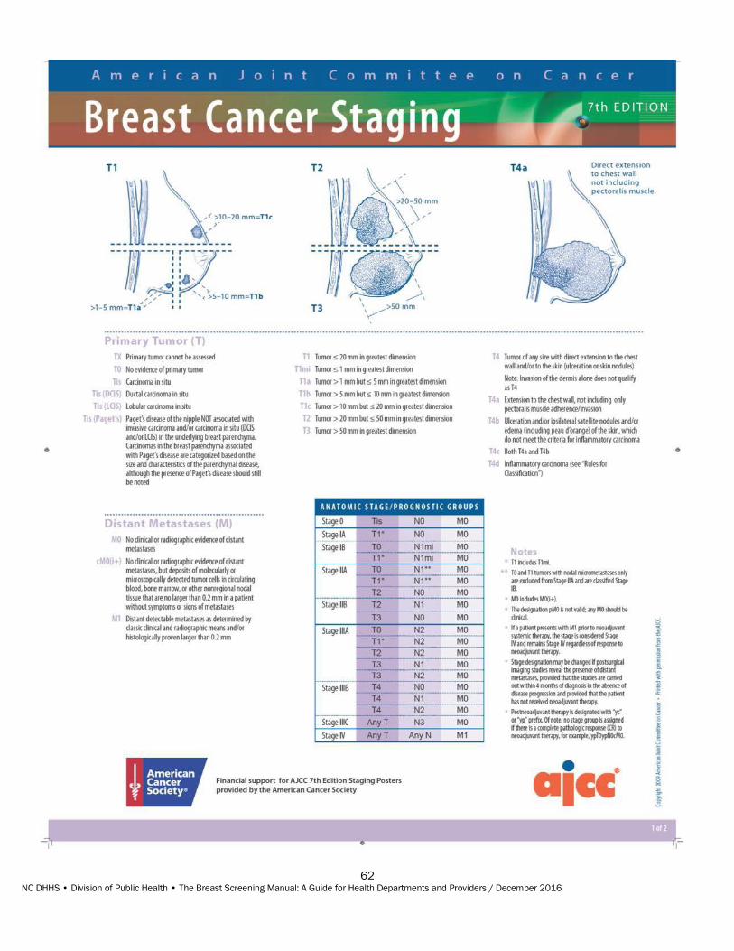
62 NC DHHS • Division of Public Health • The Breast Screening Manual: A Guide for Health Departments and Providers / December 2016

63 NC DHHS • Division of Public Health • The Breast Screening Manual: A Guide for Health Departments and Providers / December 2016

TAB 13: Appendix G

APPENDIX G: BREAST DENSITY
North Carolina requires all health care facilities that perform mammography examinations to
include information about the patient’s individual breast density in the summary of the
report.
The American College of Radiology's BI-RADS® reporting of breast composition describes
the proportion of fatty tissue to fibroglandular tissue as one of four categories:
1. The breast is almost entirely fat.
2. There are scattered fibroglandular densities.
3. The breast tissue is heterogeneously dense, which could obscure detection of small
masses.
4. The breast tissue is extremely dense. This may lower the sensitivity of
mammography.
Categories 3 (heterogeneously dense) and 4 (extremely dense) are considered “dense
breasts.”
Breast density describes the amount of normal fibrous and glandular tissue in proportion to
normal fatty tissue. Breasts are considered mammographically dense if a patient has a more
fibrous or glandular tissue than breast fat.
As many as 50% of women in the U.S. have dense breast tissue and the other half have non-
dense tissue (more fatty tissue). Of those women with dense breast tissue, 10% have
extremely dense breast tissue.
Dense breast tissue may make it more difficult for radiologists to detect cancer on
mammograms. Normal dense tissue, cancer, and benign masses may all appear white on
the mammogram. This accounts for the phenomenon called masking. The cancer/benign
lesion is obscured by the normal surrounding breast tissue. Hence, the sensitivity of
mammography is reduced by 10-20%, making screening mammograms less precise in
women with dense breasts.
The recommendations for mammography are the same for women with dense breasts as
non-dense breast women. Many cancers are still detected on mammograms even if the
patient has dense breast tissue.
A mammogram is the only screening test that has demonstrated a reduction in breast
cancer deaths. There is no recommendation that it be replaced with another screening test
at the current time.
If a patient has dense breast tissue and is interested in additional screening options, a
breast cancer risk assessment may be useful. This will allow a discussion of whether
supplemental tests will be helpful, and if so, what tests to order.
Screening breast MRI (magnetic resonance imaging) in HIGH RISK patients (>20% lifetime
risk of developing breast cancer) has been shown to increase the cancer detection rate. It is

65 NC DHHS • Division of Public Health • The Breast Screening Manual: A Guide for Health Departments and Providers / December 2016
recommended by the American Cancer Society for high risk patients in addition to a yearly
screening mammogram.
For patients at INTERMEDIATE RISK (15-20% lifetime risk), such as those with a personal
history of breast cancer or a prior biopsy with a diagnosis of atypia, the American Cancer
Society reports that there is not enough data at the current time to recommend a MRI in
these patients. However, a patient-centered shared decision-making approach is
recommended and it should be discussed with the patient that the additional test may
detect other findings (non-cancers/false positives) that may lead to follow-up testing or
biopsy.
The data on screening ultrasound is limited; therefore, there is no formal approval from the
radiology community at the current time. Screening breast ultrasound is now offered at
many centers. The patient must be made aware that if she chooses to have ultrasound
screening for dense breasts with no other abnormal finding, she will be charged additional
out of pocket expenses.
Breast tomosynthesis (3D mammography) expands the technology of conventional
mammography. Some centers are currently using it in addition to screening mammography
since preliminary results on its performance are positive. However, we do not know how well
tomosynthesis performs in women with extremely dense breasts. At the current time, your
patient must be made aware that if she chooses to have breast tomosynthesis screening for
dense breasts with no other abnormal finding, she will be charged additional out of pocket
expenses.
Insurance coverage for any supplemental breast cancer screening tests is not mandated in
North Carolina. Screening breast MRI may be covered by insurance for HIGH RISK women,
but not for the average risk patient. Consequently, women who want other types of
screening may be asked to pay out of pocket.
Please visit this website for more information: http://www.ncacr.org/breast-health.php
NOTE: NC BCCCP does not pay for screening with ultrasound or MRI as
adjunct screening solely for the finding of dense breasts. Under certain rare
circumstances, a MRI may be performed and paid for through N.C. BCCCP
only if preauthorized.

TAB 14: Appendix H

APPENDIX H: UNDERSTANDING GENETIC RISK FOR BREAST CANCER
How genes affect cancer risk
Cancer is a disease of abnormal gene function. Genes contain the instructions on how to
make proteins the body needs to function, when to destroy damaged cells, and how to keep
the cells in balance. Genes control things such as hair color, eye color, and height. They can
also affect a person’s chance of getting certain diseases, such as breast cancer.
Every cell in the body has all the genes a person was born with, but different types of cells
may use different genes. For example, muscle cells use a different set of genes than skin
cells. The genes that the cell is using are activated or turned on, while the genes that the cell
doesn't need are turned off and not used.
An abnormal change in a gene is called a mutation. The 2 types of mutations are inherited
and acquired (somatic).
• An inherited gene mutation is present in the egg or sperm that formed the child. After
the egg is fertilized by the sperm, it created one cell called a zygote that divided to
create a fetus (which became a baby). Since all the cells in the body came from this
first cell, these kinds of mutations are in every cell in the body (including eggs or
sperm) and so can be passed on to the next generation.
• An acquired (somatic) mutation is not present in the zygote, but is acquired some
time later. It occurs in one cell, and then is passed on to any new cells that are the
offspring of that cell. This kind of mutation is not present in the egg or sperm, and
cannot be passed on to the next generation. Somatic mutations are much more
common than inherited mutations. Most cancers are caused by acquired mutations.
All people have 2 copies of most genes – one from each parent. When someone has
inherited an abnormal copy of a gene, their cells already start out with one mutation. If the
other copy of the gene stops working (because of an acquired mutation, for example), the
gene can stop functioning altogether. When the gene that stops working is a cancer
susceptibility gene, cancer can develop. Some cancer susceptibility genes function as tumor
suppressor genes. Tumor suppressor genes are normal genes that slow down cell division,
repair DNA mistakes, or tell cells when to die (a process known as apoptosis or programmed
cell death). When tumor suppressor genes do not work properly, cells can grow out of
control, which can lead to cancer. Many family cancer syndromes are caused by inherited
defects of tumor suppressor genes.
Someone who is not born with a bad copy of a gene would have to acquire 2 different
mutations for that gene not to work. Acquiring 2 mutations in the same gene takes longer
than acquiring one, which is why cancers that are caused by inherited gene mutations tend
to occur earlier in life than cancers of the same type that are not. (American Cancer Society,
“Family Cancer Syndromes”, 2014)
Family Cancer Syndromes
Cancer is such a common disease that it is no surprise that many families have at least a
few members who have had cancer. Sometimes, certain types of cancer seem to run in

some families. This is because family members have certain risk factors in common, such as
smoking, which can cause many types of cancer. It can also be due in part to other factors,
like obesity, that tend to run in families and influence cancer risk.
But in some cases, cancer is caused by an abnormal gene that is being passed along from
generation to generation. Although this is often referred to as inherited cancer, what is
inherited is the abnormal gene, not the cancer itself. Only about 5% to 10% of all cancers
result directly from inherited gene mutations.
Family cancer syndromes related to breast cancer - when is it of concern?
When many cases of cancer occur in a family, it is usually due to chance or exposure to a
common toxin, such as cigarette smoking. But sometimes these cancers may be caused by
an inherited gene mutation causing a family cancer syndrome. Certain things make it more
likely cancers in a family are caused by a family cancer syndrome.
For each case of breast cancer, look at:
• Who is affected? How are they related to the patient? Breast cancer in a close
relative, like a parent or sibling, is more cause for concern than breast cancer in a
more distant relative. Even if the cancer was from a gene mutation, the chance of it
passing on gets lower with more distant relatives. It is also important to look at each
side of the family separately. Having two relatives with breast cancer is more
concerning if the two relatives with breast cancer are women on the same side of the
family.
• How old was the relative (or the patient) when they were diagnosed? The age of the
woman when the cancer was diagnosed is important. Breast cancer is rare in women
who are pre-menopausal. Having two or more cases in close relatives prior to
menopause could be a sign of an inherited cancer syndrome.
• Did the relative (or the patient) get more than one type of cancer? More than one
type of cancer in a person (like a woman with both breast and ovarian cancer) may
be a red flag for a family cancer syndrome.
• Did the relative (or the patient) get cancer in both breasts?
• Did the breast cancer occur in a male relative (or patient)?
• Did the relative or patient with cancer smoke or have other known risk factors?
Having the same type of cancer in many relatives is more concerning than if it is several
different kinds of cancer. In some family cancer syndromes, a few types of cancer seem to
go together. For example, breast cancer and ovarian cancer run together in families with
hereditary breast and ovarian cancer syndrome (HBOC).
Hereditary Breast and Ovarian Cancer syndrome
For many years, doctors noticed that in some families, many of the women developed breast
and/or ovarian cancer. Often the cancers were found at younger than usual ages, and some
of the women had more than one cancer. Some had breast cancer in both breasts, and
some got both breast and ovarian cancer. Doctors studying these families coined the term
Hereditary Breast and Ovarian Cancer Syndrome (HBOC).

Scientists studied the genes of these families, and discovered the genes BRCA1 and
BRCA2. Because some women (and families) have HBOC based on cancer history, but do
not have mutations in these genes, scientists believe that there is at least one more gene
that can cause HBOC, which they have called BRCA3. The BRCA3 gene has not yet been
identified.
In normal cells, these genes help prevent cancer by making proteins that help keep the cells
from growing abnormally. Mutated versions of these genes cannot stop abnormal growth,
and can result in a higher risk for breast cancer. In some families with BRCA1 mutations the
lifetime risk of breast cancer is as high as 80%, but on average this risk seems to be in the
range of 55% to 65%. For BRCA2 mutations the risk is lower, around 45%.
Although the risk of breast and ovarian cancer is very high with mutations in either BRCA1 or
BRCA2, tends to be higher with BRCA1 mutations.
Male breast cancer, pancreatic cancer, and prostate cancer can be seen with mutations in
either gene, but are more common in persons with BRCA2 mutations. In the US, mutations
in the BRCA genes are more common in women of Ashkenazi Jewish descent than in the
general population (American Cancer Society, “Family Cancer Syndromes”, 2014).
If someone in a family is found to have a BRCA mutation, it means that their close relatives
(parents, siblings, and children) have a 50% chance of having a mutation, too. They may
wish to be tested, or even without testing may wish to start screening for certain cancers
early or take other precautions to lower their risk of cancer.
Li-Fraumeni syndrome
Li-Fraumeni syndrome is a rare syndrome that can lead to the development of several
cancers, including sarcoma (such as osteosarcoma and soft-tissue sarcomas), leukemia,
brain (central nervous system) cancers, cancer of the adrenal cortex and breast cancer. The
cancers most often occur in childhood, although the breast cancers occur in young adults.
Women with Li-Fraumeni can also be affected by more than one cancer in their lifetime.
They also seem to be at higher risk of cancer from radiation therapy, and so doctors treating
these patients may try to avoid giving them radiation when possible.
This syndrome is most often caused by inherited mutations in the TP53 gene, a tumor
suppressor gene. A normal TP53 gene stops the growth of abnormal cells.
Li-Fraumeni syndrome can also be caused by mutations in a gene called CHEK2. A CHEK2
mutation, even when it doesn’t cause this syndrome, can increase breast cancer risk about
2-fold.
Changes in other genes
Other gene mutations can also lead to inherited breast cancers. These gene mutations are
much less common and most of them do not increase the risk of breast cancer as much as
the BRCA genes. They are only occasional causes of inherited breast cancer.
• ATM: The ATM gene normally helps repair damaged DNA. Inheriting 2 abnormal
copies of this gene causes the disease ataxia-telangiectasia. Inheriting one abnormal
copy of this gene has been linked to a high rate of breast cancer in some families.
• PTEN: The PTEN gene normally helps regulate cell growth. Inherited mutations in this
gene cause Cowden syndrome, a rare disorder that puts women at higher risk for

both non-cancerous and cancerous tumors in the breasts, as well as growths in the
digestive tract, thyroid, uterus, and ovaries. Defects in this gene can also cause a
different syndrome called Bannayan-Riley-Ruvalcaba syndrome that is not thought to
be linked to breast cancer risk. The syndromes caused by mutations in PTEN can be
grouped together as PTEN Tumor Hamartoma Syndrome.
• CDH1: Inherited mutations in this gene cause hereditary diffuse gastric cancer, a
syndrome in which people develop a rare type of stomach cancer. Women with
mutations in this gene also have an increased risk of invasive lobular breast cancer.
• STK11: Defects in this gene can lead to Peutz-Jeghers syndrome. Persons affected
with this disorder have pigmented spots on their lips and in their mouths, polyps in
the urinary and gastrointestinal tracts, and a higher risk of many types of cancer,
including breast cancer.
• PALB2: The PALB2 gene makes a protein that interacts with the protein made by the
BRCA2 gene. Mutations in this gene can lead to a higher risk of breast cancer. It is
not yet clear if PALB2 gene mutations also increase the risk for ovarian cancer and
male breast cancer. (American Cancer Society, “Family Cancer Syndromes”, 2014)
A. Genetic Counseling and Genetic Testing for Breast cancer
If the patient is thinking about genetic testing, it is strongly recommended that she first talk
to a genetic counselor who can explain and interpret the results of these tests. It is
important to understand what genetic testing can and cannot tell her, and to carefully weigh
the benefits and risks of genetic testing before these tests are done.
Cancer-related genetic tests are most commonly done as predictive genetic tests, to see if a
woman has a certain gene mutation known to increase the risk for breast cancer. They can
also be used to confirm a suspected gene mutation in a woman or her family.
Sometimes after a woman has been diagnosed with breast cancer, the doctor will do tests to
look for gene changes in the cancer cells. These tests can give information on her prognosis
and can sometimes help tell whether certain types of treatment might be useful. These types
of tests look for gene changes only in the cancer cells that are taken from the patient.
These tests are not the same as the tests used to find out about inherited cancer risk.
B. Deciding to have genetic testing – the genetic counselor
It is important to find out how useful testing may be before doing it. Talking to a primary care
provider and meeting with a genetic counselor before the actual test will help her decide
whether to have the testing done. The counselor can tell her about the pros and cons of the
test, what the results might mean, and what her options are (American Cancer Society,
“Understanding Genetic Testing for Cancer”, 2016).
C. The counselor will do a risk assessment to find out how likely the woman is to develop
breast cancer. This risk is based on things like her medical history and family history.
70 NC DHHS • Division of Public Health • The Breast Screening Manual: A Guide for Health Departments and Providers / December 2016
The woman will be asked why she wants to be tested. Family history as far back as possible
and up to the present day will be reviewed in depth. It is important to confirm family history
by medical records and/or death certificates whenever possible.
She will also be asked about her own medical history. This may include early detection
practices, gynecologic history, lifestyle factors, and exposure to known carcinogens.
The woman will want to think about how the results might affect her and her relatives, and
discuss these issues with the counselor before testing. At this point, it often helps to look at
her family’s attitudes about cancer and the possibility of a family member being “blamed”
for the cancer. Cultural beliefs, support systems, and finances may also play a role in how
her family views cancer. These topics may be discussed, too.
The counselor will explain how families inherit cancers and how genes are passed on to
children. They will also talk about the types of cancer seen in the family and estimates of the
woman’s breast cancer risk. The pros and cons, costs, and limits of testing are discussed
too, as well as who in the family should consider being tested. More than one family
member may be offered testing.
For example, if testing shows that the woman has a high breast cancer risk, the counselor
may also talk about the best ways to manage it. These may include lifestyle changes, early
detection, watching for signs and symptoms of breast cancer, medicines to reduce cancer
risk, or even preventive surgery.
Many of these issues require the skills of an expert counselor. An increased risk of cancer,
and the potential for discrimination can be frightening. The counselor will explore ways to
cope, as well as her specific fears and concerns. How to discuss the test results and what
they mean with other family members is another key topic that will be covered.
The woman’s primary care provider may be able to refer her to a genetic counselor in her
area. If not, she can find a list of certified genetic counselors on the websites of the National
Society of Genetic Counselors (www.nsgc.org) or the National Cancer Institute
(www.cancer.gov/cancertopics/genetics/directory).
D. Informed consent
After risk assessment and genetic counseling, if the woman decides to be tested she will be
asked to give her informed consent in writing. Informed consent about testing should cover:
• The purpose of the genetic test
• The reason for offering the test
• The type and nature of the genetic condition being tested for
• Test accuracy
• Pros and cons of testing (including the limits of what the results might tell)
• Other testing options
• Treatment options that might be available depending on the test results
• Further decisions that may need to be made once the results are back

71 NC DHHS • Division of Public Health • The Breast Screening Manual: A Guide for Health Departments and Providers / December 2016
• The possible consent to use the results for research purposes after the test
• Availability of counseling and support services
• The right to refuse testing
E. Specimen collection and lab testing
Once the consent form is signed, lab tests are done. Genetic tests may be done on a sample
of blood, hair, cheek swab, urine, or other body tissues. Genetic tests for cancer usually
mean giving several tubes of blood.
F. Getting the test results
After the test is done, the genetic counselor will share the results with the woman at another
counseling session. This might not happen until several weeks or even months after the
samples are taken. Some women choose to bring a family member to help share results
with other family members.
Testing does not always give clear answers, but genetic counselors are trained to interpret
and explain the test results and what they might mean. If a mutation is found, the counselor
will talk to the woman about which of her family members might also be affected. It will be
important for those family members to know exactly which mutation was found and in which
gene. They can then discuss this with their primary care providers and may decide to get
tested, too.
If the result is positive
A positive result means there is a mutated gene (or genes) that may place the woman at
risk. Her risk of developing breast cancer will be discussed because she has the mutation.
Many women are concerned or anxious after learning they are at increased risk for breast
cancer. This is normal. The results may imply risk for certain blood relatives, and lead to
strains in family relationships as well. Concern about being treated differently may become
more real.
After the test is complete, there is often a great deal of uncertainty. There is no way to know
for sure that breast cancer will develop. Her primary care provider or counselor can help sort
through options of what she might be able to do to lower her risk.
If the result is negative
If the test result is negative, the woman does not have the gene mutation she was tested
for. She will probably feel relieved that the test did not show she was at increased risk.
But it is important to understand that genetic test results cannot always guarantee she is
not at increased risk. For instance, there might still be a chance that she has a different
mutation that she was not tested for. And rarely, the test result may be a “false negative.”
This means the test reads negative but the mutation is there.
Even a result that is truly negative does not mean her risk is zero – it just means she has the
same average risk as most other women.

72 NC DHHS • Division of Public Health • The Breast Screening Manual: A Guide for Health Departments and Providers / December 2016
Family members with negative test results may feel guilty if other family members test
positive. Her primary care provider or genetic counselor can help her understand what the
test results mean for her and her family and how to deal with them.
If the result is inconclusive
If there is not enough information to know whether the genetic changes are present, the test
result is said to be inconclusive. The woman (and family) may still be at a higher risk of
developing breast cancer. Taking steps to lower her risk can be helpful for some women, but
not having a sure result can still cause anxiety and frustration. Her primary care provider or
genetic counselor can help her understand what the results might mean for her and her
family and help her cope with them. In some cases, testing blood relatives can help clarify
this result.
If the result is variance of unknown or uncertain significance
When genetic tests show that a woman has an unusual form of a gene, but doctors do not
know what this gene change means, the result is called a variance of unknown or uncertain
significance (VUS). It may be a normal variant, simply a different version of gene that is not
seen often enough to be sure, or there may be some other explanation.
For some, a VUS result can cause anxiety, frustration, and even anger because this result
gives no information to guide future decisions. Her primary care provider or genetic
counselor can help her understand what the results might mean for her and her family and
help her cope with them.
G. What if genetic testing shows an increased breast cancer risk?
If the woman’s genetic test result is positive or inconclusive for a gene mutation that could
increase her risk of breast cancer, managing the risk becomes a priority.
Some of the ways she might lower her risk include:
• Chemoprevention: Taking medicines to help reduce risk
• Preventive or prophylactic surgery: Removing the healthy breasts to try to keep
cancer from starting there
• Lifestyle changes: Making healthy choices and changing behaviors to try to help
reduce breast cancer risk
• Breast cancer screening: Doing what she can to find breast cancer early (through
testing and awareness of early signs and symptoms), when the cancer is small and
treatment is most likely to be successful
Her primary care provider may recommend one or more of these approaches, but it is
important to understand how much they could affect her risk before she decides on a
course of action. She will also want to be sure she understands her risks and downsides
before deciding on a plan. These approaches are discussed in more detail below.

73 NC DHHS • Division of Public Health • The Breast Screening Manual: A Guide for Health Departments and Providers / December 2016
Chemoprevention
Chemoprevention is the use of medicines to help keep cells from developing into certain
types of cancer. Medicines such as tamoxifen and raloxifene can be used to help reduce
breast cancer risk cancer in women known to be at high risk.
Each woman’s risk and medical situation must be considered carefully so that any harmful
effects of the drugs do not outweigh the benefits.
Prophylactic (preventive) surgery
Prophylactic (preventive) surgery is another option in some cases. Some women at high risk
for breast cancer may decide to have their healthy breasts removed.
Changes in lifestyle factors
Ask her primary care provider about lifestyle changes she can make that could help
decrease her breast cancer risk. For instance, limiting alcohol intake and exercising is
known to help lower the risk of breast cancer.
In some cases, the effect of these changes on risk might be small compared to the
increased risk from the mutation, but she may still want to ask her provider what she can
do.
Breast cancer detection tests and awareness
Early detection tests may be started at an earlier age or be done more often, or special tests
may be needed if she has a positive genetic test result. For instance, a woman with a
genetic mutation that raises her risk of breast cancer might choose to have breast MRI
scans along with her mammograms to look for early signs of the breast cancer.
It is also important to be aware of the possible signs and symptoms of breast cancer, and to
see a doctor right away if anything concerns her. Finding breast cancer early – when it is
small and has not spread – offers the best chance of treating it successfully.
H. Sharing results with family members
If the woman has a gene mutation that raises her risk, she may need to think about whether
to tell other family members who might also be at increased risk. Telling them might help
them decide if they should get tested or adopt some of the approaches to try to lower their
risk.
On the other hand, some test results may cause more anxiety than anything else, and some
family members may not want to know their own risk. This is especially true if there is not
much they can do with the results. She might want to speak with family members before she
gets tested to find out if they want to know her results. (American Cancer Society, “What
Happens During Genetic Testing for Cancer?”, 2016)
I. Advantages and Disadvantages of Genetic Testing for Breast Cancer Risk
Genetic testing is a hot topic and can be used to learn about inherited breast cancer risk.
But there are many things to think about before having it done. Talking to a primary care
provider and meeting with a genetic counselor before the actual test will help a woman

74 NC DHHS • Division of Public Health • The Breast Screening Manual: A Guide for Health Departments and Providers / December 2016
know what to expect. Here are some of the advantages and disadvantages she should
discuss before testing.
J. Advantages
The most obvious benefit of genetic testing is the chance to better understand her risk for
breast cancer.
For families at risk, a negative result may help ease anxiety.
A positive result can help her make important decisions about her health, including things
she can do that might help lower her risk. A positive result may also lead her primary care
provider to have her:
• Start breast cancer screening tests earlier
• Get screened for breast cancer more often
• Get screening tests that are used only for women known to be at increased breast
cancer risk
• Watch herself closely for signs or symptoms of breast cancer
• Learn about options to help reduce the risk of breast cancer, such as drugs or
surgery
If she does develop breast cancer, finding it early often means that treatment is more likely
to be helpful.
Disadvantages
Genetic testing results often give limited answers. Testing can only tell her if she has a
specific gene mutation, not if she will get breast cancer. A positive test result does not
always mean she will get the disease. The test can tell what might happen, but it cannot tell
what will happen. On the other hand, a negative result does not mean she has no risk of
getting breast cancer. And risk can change over time due to lifestyle choices, and simply
getting older.
As with many medical tests, genetic tests may be flawed, or test results may be read wrong.
This is not common, as many steps are taken to prevent this, but at the current time genetic
testing is not tightly regulated. Different labs may have different ways of looking for certain
changes.
Many women are anxious even before they get their test results. Learning that she or a loved
one might develop breast cancer can be frightening. The woman being tested may find it
even more upsetting if family members have already died of breast cancer. Having a gene or
passing the gene on to children can also lead to guilt or anger.
Not all family members might want to know if they might be at increased risk, especially if
there is not much they can do about it. Testing any family member might lead to anxiety and
other concerns in other family members.

75 NC DHHS • Division of Public Health • The Breast Screening Manual: A Guide for Health Departments and Providers / December 2016
Privacy may become an issue when many family members could be affected by a single
positive genetic test result. More family members may need to be tested. Sometimes family
secrets are revealed as a result – paternity, adoptions, or other difficult issues may come
up.
In some cases, more medical tests or procedures may be recommended because of genetic
testing. For example, if a gene mutation for breast cancer is found, more tests like breast
MRI may be recommended. This can be a good thing, if these other tests help keep her free
of breast cancer or if they find it early, when it is likely to be easier to treat. But the tests can
have downsides as well, such as the time and expense involved, as well as possible risks
from the tests themselves. These extra tests can also lead to more stress and anxiety.
Genetic testing can be expensive. Depending on the tests done, the final bill can be
thousands of dollars. She needs to have an idea of how much it will cost before she has
testing done.
Not all insurance companies will pay for the testing.
Although the law limits what most (not all) employers and most (not all) health insurance
companies can do with genetic information, positive results may lead to denial of coverage
or higher rates for life insurance, disability insurance, and long-term care insurance.
What about privacy?
Many women are concerned about the privacy of her genetic information. Even if her
insurance company does cover genetic testing, she may choose to pay for it herself to keep
the results as private as possible.
Most women who ask about the privacy of genetic information are worried about how the
information could be used in ways that can harm them. Before she decides on testing, it is
important to think about who might learn about her results and with whom she will share her
results. Most Americans are afraid that employers and insurance companies might get and
use their genetic information. The truth is, patients, families, and primary care providers are
not the only ones interested in genetic information. Here are some of the other groups who
might want to use this information:
Medical and pharmaceutical researchers
Medical and pharmaceutical researchers are interested in low-cost access to genetic
information and materials. Members of the pharmaceutical lobby have argued against
women owning their own genetic information, stating it would drive up drug costs, which
would be passed on to the consumer.
Today, medical researchers must get the individual’s informed consent before any studies of
tissue samples and DNA can be done.
Employers
Employers can ask for genetic testing only when it is used to monitor exposure to potentially
toxic chemicals and substances in the workplace. Discrimination and employment decisions
based on genetic information are barred at the national level for most employers.

76 NC DHHS • Division of Public Health • The Breast Screening Manual: A Guide for Health Departments and Providers / December 2016
Insurers
Federal law does not allow health insurers to use genetic information when deciding who to
cover and how much to charge for insurance. But the law does not restrict use of genetic
information for life insurance, disability insurance, or long-term care insurance.
Legal protection – what it does and does not do
GINA (Genetic Information Nondiscrimination Act of 2008)
GINA is a federal law that prohibits the use of genetic information in workplace employment
decisions for non-governmental organizations with more than 15 employees. This law also
bars health insurers from making coverage or cost decisions based on genetic information.
GINA defines genetic information as:
• A woman’s genetic test results
• The genetic test results of family members
• If one or more family members are known to have a genetic disease or disorder
Employers: GINA bars employers from discriminating based on genetic information in hiring,
firing or layoffs, pay, or other personnel actions such as promotions, classifications, or
assignments. The law applies no matter how they got the information.
Employers are not allowed to require genetic testing and cannot collect genetic information
except for very limited exceptions. For instance, it may be allowed when information is
needed to meet the requirements of family and medical leave laws or to watch for harmful
effects from hazardous workplace exposures.
Employers must keep genetic information confidential. They cannot release or share genetic
information except when:
• The employee asks them to
• Fulfilling a request from a health researcher
• Complying with medical leave law
• Disclosing or reporting to a public health agency
Health insurers: GINA bars health insurers (including group health plans, individual plans,
and Medicare supplemental plans) from turning down women or charging higher premiums
for health insurance based on genetic information or for using genetic services. This
includes genetic counseling and testing. The law also bars these insurers from asking for or
requiring genetic tests. GINA applies to all health insurance plans (including federally
regulated ERISA plans, state-regulated plans, and private individual plans).
More details about GINA: A few states have stronger laws than GINA. GINA does not replace
state laws against genetic discrimination that are broader in scope. Rather, GINA
establishes a national baseline protection while allowing states to impose stronger
protection.

77 NC DHHS • Division of Public Health • The Breast Screening Manual: A Guide for Health Departments and Providers / December 2016
What GINA does not do:
GINA’s protections do not apply to life insurance, disability insurance, or long-term care
insurance. It also doesn’t require health insurance to cover genetic testing.
GINA does not apply to very small employers (with fewer than 15 employees), nor does it
apply to military health plans, the Veterans Administration, or the Indian Health Service.
GINA does not apply to federal employees who get health coverage through the Federal
Employees Health Benefits Plans. (American Cancer Society, "Should I Get Genetic Testing
for Cancer Risk?", 2016)
REFERENCES
Family Cancer Syndromes. (2014, June 25). Retrieved October 13, 2016, from
http://www.cancer.org/cancer/cancercauses/geneticsandcancer/heredity-and-cancer.
Understanding Genetic Testing for Cancer. (2016, May 23). Retrieved October 13, 2016,
from http://www.cancer.org/cancer/cancercauses/geneticsandcancer/understanding-
genetic-testing-for-cancer.
What Happens During Genetic Testing for Cancer. (2016, May 23). Retrieved October 13,
2016, from http://www.cancer.org/cancer/cancercauses/geneticsandcancer/what-
happens-during-genetic-testing-for-cancer.
Should I Get Genetic Testing for Cancer Risk? (2016, May 23). Retrieved October 13, 2016,
from http://www.cancer.org/cancer/cancercauses/geneticsandcancer/should-i-get-genetic-
testing-for-cancer-risk.
NOTE: NC BCCCP does not pay for Genetic Testing at the current time.
Please contact your NC BCCCP Nurse Consultant for questions.

TAB 15: Appendix I

APPENDIX I: STAFF DIRECTORIES
Cancer Prevention and Control Branch
Main Branch Number (919) 707-5300 Fax Number (919) 870-4812
Breast and Cervical Cancer Program
Debi Nelson, MAEd
Branch Manager
(919) 707-5155
WC: (919) 218-2585
Vicki Deem, R.N., M.P.A.
Public Health Nurse Consultant
(919) 707-5324
WC: (919) 218-4270
Susan Smith Fuller
Administrative Assistant
(919) 707-5301
Cindy Herndon, PhD, R.N.
Public Health Nurse Consultant
(919) 707-5310
WC: (919)218-7660
Tammie Hobby Administrative Assistant
(919) 707-5302
Krystal Moody Administrative Assistant
(919) 707-5306
Terence Tumenta Shendeh (Interim) NC BCCCP Data Manager
(919) 707-5327
Terence Tumenta Shendeh Epidemiologist
(919) 707-5327
Tavonyia Thompson
Operations Supervisor
(919) 707-5326
Lavanya Gupra
NC BCCCP Evaluator
(919) 707-5336
Brittney Wooten Sala
NC BCCCP Health Educator
(919) 707-5330
Sherry Wright, R.N.
Public Health Nurse Consultant
(919) 707-5325
WC: (919) 218-0183

Women’s Health Branch (Abbreviated List) Main Branch Number (919) 707-5700
Name: Sydney Atkinson, MSSW
Job Title: Family Planning & Reproductive
Health Unit Supervior
Work #: 919-707-5693
Email: [email protected]
Name: Belinda Pettiford, MPH
Job Title: Women’s Health Branch Head
Work #: 919-707-5699
Email: [email protected]
Name: Sarah Conte, DNP, MSN, RN
Job Title: Maternal Health Nurse Consultant
Work #: 919-707-5689
Email: [email protected]
Name: Betty Cox, RN, MSHA
Job Title: Regional Nurse Consultant
Work #: 910-425-1025
Email: [email protected]
Name: Dara Dockery, MPH, BSN, RN Job Title: Regional Nurse Consultant
Work #: 919-269-4351
Email: [email protected]
Name: Debbie Farb, RN, BSN, MPH, IBCLC Job Title: Family Planning Nurse Consultant
Work #: 910-707-5719
Email: [email protected]
Name: Patty Kempton, BSN, MPH, RN
Job Title: Regional Nurse Consultant
Work #: 919-947-2464 Email: [email protected]
Name: Julie Gooding Hasty, RHEd
Job Title: Program Consultant
Work #: 919-707-5695 Email: [email protected]
Name: Kimberly Leathers-Raynor, JD
Job Title: Public Health Education Consultant
Work #: 919-707-5716
Email: [email protected]
Name: Dana Lynch, BSN, RN
Job Title: Regional Nurse Consultant
Work #: 704-922-0373
Email: [email protected]

TAB 16: References

80 NC DHHS • Division of Public Health • The Breast Screening Manual: A Guide for Health Departments and Providers / December 2016
References
American Academy of Family Physicians, [AAFP] (2016). Clinical Preventive
Service Recommendation: Breast Cancer. Retrieved on April 11, 2016 from
www.aafp.org/patient-care/clinical-recommendations/all/breast-cancer.html.
American Cancer Society. [ACS] (2015). Breast cancer prevention and early
detection. Retrieved on April 7, 2016 from www.americancancersociety.org.
American Cancer Society. (2016). Genetics and cancer. Retrieved October 13, 2016, from
http://www.cancer.org/cancer/cancercauses/geneticsandcancer/heredity-and-cancer.
American College of Obstetrics and Gynecologists [ACOG] (2015). ACOG statement on
revised American Cancer Society recommendations on breast cancer screening.
Retrieved on April 12, 2016 from http://www.acog.org.
American College of Physicians [ACP] (2016). Screening for breast cancer:
recommendations from the U.S. Preventive Services Task Force. Annals of Internal
Medicine, 164: 1-28. Retrieved on April 12, 2016 from http://annals.org.
American College of Radiology [ACR] (2016). Retrieved on April 19, 2016 from www.acr.org/.
American College of Radiology [ACR] (2015). ACR and SBI continue to recommend
regular mammography starting at age 40. Retrieved on April 12, 2016 from
www.acr.org/about-us/media-center/press-releases/20151020.
American College of Radiology [ACR] Breast Imaging Reporting and Data System, Breast
Imaging Atlas (2013). ACR BI-RADS®- Mammography (5th Ed). Reston, VA: American
College of Radiology.
American Joint Committee on Cancer, [AJCC], (2010). In Compton, C. C., Fritz, A. G., Greene,
F. L., Trotti, A. (Eds) American Joint Committee on Cancer Staging Manual, 7th
Edition. New York, NY: Springer, pp. 347-76.
American Joint Committee on Cancer, [AJCC], (2016).
https://cancerstaging.org/references-tools/quickreferences/Pages/default.aspx.
Accessed August 16, 2016.
American Society of Breast Surgeons [ASBrS] (2016). Consensus Statement in
Screening Mammography. Society’s Research Committee, approved by the
Board of Directors on October 29, 2015.
Breast Cancer Staging poster. https://cancerstaging.org/references-
tools/quickreferences/Documents/BreastSmall.pdf. Accessed August 16, 2016.

81 NC DHHS • Division of Public Health • The Breast Screening Manual: A Guide for Health Departments and Providers / December 2016
California Department of Health Services (Summer 2006). Clinical Breast
Examination: Proficiency and Risk management. Retrieved in April 18,
2016 from Breast Cancer News via
http://www.ochealthinfo.com/newsletters/bcnews/2000-summer.htm.
Cancer Dictionaries (2016) Retrieved on March 31 from
http://www.cancer.gov/publications/dictionaries/cancer-terms?cdrid=44411.
Center for Disease Control and Prevention. Retrieved on April 7, 2016 from
http://www.cdc.gov/cancer/breast/basic_info.
Center for Disease Control, National Breast and Cervical Cancer Early Detection
Program (NBCCEDP) (2016). Retrieved on April 7, 2016 from
http://www.cdc.gov/cancer/nbccedp/index.
Duffy, S. W., Yen, A. M., Chen, T. H., Chen, S. L., Chiu, S. Y., Fan, J. J., Smith, R.
A., Vitak, B., Tabar, L. (2012). Breast care management: Long-term benefits of breast
screening. Future Medicine, 1: 1, p. 31- 38. Retrieved on April 15, 2016 from
www.fururemedicine.com.
Federal Register, U.S. Federal Poverty Guidelines (2016). Annual update of the
Health and Human Services Poverty Guidelines. Retrieved on April 21, 2016 from
http://www.federalregister.gov.
Harold P. Freeman Patient Navigation Institute (2016) Retrieved on November 18, 2016
from http://www.hpfreemanpn.org.
Houssani, H. (2016). Screening with tomosynthesis or ultrasound detects more
cancers in dense breast. ECCO European Breast Cancer Conference March 2016/
accepted by the Journal of Clinical Oncology.
Mayo Clinic (2016). Retrieved on April 20, 2016 from
http://www.mayoclinic.org/diseases-conditions/mammaryductectasia.
National Breast Cancer Foundation (2016). Retrieved on April 19, 2016 from
http://www.nationalbreastcancer.org.
National Cancer Institute (NCI), (2016). Retrieved on April 7, 2016 from
http://www.cancer.gov/cancertypes/breasts.
NC Department of Public Health Breast and Cervical Cancer Control Program
(2016). BCCCP/ WISEWOMAN Agreement Addendum 2016- 2017.
NCI, (2016). Dictionary of Cancer Terms. Retrieved on April 7,
2016 from http://www.cancer.gov/publications/dictionaries.
Russell, L. C. (1989). Caffeine restriction as initial treatment for breast pain. Nurse
Practice, 14: (2): p. 36-7.
US Department of Health and Human Services (2016). National Guidelines

82 NC DHHS • Division of Public Health • The Breast Screening Manual: A Guide for Health Departments and Providers / December 2016
Clearinghouse. Retrieved on April 14, 2016 from www.guideline.gov.
US Preventive Services Task Force, (2016). Retrieved on April 7,
2016 from http://www.uspreventiveservicestaskforce.org.