Embed Size (px)
Citation preview

E. Blair Holladay, PhD, SCT(ASCP)CM
Vice President and Executive Director; BORAmerican Society for Clinical Pathology (ASCP)Chicago, Ill
Bridging Morphology with Molecules

Goals of this LectureThe intent of this lecture is to discuss assays relevant to cytopathology and anatomic pathology, and illustrate how cytologists can integrate these skills into their clinical practice.

Presentation Outline1. Introduction and Rationale
2. Hybrid Capture (Signal Amplification)
3. In situ hybridization (ISH/FISH) for nucleic acids
4. PCR
5. Invader Technology
5. Fluorescent ISH
6. Specific Oncology Probes
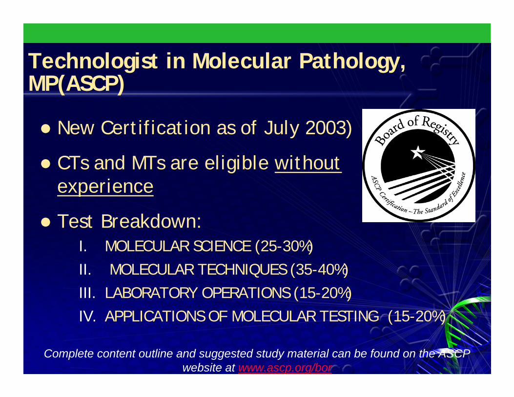
Technologist in Molecular Pathology, MP(ASCP)
New Certification as of July 2003)
CTs and MTs are eligible without experience
Test Breakdown:
Complete content outline and suggested study material can be found on the ASCP website at www.ascp.org/bor
I. MOLECULAR SCIENCE (25-30%)II. MOLECULAR TECHNIQUES (35-40%)III. LABORATORY OPERATIONS (15-20%)IV. APPLICATIONS OF MOLECULAR TESTING (15-20%)

What’s So Great About Molecular Diagnostics?
As many as 5,000 diseases have direct genetic causesHigh sensitivity and increased specificity for most tests adds diagnostic utility
Potential for simple standardized procedures and automation
rapid throughput
Increased number of techniques for infectious diseases and tumordiagnostics
A viable reflex for equivocal morphology
Even one step further- DNA with Pap
Prices are falling

Why Perform Molecular Tests?Ability to Enhance Service/Patient Care
Capacity to Enhance Profitability
Avoiding Competition
Convenience
Retain Control of Testing Process
Assure Testing Quality
Underutilization of Technical Staff
Integrate and Collect Data
Bolick, DR. Arch Pathol Lab Med, 2003;127:988

Current Molecular Techniques in Diagnostic Cytopathology
1. Hybrid Capture (HC)
2. In situ hybridization (ISH)
A. Chromogenic
B. Fluorescent
3. Polymerase Chain Reaction (PCR)
4. Invader Technology
Future Techniques (Investigational only)
1. Transcriptional Profiling (Microarrays)
- FNA (Guidance of neoadjuvant therapy)

Denature specimen
Specimen Hybridizes with RNA probe
Capture RNA probe detect hybrids on a solid
phase
Luminescence
Antibodies conjugated with thousands of enzymes attach to captured hybrids then react with a substrate to emit an amplified chemiluminescent signal.
Digene Hybrid Capture®
DML 2000 System

Digene Hybrid Capture® Sample Collection and Test Results
Liquid cytology, such as ThinPrep, facilitates Digene testing
OR

In Situ Hybridization
Morphology-Based Microscopic Diagnostics

What is in situ Hybridization ?
Technique in which a single-stranded RNA or DNA probe is used to locate a gene or an mRNA molecule in a cell or a tissue

The ISH Staining ProcessAlkaline Phospahatase conjugated Avidin
Fluorescein-Labeled probe
Biotinylated linking Antibody
Primary antibody:anti-FITC, anti-Biotin or Anti-Digoxigenin
Target
FISH (Fluorescent in situ Hybridization)Bright Field (Chromogenic detection)

Increasing temperature to denature and separate target nucleic acid strands
Addition of labeled probe
Reducing temperature to hybridize probe to target
Post hybridization stingency washes
Denaturation and Hybridization

Polymerase Chain Reaction
http://www.unlv.edu/staff/wmojica/PCR_LAB2.htm

Invader® Technology (Third Wave, Inc.)Directly detects specific nucleic acid sequences
Based on signal amplification
Isothermal reactions; no thermal cycling
MethodAn overlapping structure created with the mutant probe and the Invader® oligo. Cleavase® enzymes cleave the primary probes that form overlapping structures releasing the 5' flaps plus one nucleotide.Released flaps from the primary reaction serve as oligos in a second simultaneous reaction on a labeled, synthetic oligo (Fluorescence resonance energy transfer (FRET) probe).Cleavage of this FRET probe = fluorescent signal.
Produce 1 million to 10 million labeled cleavage products per target sequence

Invader® Technology
A
T
C
G
A
A
C
C
F1 Q F2 Q
F1
Invader® OligoProbe Probe
FRET Cassette 1 FRET Cassette 2
Released 5´ Flap
Cleavage Site
Genotype specific target Control target
Cleavage Site
Invader® Oligo
Released 5´ Flap
CleavageSite
CleavageSite
F2

Applications of Molecular Diagnostics
A. Infectious Disease Markers
1. HPV
2. CT/GC
3. CMV
B. Oncology Markers
a. Her2/neu
b. Urovision, LaVision, Provision, Pathvision
c. Kappa/Lambda
d. CK20,t(8;21), t(9;22), t(12;18), t(15;17)
e. BCRA 1 and 2
f. FNA applications

Molecular Techniques for HPV Detection
It is the second most common cancer among women worldwide500,000 women, usually in their 30s or 40s, are stricken by the disease annually (Central Am, Africa, Asia)
Estimated that by 2020 more than 1 million new cases will emerge each year
About 266,000 women in the Asian-Pacific region--more than half of the total number of women afflicted worldwide – and 143,000 die each year

Current HPV DNA Test Techniques
Hybrid Capture 2 (HC2)
In Situ Hybridization (ISH)
Invader Technology
Polymerase Chain Reaction (PCR)

HPV DNA Testing Uses
Triage of Atypical squamous cells
Primary Screening Co-test with Cytology
Quality assurance and clinical follow-up:
Monitor percent HPV positive by category
Additional tool with cytology as “test of cure” in clinical follow-up and as basis for referral to diagnostic excisional procedures (conization, leep,etc)

Hybrid Capture Method
Performance Characteristics– 83-96% Sensitivity in CIN 2+ – 98 % Negative Predictive Value– 61 % Specificity– 17.2 % Positive Predictive Value
Data is from Digene package insert and JAMA Bethesda Guidelines

Hybrid Capture Method
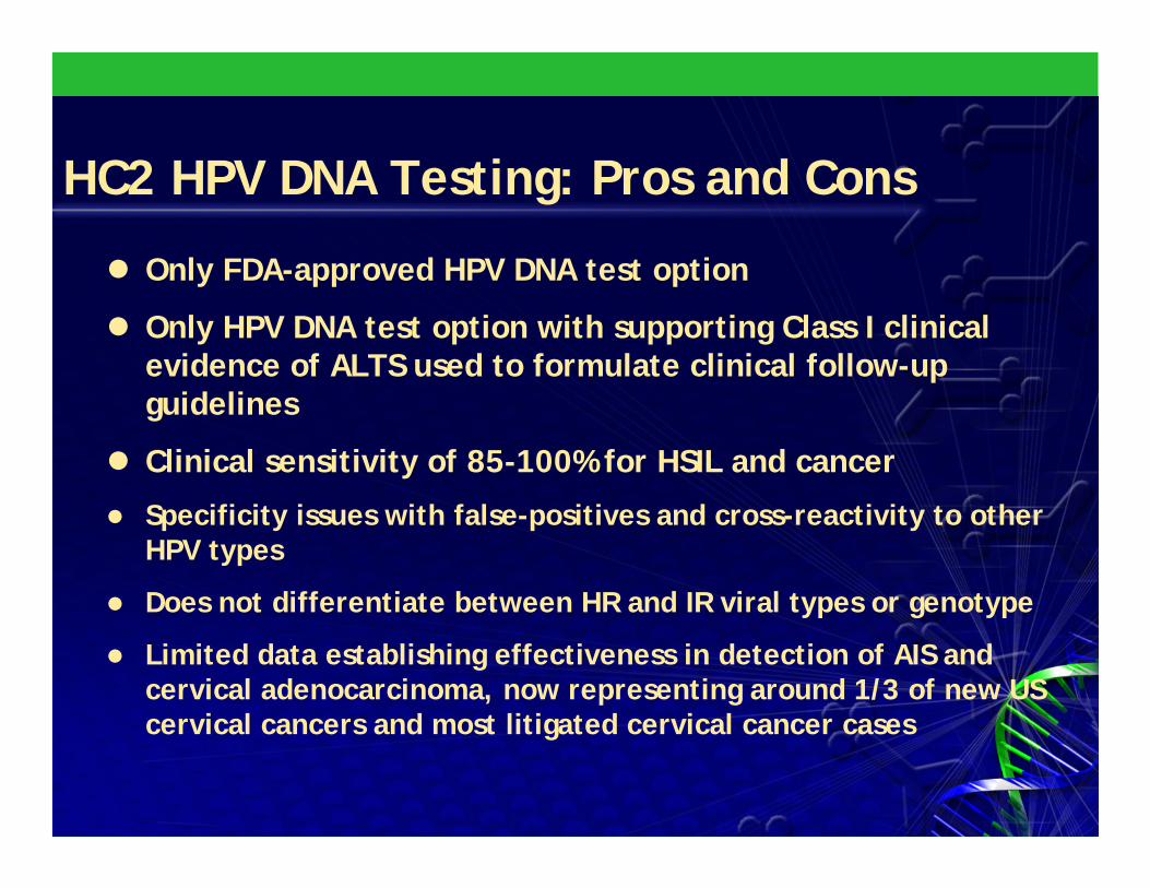
HC2 HPV DNA Testing: Pros and Cons
Only FDA-approved HPV DNA test option
Only HPV DNA test option with supporting Class I clinical evidence of ALTS used to formulate clinical follow-up guidelines
Clinical sensitivity of 85-100% for HSIL and cancer
Specificity issues with false-positives and cross-reactivity to other HPV types
Does not differentiate between HR and IR viral types or genotype
Limited data establishing effectiveness in detection of AIS and cervical adenocarcinoma, now representing around 1/3 of new US cervical cancers and most litigated cervical cancer cases

Ventana ISH Testing Method
Automated in situ hybridization (ISH) assay
Analyte Specific Reagent (ASR) for High and Low Risk types of HPV
Can be performed on Liquid Based Pap and Tissue Biopsies
Chromogenic Assay

In Situ Hybridization for HPV: Pros and Con’s
Excellent tissue or cytology localization of HPV-infected cellsGood specificity (>95%)Fully automated with walk-away capacityDifferentiates between episomal (diffused) versus integrated (punctate) HPV DNA patternsASR labeling places an additional responsibility on the medical director to assure performance characteristicsNo data on prognostic value for future diseaseHigher cost for payers and patients (could be addressed if studies establish method as reliable but more selective basis for colposcopic referral)

Invader® Technology
A
T
C
G
A
A
C
C
F1 Q F2 Q
F1
Invader® OligoProbe Probe
FRET Cassette 1 FRET Cassette 2
Released 5´ Flap
Cleavage Site
Genotype specific target Control target
Cleavage Site
Invader® Oligo
Released 5´ Flap
CleavageSite
CleavageSite
F2

Invader® Technology: Pros and ConsEase of Use
Minimal start up costs and no specialized trainingSimple procedure enhances throughput and cost-efficiencyConsistent product formatStraightforward, objective results
Very limited data to date
Sensitivity for CIN 2/3 and cancers not yet established in published studies
ASR labeling
No data on prognostic value for future disease
Use of some molecular CPT codes may need to be negotiated with payers by the laboratory

Polymerase Chain Reaction (PCR)

PCR: Commercial tests for the future?Commercially available test versus a “home brew” test ?
Home Brew
Typing done with Sequencing (Blastn) or Dot/Line Blot hybridization
Extremely sensitive – as few as 10 copies
Can be performed using Parrafin-embedded cervical tissue or liquid based Paps
Works beautifully with TP specimens

PCR - Gel Electrophoresis


HPV Genotyping Assays
Roche, Inc. Linear Array
Evaluated liquid cytology specimens (n = 534) and found a strong association with HPV risk group (HPV16 and HPV18 with increasing severity of cytology (PTrend <0.0005) and histology (PTrend < 0.0005).
Castle PE, Sadorra M, Garcia FR, Holladay EB, Kornegay J. Pilot Study of a Commercialized Human Papillomavirus (HPV) Genotyping Assay: Comparison of HPV Risk Group to Cytology and Histology. Journal of Clinical Microbiology. Vol. 44 (11). November, 2006.

HPV Genotyping Assays (TaqMan)
Roche, Inc.
COBAS TaqMan PCR Genotyping Assay
Purpose: Identify presence of 16 carcinogenic HPV types in aggregate while concurrently providing HPV16 and HPV18 genotype information from cervical specimens.Results: For detection of HPV16 and 18, there was 98.0 and 99.0% agreement between TaqManHPV testing Linear Array genotyping
Sadorra M, Castle PE, Garcia FR, Holladay EB, Kornegay J. Ability of a prototype COBAS TaqMan HPV test to simultaneously identify high risk HPV infection and provide HPV16 and HPV18 genotyping. International Papillomavirus Conference. Prague, Czech Republic. June 2006.

HPV Genotyping Assays (APTIMA)
• HPV mRNA for E6 and E7 oncoproteins may be a more specific biomarker for cervical precancer and cancer than carcinogenic HPV DNA
• Method: detected oncogenes from E6/E7 mRNA for 14 carcinogenic HPV genotypes on liquid cytology specimens (n = 531) to the presence of HPV genotypes detected by PGMY09/11 L1 consensus primer PCR assay
•GenProbe Inc.
APTIMA® Human Papillomavirus (HPV) Assay


HPV Genotyping Assays (APTIMA)
• Results: • An increasing likelihood of testing positive for
carcinogenic HPV E6/E7 mRNA with increasing severity of cytology (PTrend <0.0005) and histology (PTrend < 0.0005), with 94% of CIN3 histology cases (n = 49) and all five cancer cases testing positive for carcinogenic HPV E6/E7 mRNA
• Fewer specimens tested positive for carcinogenic HPV E6/E7 mRNA than for carcinogenic HPV DNA ((P < 0.0005, McNemar’s c2).
Castle P, Holladay EB, Garcia FR, Kolk D. A Cross sectional study of a prototype carcinogeneic HPV E6/E7 mRNA assay for Detection of Cervical Precancer and Cancer. Clinical Cancer Research. 2007;13 (9). May, 2007

HPV Testing
Take home message:
Specificity and sensitivity is clearly improving for HPV genotyping
Roche has submitted LA and Amplicor for FDA approval as of March 5, 2007

Cancer and Molecular DxCancer is a multi-step genetic disease, resulting from specific alterations in the function of one or more genes
• Molecular changes occur at the chromosome, gene & sequence level•Important to detect the full range of genetic abnormalities thatare indicative of cancer
ISH and FISH which detects chromosome copy numberPCR and FISH which can detect gene expression Sequencing for detection at the base pair level
DNA SequencesChromosomes GenesNormal Cell

Trends in Disease ManagementIndividual genetic composition will determine therapeutic selection
Will need to interpret increasingly complex data sets
– Simultaneous evaluation of multiple genetic targets
– Integration of genetic data with therapeutic effectiveness
GenomicGenomicAssessmentAssessment
Guided TherapeuticsGuided TherapeuticsDisease PreventionDisease Prevention
BioinformaticBioinformaticData Reduction Data Reduction for Clinical Utilityfor Clinical Utility

Trends in Disease ManagementTrends in Disease Management
•Pharmacogenomics a key driver — genetic variations that influence the response to therapeutic drugs.

Oncology MarkersUsed to assess genetic mutations for an
individual patient’s tumor (a.k.a. tumor profiling)
Helps guide clinicians in selecting the most appropriate therapeutic regimen (for instance, N-myc amplification in neuroblastoma as an indicator of cisplatin resistance)
Development of molecular pharmacogenetics is results in new prognostic indicators being used for the assessment of individual tumors

Oncology Markers
a. Kappa/Lambda
b. FISH (Bladder. Lung, Breast) Urovision, LaVision, Pathvision
c. Her2/neu
d. BCRA 1 and 2
e. Other
a. p53
b. p16
c. K-RAS

Kappa and Lambda for Lymphoma•Detection of Kappa and Lambda light chain mRNA in plasma cells and B-lymphocytes•Each immunoglobulin molecule contains either two •Copies of Kappa or lambda light chains
K/L ratio 2:1 = Reactive Lymphoid HyperplasiaK/L or L/K ratio 3:1 or greater: B cell Lymphoma *

Urothelial Carcinoma
340,000 cases/year
130,000 deaths
>95% of the bladder are Urothelial Carcinomas in Europe, North America, Australia
Bilharzial bladder cancer (squamous cell carcinoma)- most common in Northern Africa

Sensitivity of cytology is good for establishing the diagnosis of high-grade urothelial tumors, low-grade tumors are difficult and often impossible to distinguishfrom benign urothelial cells.
Most bladder cancer recurs, and early recurrence is often difficult to detect cytologically with cystoscopic specimens.
Urothelial Carcinoma

Laboratory Detection of UC
•*Antigen based methods BTA-Stat, Immunocyt, NMP22, FDP
•Cytology•Molecular genetic methods
Telomerase, microsatellite analysis•Flow cytometry/Digital Image Analysis •Cytogenetic methods
FISH (Vysis, Inc. (UroVision)) multi-color test that detects aneuploidy for chromosomes 3,7, 9p21, and 17
*not shown utility for confirming the primary diagnosis of low-grade urothelial neoplasms.
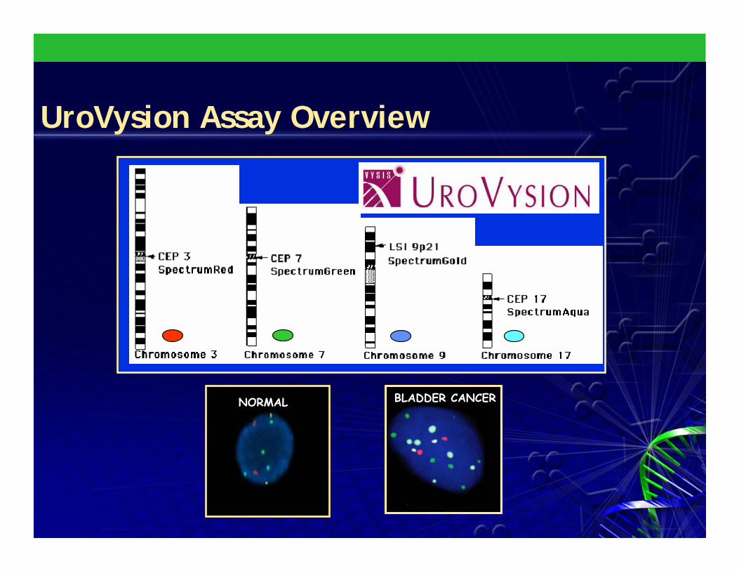
BLADDER CANCERNORMAL
UroVysion Assay Overview

Fine Needle Aspiration Biopsy (Applications)
Growing trend in FNA cytology to better classify genetic mutations that can aid the cytologist in stratifying aggressive versus less aggressive lesions.
a.Neuroblastoma: N-Myc characterization using fluorescent in situ hybridization (FISH)
b. Thyroid (papillary) cancer: – the RET tyrosine kinase domain (RETTK) using
PCR– BRAF mutations by PCR; preoperative dx for
PTC; not seen in benign nodules– Telomerase activity: limited use due to +
thyroiditisC. Ewings sarcoma: FISH; t(11:22) (q24;q12) chromosomal translocation using the SLI-EWSRI (22q12) dual color probe

Breast Cancer: Her2/Neu•25% of breast cancer patients have tumors which overexpress Her2/neu gene (chromosome 17 (17q11.2-q12)
•ER/PR negativity and a poor prognosis. Patients are eligible for Herceptin (trastuzumab) therapy
•Immunohistochemistry (IHC) or protein overexpression for Her2/neu often is difficult to discern (due to weak positive signals or background staining)
•* Large percentage of tumors considered positive by IHC (>=2) for Her2/neu by IHC show no gene amplification (presence of detectable mRNA); therefore, ICH was falsely positive.

Differences in Interpretation: HER-2 Assessment by FISH and IHC
1+1+2+2+ Negative/0Negative/03+3+
High High amplificationamplification NormalNormalLow Low
amplificationamplification

Her2/neuFISH testing may have a sensitivity and specificity of 96-100% for assessing over expression of the Her2/neu gene
Enhances the opportunity for more effective therapeutic triage
Vysis, Inc. (PathVision) – (ratio of Her2/neu to CEP 17) prevents misdiagnosis
in cases with polysomy for chromosome 17)
Oncor, Ventana (ISH)– Her2/neu only
* Ventana Benchmark– Her2/neu only
* Not FDA approvedVentana Benchmark (ISH)
*Single Colormetric k
Vysis (Pathvision)
*Dual Colormetric marker

FNA Applications
Breast cancer:
Human androgen receptor monoclonality has been linked to breast cancer and reflex testing for equivocal or “atypical”breast aspirates (as well as ductal lavage)
Loss of heterozygosity (LOH) of p53 oncogene mutations are found in 50% of all human cancers, including up to 80% of breast cancer (17p13.1) and colon cancer

FNA Applications
Pulmonary Neoplasms– Rationale: differentiation between “atypical”,
“suspicious”, or “indeterminate”– CNB could not be performed or because of the
development of pneumothorax– LaVision (Vysis) multitarget FISH assay for 6p11-
q11, 7p12 (EGFR), 8q24 (myc), and 5p15.2-chromosomal loci affected in non-small cell lung carcinoma
RenalRT-PCR of MN/CA9 gene expression response to hypoxic conditions
– Clear RCC (100%) and papillary RCC (56%) . Not found in chromophobe RCC, oncocytoma, or normal tissue

FNA ApplicationsPancreatic cancer:
Paucity of diagnostic material, overlapping features of low grade malignancies, chronic pancreatitis
K-ras mutations are seen in 90% of pancreatic carcinomas and up to 80% of cholangiocarcinomas
Teleomerase activity: limited use: not expressed in all PN
Effusions: FISH analysis for chromosomes 3, 8, 10, and 12 are being used to determine hyperdiploidy for equivocal or “atypical” cells

FNA Applications
Metastatic Tumors in lymph nodesMicrometastasis in EUS-FNAB to r/ogastrointestinal vs lung: hypermethylationof CpP islands in promoter regions of MGMT, p16 and p14
Infectious disease:HPV: differentiate anogenital or head and neck SCC versus espohagus, lung and skin SCC

Future FNA Applications
Colon Cancer
Hereditary nonpolyposis colorectal cancer, an inherited syndrome and analysis of mutations associated with familial polyposis (FAP) and attenuated FAP is available with COLARIS APSM.
– analysis is a PCR-based assay (Myriad Genetics).
Melanoma and Pancreatic Cancer: the p16 gene mutation can be determined with PCR using MELARISSM ( Myriad Genetics).

New Technologies: Not all Molecular

P16 as a Marker for Cervical Cancer?
Overexpression of p16INK4a protein indicates infection and genomic integration of high-risk human papillomavirus (HR HPV) and predicts progression to cervical high-grade squamousintraepithelial lesions (HSILs) and carcinoma.
p16INK4a protein was immunolocalized using a specific monoclonal antibody, and the detection of HR HPV in all 400 specimens was determined using HC2.
p16INK4a was found to be positive in only 78% of HSIL
Holladay et al. Cancer (Cytopathology) Vol 108, (6) October 2006

p16 as a Screening Test
HSIL-3110 HPV DNA positive, p16 true positive
HSIL-3118HPV DNA positive p16 true positive
Holladay et al. Cancer (Cytopathology) Vol 108, (6) October 2006

p16 as a Screening Test
WNL-3027HPV DNA negative, p16 false positive metaplastic cell
HSIL-3127 HPV DNA positive, p16 false negative
Holladay et al. Cancer (Cytopathology) Vol 108, (6) October 2006
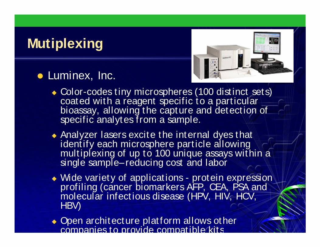
Mutiplexing
Luminex, Inc. Color-codes tiny microspheres (100 distinct sets) coated with a reagent specific to a particular bioassay, allowing the capture and detection of specific analytes from a sample.
Analyzer lasers excite the internal dyes that identify each microsphere particle allowing multiplexing of up to 100 unique assays within a single sample– reducing cost and labor
Wide variety of applications - protein expression profiling (cancer biomarkers AFP, CEA, PSA and molecular infectious disease (HPV, HIV, HCV, HBV)
Open architecture platform allows other companies to provide compatible kits

Glass slide microarray of thousands of genes for evaluation as new biomarkers for cancer prognostic assessment
Eleftherios P. Diamandis, M.D., Ph.D., FRCP(C)Mt. Sinai Hospital and Univ. of Toronto
The Future of Molecular Pathology

Genome: 12,800 points
Diameter: 120 microns
Slide size: 170 mm x 340 mm
Eleftherios P. Diamandis, M.D., Ph.D., FRCP(C)
Eleftherios P. Diamandis, M.D., Ph.D., FRCP(C)
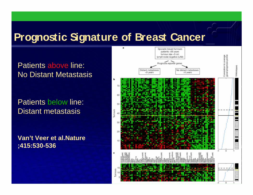
Prognostic Signature of Breast Cancer
Patients aboveabove line:No Distant Metastasis
Patients belowbelow line:Distant metastasis
Van’t Veer et al.Nature ;415:530-536

The Future??
Cancer Patient
↓ Surgery/BiopsySurgery/Biopsy
Cancerous Tissue
↓ Array AnalysisArray Analysis
Tumor Fingerprint
↓Individualized Treatment
Eleftherios P. Diamandis, M.D.,

Thank You