Embed Size (px)
Citation preview

Bronchial aspirated samples rPCR increases pathogen identification rate in critically ill patients with pneumonia: a pilot experience
DV. Pérez Civantos*,1,2, M. Fajardo Olivares2,3, P. Nieto Sánchez1, M. Robles Marcos1, V. Jerez Gómez-Coronado1,2 and H. Fariñas Seijas4 1 ICU; Department of Intensive Care Medicine. University Hospital Infanta Cristina. University of Extremadura. Avda de
Elvas s/n. 06071 Badajoz, Spain 2 Department of Biomedical Sciences. Faculty of Medicine, University of Extremadura. Avda. Elvas s/n, 06071 Badajoz,
Spain 3 Department of Microbiology and Parasitology. University Hospital Infanta Cristina. Faculty of Medicine. University of
Extremadura. Avda de Elvas s/n. 06071 Badajoz. Spain 4 CICAB (Center of Clinical Investigation of the Area of Badajoz). SES-University of Extremadura. Avda de Elvas s/n.
06071 Badajoz. Spain * Corresponding author: email: [email protected]
Community acquired pneumonia (CAP) and ventilator associated pneumonia (VAP) are two medical entities that carry high elevated health-care costs and mortality. A rapid an accurate diagnosis with a prompt and correct antibiotic treatment are the mainstay for good outcome. Knowledge of etiologic microbiological involvement is a cornerstone for antibiotic treatment, further de-escalation and avoids the appearance of antibiotic resistance pathogens. Molecular base techniques as real time polymerase chain reaction (r PCR) can improve microbiological results.
Keywords: Community acquired pneumonia; Health-care acquired pneumonia; Ventilator-associated pneumonia; Real time PCR; microbiological diagnosis of severe pneumonia; Pneumonia in ICU.
1. Introduction and Justification
Ventilator-associated pneumonia (VAP) is defined as pneumonia that occurs 48–72 hours or thereafter following endotracheal intubation, characterized by the presence of a new or progressive infiltrate, signs of systemic infection (fever, altered white blood cell count), changes in sputum characteristics, and detection of a causative agent (1). VAP contributes to approximately half of all cases of hospital-acquired pneumonia (1, 2). VAP is estimated to occur in 9–27 % of all mechanically ventilated patients, with the highest risk being early in the course of hospitalization (1-3). It is the second most common nosocomial infection in the intensive care unit (ICU) and the most common in mechanically ventilated patients (4, 5). VAP rates range from 1.2 to 8.5 per 1,000 ventilator days and are reliant on the definition used for diagnosis (6). Risk for (VAP) is greatest during the first 5 days of mechanical ventilation (3 %) with the mean duration between intubation and development of VAP being 3.3 days (1-6). Mortality rate is variable and relies heavily on the underlying medical illness (1). The attributable risk of death has decreased and is more recently estimated at 9–13 % (5, 9, 10), largely because of implementation of preventive strategies. Approximately 50 % of all antibiotics administered in ICUs are for treatment of VAP (2, 4). Early onset VAP is defined as pneumonia that occurs within 4 days and this is usually attributed to antibiotic sensitive pathogens whereas late onset VAP is more likely caused by multidrug resistant organisms (MDRO) and emerges after 4 days of intubation (1, 4, 5). Thus, VAP poses severe implications in endotracheally intubated adult patients in ICUs worldwide and leads to increased adverse outcomes and healthcare costs. Independent risk factors for development of VAP are male sex, admission for trauma and intermediate underlying disease severity, with odds ratios (OR) of 1.58, 1.75 and 1.47–1.70, respectively (6). Most community-acquired pneumonia (CAP) now occurs in elderly patients (> 65 yr), but little is known about it in younger adults (18 to 65 yr). Using a database on 7,803 German patients with CAP, Klapdor and colleagues compared those age groups and identified significant differences between their comorbidities, clinical presentations, severities, etiologies, and mortality rates (7). Only half of the younger patients had comorbidities (compared with 88% of older patients), their CAP was less severe, Streptococcus pneumoniae and Mycoplasma pneumoniae represented the most frequent causative microorganisms identified (respectively, 25.1 and 4.5%) (7). Health-care contact before pneumonia onset, including admission from a nursing home, recent hospitalization, antibiotic exposure, immunosuppression, and/or chronic hemodialysis, may constitute a risk factor for MDRO, thereby substantially changing the microbes expected to be causing CAP, and, these patients may require more aggressive therapeutic management to prevent excess mortality. A major challenge for patients hospitalized with CAP is the early identification of those at risk of deterioration who could benefit from ICU admission. Hypocapnia and hypercapnia should be considered for severity stratification and to
1013© FORMATEX 2015
The Battle Against Microbial Pathogens: Basic Science, Technological Advances and Educational Programs (A. Méndez-Vilas, Ed.)

identify patients at higher risk of mortality who require intensive care (8). Adding arterial pH less than 7.30 to the 2007 Infectious Disease Society of America/American Thoracic Society major criteria, improved identification sensitivity and area under the receiver operating characteristic curve for those who would require ICU (11).
1.1 Pathogenesis
The complex interplay between the endotracheal tube, presence of risk factors, virulence of the invading bacteria and host immunity largely determine the development of VAP. The presence of an endotracheal tube is by far the most important risk factor, resulting in a violation of natural defense mechanisms (the cough reflex of glotis and larynx) against microaspiration around the cuff of the tube (4, 12). Infectious bacteria obtain direct access to the lower respiratory tract via: (a) microaspiration, which can occur during intubation itself; (b) development of a biofilm laden with bacteria (typically Gram-negative bacteria and fungal species) within the endotracheal tube; (c) pooling and trickling of secretions around the cuff; and (d) impairment of mucociliary clearance of secretions with gravity dependence of mucus flow within the airways (4, 5, 13, 14). Host factors such as the severity of underlying disease, previous surgery and antibiotic exposure have all been implicated as risk factors for development of VAP (1). In addition, it has recently been noted that critically ill patients may have impaired phagocytosis and behave as functionally immunosuppressed even prior to emergence of nosocomial infection (4, 5, 13, 14). This effect is attributed to the detrimental actions of the anaphylatoxin, C5a, which impairs neutrophil phagocytic activity and impairs phagocytosis by neutrophils (15). More recently, a combined dysfunction of T-cells, monocytes, and neutrophils has been noted to predict acquisition of nosocomial infection (15).
1.2 Microbiology
The type of organism that causes VAP usually depends on the duration of mechanical ventilation. In general, early VAP is caused by pathogens that are sensitive to antibiotics, whereas late onset VAP is caused by MDRO and more difficult to treat bacteria. However, this is by no means a rule and merely a guide to initiate antibiotic therapy until further clinical infor mation is available. Typically, bacteria causing early-onset VAP incluye S. pneumoniae (as well as other streptococcus spp.), Hemophilus influenzae, methicillin-sensitive Staphylococcus aureus (MSSA), antibiotic-sensitive enteric Gram-negative bacilli, Escherichia coli, Klebsiella pneumonia, Enterobacter spp., Proteus spp. and Serratia marcescens. Culprits of late VAP are typically MDRO, such as methicillin-resistant S. aureus (MRSA), Acinetobacter baumanii, Pseudomona aeruginosa, and extended-spectrum beta-lactamase producing bacteria (ESBL) (4). The exact prevalence of MDRO is variable between institutions (1). Patients with a history of hospital admission for � 2 days in the past 90 days, nursing home residents, patients receiving chemotherapy or antibiotics in the last 30 days and patients undergoing hemodialysis at outpatient centers are susceptible to MDRO (1, 4). Frequently, VAP is due to polymicrobial infection. VAP from fungal and viral causes has a very low incidence, especially in the immunocompetent host (1). Contemporary standards for high-quality microbiological analysis for CAP include 4 components: specimen source, pathogenic potential of various organisms, concentrations of organisms recovered, and the influence of prior antibiotics. The most readily available specimen and diagnostic standard in most cases is expectorated sputum. This specimen must traverse the upper airways, which are colonized with large concentrations with multiple bacteria (109–1010 colony-forming units [CFU]/mL saliva), including some that are potentially agents of pneumonia, such as S. pneumoniae, H. influenzae, and, in patients with prior antibiotics or underlying diseases, enteric gram-negative bacilli (11, 16). With respect to the problem of contamination, there have been numerous attempts to decrease it. Neither of these procedures attracted much attention. The one system that survived this period of attempts at quality improvement is the molecular analysis of cellular constituents of sputum, as initially reported from Murray and Washington (17). This report recommended discarding specimens that contained >10 squamous epithelial cells (SECs) per low-power field (LPF) with a 100x magnification of the gram-stained smear. Particularly important among the multitude of studies examining the various criteria was the report by Geckler et al (18), who compared the results from various methods of expectorated sputum screening with results from transtracheal aspiration. This supported the use of ,25 SECs/LPF, which became the criterion that was subsequently endorsed by Washington (19). It is important to pose that an etiologic agent can rarely be identified in more than 50% of patients with community-acquired pneumonia (CAP) (20), further development of diagnostic methods has been encouraged. An important development for detection of this specific pathogen is the urinary antigen assay, which has the advantages of ease of getting a diagnostic specimen and substantial improvement over sputum in terms of diagnostic yield and ability to establish this diagnosis after antibiotic treatment, sensitivity of 82% and a specificity of 97% (21, 22). The urinary antigen test has also been developed and is now the favored and the most practical test for detection of Legionnaires disease, which accounts for 2%–6% of CAP cases (21-26, 47).
1014 © FORMATEX 2015
The Battle Against Microbial Pathogens: Basic Science, Technological Advances and Educational Programs (A. Méndez-Vilas, Ed.)

1.3 Diagnosis
At the present time, there is no universally accepted, gold standard diagnostic criterion for VAP. Several clinical methods have been recommended but none have the needed sensitivity or specificity to accurately identify this disease (27). Daily bedside evaluation in conjunction with chest radiography can only be suggestive of the presence or absence of VAP, but not define it (28). Clinical diagnosis of VAP can still miss about a third of VAPs in the ICU compared to autopsy findings and can incorrectly diagnose more than half of patients, likely due to poor interobserver agreement between clinical criteria (8, 28, 29). Postmortem studies comparing VAP diagnosis with clinical criteria showed 69 % sensitivity and 75 % specificity, in comparison to autopsy findings (30). The American Thoracic Society (ATS) and the Infectious Diseases Society of America (IDSA) guidelines recommend obtaining lower respiratory tract samples for culture and microbiology (1). Analysis of these samples can be quantitative or qualitative. This guideline also allows use of tracheal aspirates for their negative predictive value (94 % for VAP) (31). Pugin et al (31) described clinical criteria for diagnosis of VAP as the clinical pulmonary infection score (CPIS) that takes into account clinical, physiological, microbiological and radiographic evidence to allow a numerical value to predict the presence or absence of VAP (Table 1) (28, 31). Scores can range between zero and 12 with a score of � 6 showing good correlation with the presence of VAP (31). Despite the clinical popularity of the CPIS, debate continues regarding its diagnostic validity. One metanálisis of 13 studies evaluating the accuracy of CPIS in diagnosing VAP reported pooled estimates for sensitivity and specificity for CPIS as 65 % (95 % CI 61–69 %) and 64 % (95 % CI 60–67 %), respectively (31). Despite its apparent straightforward calculation, the inter-observer variability in CPIS calculation remains substantial, jeopardizing its routine use in clinical trials (32). Of all the criteria used to calculate the CPIS, only time-dependent changes in the PaO2/FiO2 ratio early in VAP may provide some predictive power for VAP outcomes in clinical trials, namely clinical failure and mortality (33). However, a trial by Singh and colleagues (34) demonstrated that the CPIS is an effective clinical tool for determining whether to stop or continue antibiotics for longer than 3 days. Standard criteria for dignosis of VAP are:
1. New or progressive radiographic consolidation or infiltrate. In addition, at least 2 of the following: 2. Temperature > 38º C 3. Leukocytosis (white blood cell count � 12,000 cells/ mm3) or leukopenia (white blood cell count < 4,000 cells/mm3) 4. Presence of purulent secretions
Respiratory samples can be obtained using several techniques: The ATS/IDSA guidelines note that use of a bronchoscopic bacteriologic strategy has been shown to reduce 14-day mortality when compared with a clinical strategy (16.2 % vs. 25.8 %, p = 0.02) (1, 35). When samples are obtained by BAL techniques (BAL, mini-BAL or PSB), the diagnostic threshold is 103 colony forming units (cfu)/ml for protected specimen brushing and 104 cfu/ml for BAL. More recent evidence from the Canadian Clinical Trials study of 740 suspected VAP patients randomized to BAL or tracheal suctioning (BAS) suggests that (excluding patients known to be colonized/infected with Pseudomona spp. or MRSA) similar clinical outcomes and overall use of antibiotics is observed when either BAL with quantitative culture or BAS with non-quantitative culture is used for diagnosis (36). This finding was confirmed by a Cochrane meta-analysis (37). Culture results can be reported as semi-quantitative and/or quantitative values. Once specimens are obtained, the sample is sent for Gram stain (DE), culture and sensitivity (C). The Gram stain can provide crucial initial clues to the type of organism(s) and whether or not the material is purulent (defined as � 25 neutrophils and � 10 squamous epithelial cells per low power field) (1). Mechanically ventilated patients in the ICU receive frequent chest X-rays and presence of infiltrate(s) and/or consolidation is considered part of diagnostic criteria and is widely used. However, there are several clinical conditions that have radiographic appearances similar to VAP. There is poor correlation between radiographic signs (alveolar infiltrates, air bronchograms) and histopathological diagnosis of pneumonia (38). The sensitivity and specificity of presence of infiltrates on chest X-ray is also not encouraging (38). On the flip-side, the negative predictive value of infiltrates may have clinical utility. In a meta-analysis by Klompas, the presence or absence of fever, elevated white blood cell count, or purulent secretions did not substantively predict the probability of infection; however, the absence of a new infiltrate on a plain radiograph lowered the likelihood of VAP (28). VAP must be distinguished from tracheo-bronchitis. Clinical features of these diseases can overlap, but only VAP will demonstrate the presence of hypoxia and the presence of infiltrate/consolidation on chest radiography (38). CAP should be suspected in patients with newly acquired lower respiratory symptoms (cough, sputum production, and/or dyspnea), especially if accompanied by fever, altered breath sounds, and rales. The importance of establishing the diagnosis of pneumonia and its cause is heightened with the increasing concern about antibiotic overuse (14, 39). The diagnosis of CAP is based on a combination of clinical and laboratory (including microbiological) data. The differential diagnosis of lower respiratory symptoms is extensive and includes upper and lower respiratory tract infections, as well as noninfectious causes. Most cases of upper respiratory tract infection are of viral origin, do not
1015© FORMATEX 2015
The Battle Against Microbial Pathogens: Basic Science, Technological Advances and Educational Programs (A. Méndez-Vilas, Ed.)

require antimicrobial therapy, and are the source of great antibiotic abuse (8, 40, 41). A chest radiography is usually necessary to establish the diagnosis of pneumonia (42, 43). Physical examination to detect rales or bronchial breath sounds is neither sensitive nor specific for detecting pneumonia (8, 42, 44, 45, 46). Chest radiography is considered sensitive and, occasionally, is useful for determining the etiologic diagnosis, the prognosis, and alternative diagnoses or associated conditions (8, 45). One study showed spiral CT scans are significantly more sensitive in detecting pulmonary infiltrates (47), but the IDSA panel does not endorse the routine use of this technology because of the preliminary nature of the data and high cost of the procedure (8). The emphasis on microbiological studies (Gram staining and culture of expectorated sputum) in the IDSA guidelines represents a difference from the guidelines of the American Thoracic Society (48).
1.4 Treatment
For VAP selecting the appropriate antibiotic depends on the duration of mechanical ventilation. Late onset VAP (> 4 days) requires broad spectrum antibiotics whereas early onset (� 4 days) can be treated with limited spectrum antibiotics (1). An updated local antibiogram for each hospital and each ICU based on local bacteriological patterns and susceptibilities is essential to guide optimally dosed initial empiric therapy (1). With any empiric antibiotic regimen, de-escalation is the key to reduce emergence of resistance (49). Delays in initiation of antibiotic treatment may add to the excess mortality risk with VAP (1). Owing to the high rate of resistance to monotherapy observed with P. aeruginosa, combination therapy is recommended. Acinetobacter spp. respond best to carbapenems (also active against ESBL positive Enterobacteriaceae), colistin, polymyxin B and ampicillin/sulbactam (50, 51). Although MDRO are usually associated with late-onset VAP, recent evidence suggests that they are increasingly associated with early onset VAP as well (52, 53). The role of inhaled antibiotics in the setting of failure of systemic antibiotics is unclear (1). The usual duration of treatment for early onset VAP is 8 days and longer in the case of late-onset VAP or if MDRO are suspected or identified (53–55). Despite therapy, if no response is observed, it may be prudent to reconsider the diagnosis (65). Because of the challenges associated with diagnosing VAP, especially early in the course, the IDSA/ATS guidelines highlight the importance of reassessing patients at 48–72 hours (65). In one study, Swoboda et al. (56) found that half of the empiric antibiotic use for VAP in two surgical ICUs was prescribed for patients without pneumonia. Recommendations for severe CAP empirical therapy a regimen with a b-lactam plus a macrolide or monotherapy with a fluoroquinolone is preferred. The rationale for recommending these regimens is based on studies showing that these regimens were associated with a significant reduction in mortality, compared with that associated with administration of cephalosporin alone (57, 58). In critically ill patients with pneumonia, antibiotic therapy is mandatory to improve prognosis and should be administered as soon as possible. In the case of VAP, inappropriate antibiotic therapy might increase the length of stay in the ICU and even double mortality (59-61). The spectrum of antibiotic therapy should be adapted as soon as microbial identification is available (45). Gram staining of respiratory samples obtained with protected distal sampling, or BAL is rapid but its sensitivity is low (62, 63). The results of quantitative culture of these specimens are available only 24 to 48 h later. Furthermore, the sensitivity of culture is decreased by prior antimicrobial therapy, especially if antibiotics have been introduced recently (49).
1016 © FORMATEX 2015
The Battle Against Microbial Pathogens: Basic Science, Technological Advances and Educational Programs (A. Méndez-Vilas, Ed.)
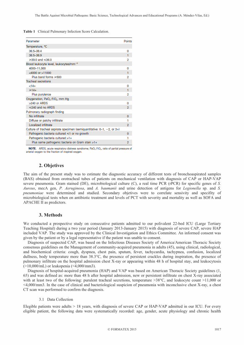
Table 1 Clinical Pulmonary Infection Score Calculation.
2. Objetives
The aim of the present study was to estimate the diagnostic accuracy of different tests of bronchoaspirated samples (BAS) obtained from orotracheal tubes of patients on mechanical ventilation with diagnosis of CAP or HAP-VAP severe pneumonia. Gram stained (DE), microbiological culture (C), a real time PCR (rPCR) for specific genes of S. Aureus, mecA gen, P. Aeruginosa, and A. baumanii and urine detection of antigens for Legionella sp. and S. pneumoniae were determined and studied. Secondary objetives were to correlate sensivity and specifity of microbiological tests when on antibiotic treatment and levels of PCT with severity and mortality as well as SOFA and APACHE II as predictors.
3. Methods
We conducted a prospective study on consecutive patients admitted to our polivalent 22-bed ICU (Large Tertiary Teaching Hospital) during a two year period (January 2013-January 2015) with diagnosis of severe CAP, severe HAP included VAP. The study was approved by the Clinical Investigation and Ethics Committee. An informed consent was given by the patient or by a legal representative if the patient was unable to consent. Diagnosis of suspected CAP, was based on the Infectious Diseases Society of America/American Thoracic Society consensus guidelines on the Management of community-acquired pneumonia in adults (45), using clinical, radiological, and biochemical criteria: cough, dyspnea, chest pain, sputum, fever, tachycardia, tachypnea, confusion, localized dullness, body temperature more than 38.3°C, the presence of persistent crackles during inspiration, the presence of pulmonary infiltrate on the hospital admission chest X-ray or appearing within 48 h of hospital stay, and leukocytosis (>10,000/mL) or leukopenia (<4,000/mm3). Diagnosis of hospital-acquired pneumonia (HAP) and VAP was based on American Thoracic Society guidelines (1, 65) and was defined as: more than 48 h after hospital admission, new or persistent infiltrate on chest X-ray associated with at least two of the following: purulent tracheal secretions, temperature >38°C, and leukocyte count >11,000 or <4,000/mm3. In the case of clinical and bacteriological suspicion of pneumonia with inconclusive chest X-ray, a chest CT scan was performed to confirm the diagnosis.
3.1 Data Collection
Elegible patients were adults > 18 years, with diagnosis of severe CAP or HAP-VAP admitted in our ICU. For every eligible patient, the following data were systematically recorded: age, gender, acute physiology and chronic health
1017© FORMATEX 2015
The Battle Against Microbial Pathogens: Basic Science, Technological Advances and Educational Programs (A. Méndez-Vilas, Ed.)

evaluation II (APACHE II), sepsis related organ failure assessment (SOFA), type of pneumonia (CAP or HAP (including VAP)), underlying immunodeficiency [defined as one of the following: AIDS, cancer or hematological disease with chemotherapy administered less than 30 days before admission, recipient of solid organ (liver) or bone marrow transplant, corticosteroids more than 1 mg/kg/day equivalent prednisone for more than 1 month, and other immunosuppressive therapies], septic shock according to international definitions (64), procalcitonine (PCT) levels ( expressed as mg.L-1 ) on first 12 hours after admission, cytology and Gram stained (DE) of the BAS, cultured results (C) of BAS, rPCR results on BAS, urine antigens detection, administration of a recent antibiotic treatment (defined as a new antibiotic therapy initiated within 48 h before BAS), and results of all microbiological specimens obtained during 2 days befote or after BAS and ICU mortality.
3.2 Procedures and analisis
The BAS is sent to the microbiology laboratory in sterile wide-mouth container, double lid and screw cap. The sample is used for a Gram stain, culture on non-selective agar medium for common bugs, and polymerase chain reaction (PCR) for multiresistant pathogens, by RealCycler (Progenien Molecular). In case of delayed simple procedure, storage frozen at -20ºC until used. This issue is an in vitro diagnostic kit of reagents that allows real-time PCR (rPCR) qualitative detection of Staphylococcus aureus, Pseudomonas aeruginosa and Acinetobacter baumannii DNA simultaneously. The amplification is done by SmartCycler� (Cepheid�). The PCR is based on the amplification of a specific región of the DNA/RNA by using complementary primers to the target sequence. This rPCR uses marked probes with fluorophores that emit fluorescence in the case of amplification. P. aeruginosa is detected in the corresponding channel of FAM fluorophore, while A. baumannii is detected in TxR fluorophore and S. aureus in Alx647 fluorophore. Finally, an internal control is detected in Alx532 fluorophore. The system includes an internal control of amplification to prevent false negatives due to reaction inhibition. The interpretation of the results is cualitative (positive, not detected or not assessable). Samples for rPCR were processed on their arrival to the microbiology department daily from 8.00-15.00 hour and from Monday till Friday; out of daily work-time hours samples were kept frozen until next workable day. One week after ICU admission, according to the clinical evolution, chest radiograph, and results of DE, C, and other conventional microbiological samples, all medical files were assessed in a blinded manner by two senior physicians to rule in or not the diagnosis of infectious pneumonia. In the case of discrepant conclusions, a third senior intensivist assessed the data until a consensus was obtained. Physicians analyzing the data were blinded to the results of the rPCR. Based on the clinical history, clinical examination, radiological file, biological results, and of other microbiological samples (blood cultures, DE, C, Streptococcus pneumoniae, and antigenuria), the diagnosis of infectious pneumonia was ruled in or ruled out and the rPCR results were assessed and compared with the results of DE and C.
3.3 Statistical analysis
Continuous variables were expressed as mean ± SD. Categorical variables were expressed as proportions and compared using the Fisher´s exact test for independent samples and the McNemar chi-square test for matched pairs. ROC curve analysis was performed to determine optimal cutoff values. Sensitivity and specificity and their 95% confidence interval were calculated. Two-tailed probability values less than 0.05 were considered statistically significant. Statistical analysis was performed using the SPSS 19.0 software (SPSS, Inc., Chicago, IL, USA).
4. Results
During the study period, pneumonia was suspected in 65 patients and finally confirmed in only 50 of them (age: 60±17 years (age range, 22 to 78 years), 35 males (70%), SOFA 7.2 ± 2.8 and APACHE II: 17.6 ± 5, invasive mechanical ventilation requirement: 100%, septic shock: 32%, immunosuppression: 18%. The chest X-ray showed the pneumonia in 84% of patients. In eight patients (16%), the pneumonia was visible only in the chest CT scan. The types of pneumonia broke as follows: CAP (n = 12, 24%) and HAP/VAP (n = 38, 76%). Among the HAP, 30 were VAP. Demographics and clinical characteristics of the 50 patients with suspected pneumonia are summarized in Table 2. Among the 15 patients for whom pneumonia was finally ruled out, alternative diagnosis was: intra-alveolar hemorrhage (n = 3), hemodynamic pulmonary edema (n = 3), a vacuo edema complicating pneumothorax exsufflation (n = 1), pulmonary fibrosis (n = 1), and acute respiratory distress syndrome or acute lung injury related to extrapulmonary sepsis (n = 7). Pathogen identification rate with rPCR (37/50; 74%, sensibility 89%, specifity 35%) was greater than with culture (19/50; 38%) (p <0.001 for the three pathogens included in the rPCR test. Pathogen identification rate provided by rPCR was not modified in the case of previous antibiotic treatment (28/38; 74%) and was still better than with culture (19/38; 50%, p <0.01) irrespective of whether pneumonia was CAP or HAP-VAP.
1018 © FORMATEX 2015
The Battle Against Microbial Pathogens: Basic Science, Technological Advances and Educational Programs (A. Méndez-Vilas, Ed.)

At the time of the BAS, 38 patients (76%) were receiving recent antibiotic therapy (patients with CAP: 8 (66%), patients with HAP-VAP: 21 (55%)). The reasons for this antibiotic treatment were: pneumonia (n = 17), bronchitis (n = 4), bactaeremia associated with febrile neutropenia (n = 2), febrile neutropenia without bactaeremia (n = 2), urinary tract infection (n = 2), catheter-associated bactaeremia (n = 1), and intra-abdominal infection (n = 1). The results of pathogen identification provided by DE, C, and rPCR are depicted in Table 3 and 4. Using data provided by other microbiological tests (as urinary pneumococcal antigen), a pathogen identification was obtained in one supplemental case, resulting in a final pathogen identification in 48/50 cases (96%). These two patients, for whom all microbiological examinations were negative, were receiving antibiotic therapy when BAS and the rest of microbiological determinations were performed. Bacterial species identified by DE, C, and rPCR are depicted on Table 4. Urinary pneumococcal antigen was positive in only one case (CAP) (n = 1). In patients for whom pneumonia diagnosis was finally ruled out, rPCR detected a pathogen in 4/15 patients (26%). This was considered as colonization. Receiver operating curve (ROC) analysis indicated: a) a critical value 2.2 as the optimal cut-off point for assessing PCT and mortality (sensitivity of 84% [CI95%: 68-97%] and specificity of 65% [48%-81%]); b) a critical value 7.5 as the optimal cut-off point for assessing SOFA and mortality (sensitivity of 84% [ 68-97%] and specificity of 81% [67%-95%]); and c) a critical value 17.5 as the optimal cut-off point for assessing APACHE II and mortality (sensitivity of 95% [CI95%: 85-100%] and specificity of 68% [51%-84%]) (Figure 1,2 and 3).
1019© FORMATEX 2015
The Battle Against Microbial Pathogens: Basic Science, Technological Advances and Educational Programs (A. Méndez-Vilas, Ed.)

5. Discussion
Recently published guidelines recommend antibiotic administration within 1 h for patients suspected of having septic shock or severe sepsis (66). During severe infection, empirical antibiotic therapy is inappropriate in roughly one-third of cases, and this substantially increases mortality and hospital length of stay (65, 67). In the setting of infection, any tool enabling prompt and accurate documentation of pathogen might theoretically reduce mortality and may serve to improve hospital resource use (43, 65). Our study was carried out during two consecutive years and all patients were correlative included if they met the inclussion criteria. Final diagnosis of pneumonia relied on a posteriori analysis of clinical, radiological, and microbiological data. This analysis was conducted by two senior physicians who were blinded to rPCR results. This study shows an increase of the spectra of microbiological studies to carry out on biological samples with rapid and reliable results that can help the clinician to discard pathogens supposedly involved as causing pneumonia. Then our study suggest that rPCR test significantly increased the identification rate of pathogen causing pneumonia, compared with usual culture gram stain (C), culture (DE) and urinary antigen detection. This was also already published by several authors (68, 69, 70, 71). Increased pathogen identification rate provided by rPCR compared to conventional cultures has already been reported during the course of bloodstream infections (72-75), febrile neutropenia (76), and persistent fever (77). The present data also suggest that rPCR can be done on samples having been kept frozen for several days until availability of microbiological technician, so that its profitability is clearly improved.
Fig. 1 ROC for PCT; SOFA and APACHE II.
The excellent rPCR negative predictive value (even on patients already on antibiotics) suggests that antibiotics directed against MRSA may not be used in most patients with a negative test. This finding is in agreement with that already published in previous studies (69, 71). Importantly, the rPCR test cannot confirm the presence or absence of VAP. The diagnosis of VAP is based on clinical, radiological and microbiological features (43, 45, 46, 65). In this study, microbiological diagnoses were achieved in 96% of patients, though 76% of then were already on antibiotic treatment; in this group of patients rPCR greatly helped to reach a final microbiological diagnosis. Rello et al. (78) claimed that microbiological diagnosis was fully justified in cases of severe community-acquired pneumonia because it had an impact on patient outcome, but others have found that diagnosis has had no impact (79, 80). However, rPCR provides the opportunity to identify the etiological agent in a clinically relevant time period. This could result in important changes in antimicrobial therapy in order to fit an early proper treatment and to lessen the appearance or increase of MDRO. Early detection of additional causative pathogens by a sensitive rPCR-based method has the potential to reduce the proportion of patients with initial inappropriate treatment (78). In contrast, detection of non-causative microorganisms may promote antibiotic overuse. Clinical judgment and consideration that some microrganisms are not part of the normal flora of the LRT and when detected must be considered as infection even when present in trace amounts (82). In the present study, pathogen identification was high and could have been better if the panel of the rPCR had included other microorganisms frequently causing pneumonia. The striking finding of our study is a prevalence rate of MRSA below 2%. This prevalence is lower than that reported in previous studies (69, 71). This is in line with data regarding the decreased number of bacteremias due to MRSA in Europe (72). It is very impressive the high number of A. baumanii detected whether in HAP-VAP or in CAP. This could be related to the high prescription pressure of antibiotics on patients that probably contributed to make a selection of the microbiological flora involved in pneumonia to MDRO.
1020 © FORMATEX 2015
The Battle Against Microbial Pathogens: Basic Science, Technological Advances and Educational Programs (A. Méndez-Vilas, Ed.)

The contribution of rPCR to pathogen identification was of particular interest in patients who had received antibiotics, that patients’ population showed low positive results (50%) in DE and C and determinations. It is known that the culture has a lower sensitivity in comparison with the PCR assay (lower limit of detection from 48 to 109.4 CFU/test) (69, 81) and that prior antibiotic therapy can lead to a negative culture. In addition, a positive test result does not necessarily indicate the presence of viable organisms but rather the presence of amplifiable DNA from dead bacteria in treated patients. This fact must be kept in mind, since bacterial DNA can be detected in clinical samples for more than 10 days after successful treatment (81, 82). The Infectious Diseases Society of America (IDSA) does not believe that regulatory clearance of a new diagnostic molecular test for VAP by the FDA should require that the test validates the presence of pneumonia or another variant of respiratory infection (69). Instead, the focus should be the accuracy of the new molecular method in the detection of a given bacterial or viral organism that will reduce antibiotic overuse (1), and in our study the Xpert assay proved to be useful for the detection of MRSA, MSSA, Pseudomona Aeruginosa and Acinetobacter baumaniii. Molecular analysis as rPCR, due to the ability to provide high pathogen identification more frequently than direct examination and culture and in a rapid fashion so that antibiotic treatment could be initiated early and secure or adding valid information if the idea is to stop them. Direct examination provides information in less than 2 h, but the diagnostic value of direct examination performed on BAS fluid was very good in our study as it identified pathogens in only 50% of patients. In routine, Gram stain is the first microbiological result available for the clinician. Its role remains a matter of debate. A meta-analysis showed that Gram stain is not reliable, with the exception of negative findings (83). PCT could help to identify those patients at high risk of mortality (if ranges > 2 mg.L-1 ) and probably also as soon it clearly descends or normalizes, when to stop antibiotics, in case the course of illness were satisfactory. SOFA and APACHE II were good predictors of mortality when puntuation was over 7 and 17 respectively. Our study has nevertheless several limitations. First, this was a monocenter study performed on a limited number of consecutive patients. These interesting data deserve to be confirmed on more large ICU population. Second, due to logistical and economical consideration, rPCR analysis was pooled and was not performed in real time in several patients. Finally, the time required by the technique (3 to 4 h) to provide microbiological results could be considered excessive, since currently available new fully automated PCR platforms provide results in 1 h.
6. Conclusions
The results of this pilot experience suggest that in critically ill patients with pneumonia, rPCR performed on BAS fluid could provide higher identification rate of pathogens involved in pneumonia than direct examination and culture, especially in patients having received antimicrobial treatment. Conflict of interest: None.
References [1] American Thoracic Society, Infectious Diseases Society of America: Guidelines for the management of adults with hospital-
acquired, ventilator-associated, and healthcare-associated pneumonia. Am J Respir Crit Care Med 2005, 171:388–416. [2] Vincent JL, Bihari DJ, Suter PM, Bruining HA, White J, Nicolas-Chanoin MH, Wolff M, Spencer RC, Hemmer M: The
prevalence of nosocomial infection in intensive care units in Europe. JAMA 1995, 274:639–644. [3] Chastre J, Fagon JY: State of the art: ventilator-associated pneumonia. Am J Respir Crit Care Med 2002, 165:867–903. [4] Hunter JD: Ventilator associated pneumonia. BMJ 2012, 344(e3325):e3325. [5] Kalanuria A, Zai W, Mirski M. Ventilator-associated pneumonia in the ICU. Critical Care 2014, 18:208
http://ccforum.com/content/18/2/208. [6] Rello J, Ollendorf D, Oster G, Vera-Llonch M, Bellm L, Redman R, Kollef MH. VAP Outcomes Scientific Advisory Group:
Epidemiology and outcomes of ventilator-associated pneumonia in a large US database. Chest 2002, 122:2115–2121. [7] Klapdor B, Ewig S, Pletz MW, Rohde G, Schütte H, Schaberg T,Welte T; CAPNETZ Study Group. Community-acquired
pneumonia in younger patients is an entity on its own. Eur Respir J 2012;39:1156–1161. [8] Sibila O, Meduri GU, Mortensen EM, Anzueto A, Laserna E, Fernandez JF, El-Sohl A, Restrepo MI. Improving the 2007
Infectious Disease Society of America/American Thoracic Society severe community acquired pneumonia criteria to predict intensive care unit admission. J Crit Care 2013 Jun;28(3):284-90. doi: 10.1016/j.jcrc.2012.09.010. Epub 2012 Dec 21.
[9] Melsen WG, Rovers MM, Koeman M, Bonten MJM: Estimating the attributable mortality of ventilator-associated pneumonia from randomized prevention studies. Crit Care Med 2011, 39:2736–2742.
[10] Melsen WG, Rovers MM, Groenwold RH, Bergmans DC, Camus C, Bauer TT,Hanisch EW, et al: Attributable mortality of ventilator-associated pneumonia: a metaanalysis of individual patient data from randomised prevention studies. Lancet Infect Dis 2013, 13:665–671.
[11] Mandell LA, Wunderink RG, Anzueto A, et al. Infectious Diseases Society of America/American Thoracic Society consensus guidelines on the Management of community-acquired pneumonia in adults. Clin Infect Dis 2007;44 (Suppl 2): S27–72.
1021© FORMATEX 2015
The Battle Against Microbial Pathogens: Basic Science, Technological Advances and Educational Programs (A. Méndez-Vilas, Ed.)

[12] Zolfaghari PS, Wyncoll DL: The tracheal tube: gateway to ventilatorassociated pneumonia. Crit Care 2011,15:310–317. [13] Mietto C, Pinciroli R, Patel N, Berra L: Ventilator associated pneumonia: evolving definitions and preventive strategies. Respir
Care 2013, 58:990–1007. [14] Morris AC, Brittan M, Wilkinson TS, McAuley DF, Antonelli J, McCulloch C, Barr LC, McDonald NA, Dhaliwal K, Jones
RO, Mackellar A, Haslett C, Hay AW, Swann DG, Anderson N, Laurenson IF, Davidson DJ, Rossi AG, Walsh TS, Simpson AJ: C5a-mediated neutrophil dysfunction is RhoA-dependent and predicts infection in critically ill patients. Blood 2011, 117:5178–5188.
[15] Morris AC, Anderson N, Brittan M, Wilkinson TS, McAuley DF, Antonelli J, McCulloch C, Barr LC, Dhaliwal K, Jones RO, Haslett C, Hay AW, Swann DG, Laurenson IF, Davidson DJ, Rossi AG, Walsh TS, Simpson AJ: Combined dysfunctions of immune cells predict nosocomial infection in critically ill patients. Br J Anaesth 2013, 3:1–10.
[16] Bartlett JG, Finegold SM. Bacteriology of expectorated sputum with quantitative culture and wash technique compared to transtracheal aspiration. Am Rev Respir Dis 1978; 117:1019–27.
[17] Murray PR, Washington JA II. Microscopic and bacteriologic analysis of expectorated sputum. Mayo Clin Proc 1975; 50:339–44.
[18] Geckler RW, Gremillion DH, McAllister CK, Ellenbogen C. Microscopic and bacteriological comparison of paired sputa and transtracheal aspirates. J Clin Microbiol 1977; 6:396–9.
[19] Washington JA. Noninvasive diagnostic techniques for lower respiratory infections. In: Pennington J, ed. Respiratory infections: diagnosis, management. 3rd ed. New York: Raven Press, Ltd, 1994: 55–71.
[20] Musher DM, Roig DL, Cazares G, Stager CE, Logan N, Safar H. Can an etiologic agent be identified in adults who are hospitalized for community-acquired pneumonia; results of a one-year study. J. Infect 2013;67:11-18. doi: 10.1016/j.jinf.2013.03.003.
[21] Smith MD, Sheppard CL, Hogan A, et al; Diagnosis of Streptococcus pneumoniae infections in adults with bacteremia and community- acquired pneumonia: clinical comparison of pneumococcal PCR and urinary antigen detection. J Clin Microbiol 2009; 47:1046–9.
[22] Smith MD, Derrington P, Evans R, et al; Rapid diagnosis of bacteremic pneumococcal infections in adults by using the Binax NOW Streptococcus pneumoniae urinary antigen test: a prospective, controlled clinical evaluation. J Clin Microbiol 2003; 41:2810–3.
[23] Joseph JA. Legionnaires’ disease in Europe 2000–2002. Epidemiol Infect 2004; 132:417–24. [24] Diederen BM. Legionella spp. and Legionnaires’ disease. J Infect 2008; 56:1–12. [25] Murdoch DR. Diagnosis of Legionella infection. Clin Infect Dis 2003; 36:64–9. [26] Dowell SF, Peeling RW, Boman J, et al; Standardizing Chlamydia pneumoniae assays: recommendations from the Centers for
Disease Control and Prevention (USA) and the Laboratory Centre for Disease Control (Canada). Clin Infect Dis 2001; 33:492–503.
[27] National Healthcare Safety Network (NHSN) July 2013 CDC/NHSN Protocol Clarifications 2013, Available at: http://www.cdc.gov/nhsn/PDFs/ pscManual/10-VAE_FINAL.pdf Accessed Oct 2013.
[28] Klompas M: Clinician’s Corner: Does this patient have ventilator-associated pneumonia? JAMA 2013, 297:1583–1593. [29] Petersen IS, Aru A, Skødt V, Behrendt N, Bols B, Kiss K, Simonsen K: Evaluation of pneumonia diagnosis in intensive care
patients. Scand J Infect Dis 1999, 31:299–303. [30] Fàbregas N, Ewig S, Torres A, Al-Abiary M, Ramirez J, de La Bellacasa JP, Bauer T, Cabello H: Clinical diagnosis of
ventilator associated pneumonia revisited: comparative validation using immediate post-mortem lung biopsies. Thorax 1999, 54:867–873.
[31] Pugin J, Auckenthaler R, Mili N, Janssens JP, Lew PD, Suter PM: Diagnosis of ventilator-associated pneumonia by bacteriologic analysis of bronchoscopic and nonbronchoscopic “blind” bronchoalveolar lavage fluid. Am Rev Respir Dis 1991, 143:1121–1129.
[32] Zil berberg MD, Shorr AF: Ventilator-associated pneumonia: the clinical pulmonary infection score as a surrogate for diagnostics and outcome. Clin Infect Dis 2010, 1:S131–S135.
[33] Shorr AF, Cook D, Jiang X, Muscedere J, Heyland D: Correlates of clinical failure in ventilator-associated pneumonia: insights from a large, randomized trial. J Crit Care 2008, 23:64–73.
[34] Singh N, Rogers P, Atwood CW, Wagener MM, Yu VL: Short-course empiric antibiotic therapy for patients with pulmonary infi ltrates in the intensive care unit. A proposed solution for indiscriminate antibiotic prescription. Am J Respir Crit Care Med 2000, 162:505–511.
[35] Fagon JY, Chastre J, Wolff M, Gervais C, Parer-Aubas S, Stéphan F, Similowski T, Mercat A, Diehl JL, Sollet JP, Tenaillon A: Invasive and noninvasive strategies for management of suspected ventilator-associated pneumonia. A randomized trial. Ann Intern Med 2000, 132:621–630.
[36] Canadian Critical Care Trials Group: A randomized trial of diagnostic techniques for ventilator-associated pneumonia. N Engl J Med 2013, 355:2619–2630.
[37] Bert on DC, Kalil AC, Cavalcanti M, Teixeira PJ (2012) Quantitative versus qualitative cultures of respiratory secretions for clinical outcomes in patients with ventilator-associated pneumonia Chocrane Database Syst Rev CD006482.
[38] Grgurich PE, Hudcova J, Lei Y, Sarwar A, Craven DE: Diagnosis of ventilator associated pneumonia: controversies and working toward a goldstandard. Curr Opin Infect Dis 2013, 26:140–150.
[39] Longo DL. Community acquired pneumonia. N Engl J Med 2014; 371:1619-28. doi:10.1056/NEJMra1312885 [40] Gonzales R, Steiner JF, Sande MA. Antibiotic prescribing for adults with colds, upper respiratory tract infections and bronchitis
by ambulatory care physicians. JAMA 1997; 278:901–4. [41] Gonzales R, Steiner JF, Lum A, Barrett PH Jr. Decreasing antibiotic use in ambulatory practice: impact of a multidimensional
intervention on the treatment of uncomplicated acute bronchitis in adults. JAMA 1999; 281:1512–9.
1022 © FORMATEX 2015
The Battle Against Microbial Pathogens: Basic Science, Technological Advances and Educational Programs (A. Méndez-Vilas, Ed.)

[42] Wipf JE, Lipsky BA, Hirschmann JV, et al. Diagnosing pneumonia by physical examination: relevant or relic? Arch Intern Med 1999; 159:1082.
[43] Bartlett JG, Dowell SF, Mandell LA, File TM Jr, Musher DM, Fine MJ. Practice guidelines for the management of community acquired pneumonia in adults. Clin Infect Dis 2000; 31:347–82.
[44] W S Lim, S V Baudouin, R C George, A T Hill, C Jamieson, I Le Jeune, J T Macfarlane, R C Read, H J Roberts, M L Levy, M Wani, M A Woodhead. Guidelines for the management of community acquired pneumonia in adults: update 2009. Pneumonia Guidelines Committee of the British Thoracic Society Standards of Care Committee.Thorax 2009;64(Suppl III):iii1–iii55. doi:10.1136/thx.2009.121434
[45] Mandell LA, Wunderink RG, Anzueto A, et al. Infectious Diseases Society of America/American Thoracic Society consensus guidelines on the Management of community-acquired pneumonia in adults. Clin Infect Dis 2007;44 (Suppl 2): S27–72.
[46] Sligl WI, Marrie TJ, MD. Severe Community-Acquired Pneumonia. Crit Care Clin 29 (2013) 563–601. http://dx.doi.org/10.1016/j.ccc.2013.03.009.
[47] Hayden GE, Wrenn KW. Chest radiograph vs. computed tomography scan in the evaluation for pneumonia. J Emerg Med. 2009;36(3):266-70.
[48] Niederman MS, Bass JB, Campbell GD, et al. Guidelines for the initial empiric therapy of community-acquired pneumonia: proceedings of an American Thoracic Society Consensus Conference. Am Rev Resp Dis1993;148:1418–26.
[49] Masterton RG: Antibiotic de-escalation. Crit Care Clin 2011, 27:149–162. [50] Munoz-Price LS, Weinstein RA: Acinetobacter Infection. N Engl J Med 2008, 358:1271–1281. [51] Martin-Loeches I, Deja M, Koulenti D, Dimopoulos G, Marsh B, Torres A, Niderman MS, Rello J, EU-VAP Study
Investigators: Potentially resistant microorganisms in intubated patients with hospital-acquired pneumonia: the interaction of ecology, shock and risk factors. Intensive Care Med 2013, 39:672–681.
[52] Pasquale TR, Jabrocki B, Salstrom SJ, Wiemken TL, Peyrani P, Hague NZ, Scerpella EG, Ford KD, Zervos MJ, Ramirez JA, File TM Jr, IMPACT-HAP Study Group: Emergence of methicillin-resistant Staphylococcus aureus USA300 genotype as a major cause of late-onset nosocomial pneumonia in intensive care patients in the USA. Int J Infect Dis 2013, 17:e398–e403.
[53] Capellier G, Mockly H, Charpentier C, Annane D, Blasco G, Desmettre T, Roch A, Faisy C, Cousson J, Limat S, Mercier M, Papazian L: Early-onset ventilator associated pneumonia in adults randomized clinical trial: comparison of 8 versus 15 days of antibiotic treatment. PloS one 2012, 7:e41290.
[54] Chastre J, Wolff M, Fagon J-Y, Chevret S, Thomas F, Wermert D, Clementi E, Gonzalez J, Jusserand D, Asfar P, Perrin D, Fieux F, Aubas S. PneumA Trial Group: Comparison of 8 vs 15 days of antibiotic therapy for ventilator associated pneumonia in adults: a randomized trial. JAMA 2003, 290:2588–2598.
[55] Dimopoulos G, Poulakou G, Pneumatikos IA, Armaganidis A, Kollef MH, Matthaiou DK: Short-versus long-duration antibiotic regimens for ventilator-associated pneumonia: a systematic review and meta-analysis. Chest 2013, 144:1759–1767.
[56] Swoboda SM, Dixon T, Lipsett PA: Can the clinical pulmonary infection score impact ICU antibiotic days? Surg Infect (Larchmt) 2006, 7:331–339.
[57] Gleason PP, Meehan TP, Fine JM, et al. Associations between initial antimicrobial regimens and medical outcomes for elderly patients with pneumonia. Arch
[58] Guglielmo BJ, Dudas V, Tran S, et al. Treatment outcomes associated with community-acquired pneumonia (CAP) in US hospitals: a 3000 patient survey [abstract K146]. In: Program and abstracts of the International Conference on Antimicrobial Agents and Chemotherapy (Toronto). Washington, DC: American Society of Microbiology, 1997.
[59] Bodmann KF: Current guidelines for the treatment of severe pneumonia and sepsis. Chemotherapy 2005, 51(5):227–233. [60] Zilberberg MD, Shorr AF, Micek ST, Mody SH, Kollef MH: Antimicrobial therapy escalation and hospital mortality among
patients with health-care-associated pneumonia: a single-center experience. Chest 2008, 134(5):963–968. [61] Dupont H, Mentec H, Sollet JP, Bleichner G: Impact of appropriateness of initial antibiotic therapy on the outcome of
ventilator-associated pneumonia. Intensive Care Med 2001, 27(2):355–362. [62] Blot F, Raynard B, Chachaty E, Tancrede C, Antoun S, Nitenberg G: Value of gram stain examination of lower respiratory tract
secretions for early diagnosis of nosocomial pneumonia. Am J Respir Crit Care Med 2000, 162(5):1731–1737. [63] Pham LH, Brun-Buisson C, Legrand P, Rauss A, Verra F, Brochard L, Lemaire F: Diagnosis of nosocomial pneumonia in
mechanically ventilated patients. Comparison of a plugged telescoping catheter with the protected specimen brush. Am Rev Respir Dis 1991, 143(5 Pt 1):1055–1061.
[64] Calandra T, Cohen J. for the International Sepsis Forum Definition of Infection in the ICU Consensus Conference. The International Sepsis Forum Consensus Conference on Definitions of Infection in the Intensive Care Unit. Crit Care Med. 2005;33(7):1538-1548.
[65] American Thoracic Society and the IDSA. Guidelines for the Management of Adults with Hospital-acquired, Ventilator-associated, and Healthcare-associated Pneumonia. Am J Respir Crit Care Med Vol 171. pp 388–416, 2005 DOI: 10.1164/rccm.200405-644STAm J Respir Crit Care Med Vol 171. pp 388–416, 2005 DOI: 10.1164/rccm.200405-644ST
[66] Dellinger RP, and the Surviving Sepsis Campaign Guidelines Committee including the Pediatric Subgroup. Surviving sepsis campaign: international guidelines for management of severe sepsis and septic shock: 2012. Crit Care Med. 2013;41(2):580.
[67] Shorr AF, Micek ST, Welch EC, Doherty JA, Reichley RM, Kollef MH: Inappropriate antibiotic therapy in gram-negative sepsis increases hospital length of stay. Crit Care Med 2011, 39(1):46–51.
[68] Baudel JL, et al. Multiplex PCR performed of bronchoalveolar lavage fluid increases pathogen identification rate in critically ill patients with pneumonia: a pilot study. Annals of Intensive Care 2014, 4:35. http://www.annalsofintensivecare.com/content/4/1/35
[69] Cercenado E, Marín M, Burillo A, Martín-Rabadán P, Rivera M, Bouza E: Rapid detection of Staphylococcus aureus in lower respiratory tract secretions from patients with suspected ventilator-associated pneumonia: evaluation of the Cepheid Xpert MRSA/SA SSTI assay. J Clin Microbiol 2012, 50:4095-4097.
1023© FORMATEX 2015
The Battle Against Microbial Pathogens: Basic Science, Technological Advances and Educational Programs (A. Méndez-Vilas, Ed.)
[70] Oh AC, Lee JK, Lee HN, Hong YJ, Chang YH, Hong SI, Kim DH: Clinical utility of the Xpert MRSA assay for early detection of methicillin-resistant Staphylococcus aureus. Mol Med Rep 2012.
[71] Leone M, Malavieille F, Papazian L, Meyssignac B, Cassir N, Textoris J, et al. Routine use of Staphylococcus aureus rapid diagnostic test in patients with suspected ventilator-associated pneumonia. Critical Care 2013, 17:R170 http://ccforum.com/content/17/4/R170.
[72] Mancini N, Clerici D, Diotti R, Perotti M, Ghidoli N, De Marco D, Pizzomo B,Emrich T, Burioni R, Ciceri F, Clementi M: Molecular diagnosis of sepsis in neutropenic patients with haematological malignancies. J Med Microbiol 2008, 57(Pt 5):601–604.
[73] Yanagihara K, et al: Evaluation of pathogen detection from clinical samples by real-time polymerase chain reaction using a sepsis pathogen DNA detection kit. Crit Care 2010, 14(4):R159.
[74] Lodes U, Meyer F, Konig B, Lippert H: Microbiological sepsis screening in surgical ICU patients with the “lightCycler” septifast test–a pilot study. Zentralbl Chir 2009, 134(3):249–253.
[75] Wallet F, Nseir S, Baumann L, Herwegh S, Sendid B, Boulo M, Roussel-Delvallez M, Durocher AV, Courcol RJ: Preliminary clinical study using a multiplex real-time PCR test for the detection of bacterial and fungal DNA directly in blood. Clin Microbiol Infect 2010, 16(6):774–779.
[76] von Lilienfeld-Toal M, Lehmann LE, Raadts AD, Hahn-Ast C, Orlopp KS, Marklein G, Purr I, Cook G, Hoeft A, Glasmacher A, Stüber F: Utility of a commercially available multiplex real-time PCR assay to detect bacterial and fungal pathogens in febrile neutropenia. J Clin Microbiol 2009, 47(8):2405–2410.
[77] Lamoth F, Jaton K, Prod’hom G, Senn L, Bille J, Calandra T, Marchetti O: Multiplex blood PCR in combination with blood cultures for improvement of microbiological documentation of infection in febrile neutropenia. J Clin Microbiol 2010, 48(10):3510–3516.
[78] Rello J, Chastre J. Update in Pulmonary Infections 2012. Am J Respir Crit Care 2013.185 (10):1061–1066. [79] Chalmers JD, Taylor JK, Singanayagam A, Fleming GB, Akram AR, Mandal P, Choudhury G, Hill AT. Epidemiology,
antibiotic therapy, and clinical outcomes in health care–associated pneumonia: a UK cohort study. Clin Infect Dis 2011;53:107–113.
[80] Shorr AF, Zilberberg MD, Micek ST, Kollef MH. Prediction of infection due to antibiotic-resistant bacteria by select risk factors for health care–associated pneumonia. Arch Intern Med 2008;168:2205–2210.
[81] Schulte B, Eickmeyer H, Heininger A, Juretzek S, Karrasch M, et al. (2014) Detection of Pneumonia Associated Pathogens Using a Prototype Multiplexed Pneumonia Test in Hospitalized Patients with Severe Pneumonia. PLoS ONE 9(11): e110566. doi:10.1371/journal.pone.0110566
[82] Rupp J, Fenner I, Solbach W, Gieffers J. 2006. Be aware of the possibilityof false-positive results in single-locus PCR assays for methicillin-resistant Staphylococcus aureus. J. Clin. Microbiol. 44:2317.
[83] Duflo F, Allaouchiche B, Debon R, Bordet F, Chassard D: An evaluation of the Gram stain in protected bronchoalveolar lavage fluid for the early diagnosis of ventilator-associated pneumonia. Anesth Analg 2001, 92:442-447.
1024 © FORMATEX 2015
The Battle Against Microbial Pathogens: Basic Science, Technological Advances and Educational Programs (A. Méndez-Vilas, Ed.)