Embed Size (px)
Citation preview
16
H. Hyakusoku et al. (eds.), Color Atlas of Burn Reconstructive Surgery, DOI: 10.1007/978-3-642-05070-1_4, © Springer-Verlag Berlin Heidelberg 2010
C H A P T E R 4 Application of VAC Therapy in Burn Injury
joseph a. molnar
Burn Wound Paradigm
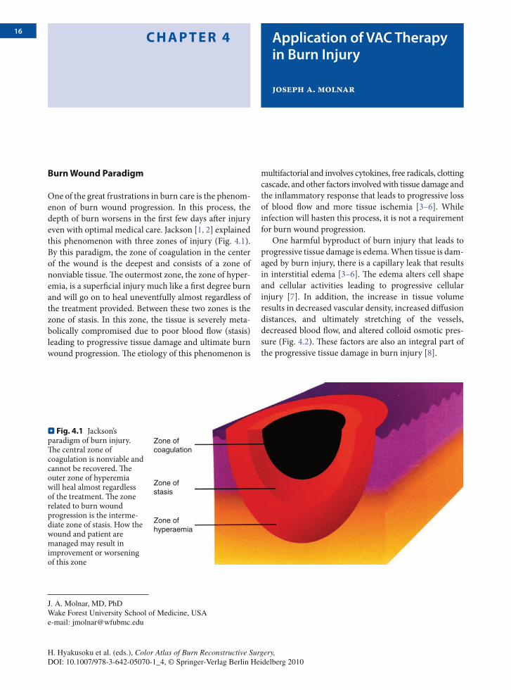
One of the great frustrations in burn care is the phenom-enon of burn wound progression. In this process, the depth of burn worsens in the first few days after injury even with optimal medical care. Jackson [1, 2] explained this phenomenon with three zones of injury (Fig. 4.1). By this paradigm, the zone of coagulation in the center of the wound is the deepest and consists of a zone of nonviable tissue. The outermost zone, the zone of hyper-emia, is a superficial injury much like a first degree burn and will go on to heal uneventfully almost regardless of the treatment provided. Between these two zones is the zone of stasis. In this zone, the tissue is severely meta-bolically compromised due to poor blood flow (stasis) leading to progressive tissue damage and ultimate burn wound progression. The etiology of this phenomenon is
multifactorial and involves cytokines, free radicals, clotting cascade, and other factors involved with tissue damage and the inflammatory response that leads to progressive loss of blood flow and more tissue ischemia [3–6]. While infection will hasten this process, it is not a requirement for burn wound progression.
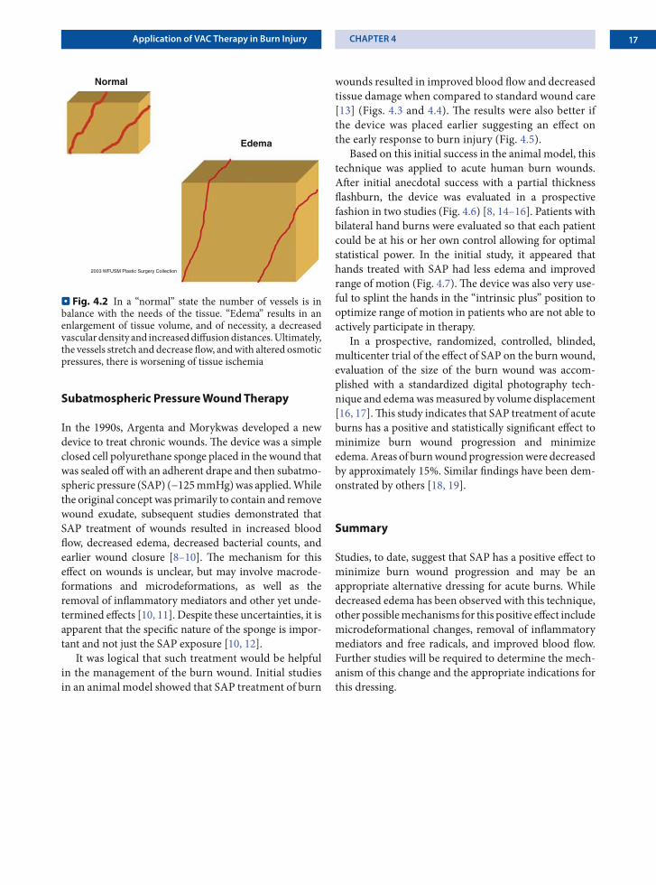
One harmful byproduct of burn injury that leads to progressive tissue damage is edema. When tissue is dam-aged by burn injury, there is a capillary leak that results in interstitial edema [3–6]. The edema alters cell shape and cellular activities leading to progressive cellular injury [7]. In addition, the increase in tissue volume results in decreased vascular density, increased diffusion distances, and ultimately stretching of the vessels, decreased blood flow, and altered colloid osmotic pres-sure (Fig. 4.2). These factors are also an integral part of the progressive tissue damage in burn injury [8].
Zone ofcoagulation
Zone ofstasis
Zone ofhyperaemia
⊡⊡ Fig. 4.1 Jackson’s paradigm of burn injury. The central zone of coagulation is nonviable and cannot be recovered. The outer zone of hyperemia will heal almost regardless of the treatment. The zone related to burn wound progression is the interme-diate zone of stasis. How the wound and patient are managed may result in improvement or worsening of this zone
J. A. Molnar, MD, PhD Wake Forest University School of Medicine, USA e-mail: [email protected]
17Application of VAC Therapy in Burn Injury CHAPTER 4
Subatmospheric Pressure Wound Therapy
In the 1990s, Argenta and Morykwas developed a new device to treat chronic wounds. The device was a simple closed cell polyurethane sponge placed in the wound that was sealed off with an adherent drape and then subatmo-spheric pressure (SAP) (−125 mmHg) was applied. While the original concept was primarily to contain and remove wound exudate, subsequent studies demonstrated that SAP treatment of wounds resulted in increased blood flow, decreased edema, decreased bacterial counts, and earlier wound closure [8–10]. The mechanism for this effect on wounds is unclear, but may involve macrode-formations and microdeformations, as well as the removal of inflammatory mediators and other yet unde-termined effects [10, 11]. Despite these uncertainties, it is apparent that the specific nature of the sponge is impor-tant and not just the SAP exposure [10, 12].
It was logical that such treatment would be helpful in the management of the burn wound. Initial studies in an animal model showed that SAP treatment of burn
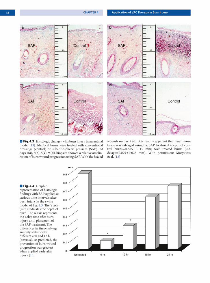
wounds resulted in improved blood flow and decreased tissue damage when compared to standard wound care [13] (Figs. 4.3 and 4.4). The results were also better if the device was placed earlier suggesting an effect on the early response to burn injury (Fig. 4.5).
Based on this initial success in the animal model, this technique was applied to acute human burn wounds. After initial anecdotal success with a partial thickness flashburn, the device was evaluated in a prospective fashion in two studies (Fig. 4.6) [8, 14–16]. Patients with bilateral hand burns were evaluated so that each patient could be at his or her own control allowing for optimal statistical power. In the initial study, it appeared that hands treated with SAP had less edema and improved range of motion (Fig. 4.7). The device was also very use-ful to splint the hands in the “intrinsic plus” position to optimize range of motion in patients who are not able to actively participate in therapy.
In a prospective, randomized, controlled, blinded, multicenter trial of the effect of SAP on the burn wound, evaluation of the size of the burn wound was accom-plished with a standardized digital photography tech-nique and edema was measured by volume displacement [16, 17]. This study indicates that SAP treatment of acute burns has a positive and statistically significant effect to minimize burn wound progression and minimize edema. Areas of burn wound progression were decreased by approximately 15%. Similar findings have been dem-onstrated by others [18, 19].
Summary
Studies, to date, suggest that SAP has a positive effect to minimize burn wound progression and may be an appropriate alternative dressing for acute burns. While decreased edema has been observed with this technique, other possible mechanisms for this positive effect include microdeformational changes, removal of inflammatory mediators and free radicals, and improved blood flow. Further studies will be required to determine the mech-anism of this change and the appropriate indications for this dressing.
Normal
Edema
2003 WFUSM Plastic Surgery Collection
⊡⊡ Fig. 4.2 In a “normal” state the number of vessels is in balance with the needs of the tissue. “Edema” results in an enlargement of tissue volume, and of necessity, a decreased vascular density and increased diffusion distances. Ultimately, the vessels stretch and decrease flow, and with altered osmotic pressures, there is worsening of tissue ischemia
18 CHAPTER 4 Application of VAC Therapy in Burn Injury
Untreated
0.9
mm
0.8
0.7
0.6
0.5
0.4
0.3
0.2
0.1
00 hr
*
*
12 hr 24 hr18 hr
⊡⊡ Fig. 4.4 Graphic representation of histologic findings with SAP applied at various time intervals after burn injury in the swine model of Fig. 4.3. The Y axis (mm) indicates the depth of burn. The X axis represents the delay time after burn injury until placement of the SAP treatment. The differences in tissue salvage are only statistically different at 0 and 12 h (asterisk). As predicted, the prevention of burn wound progression was greatest when applied early after injury [13]
a b
c d
SAP Control SAP Control
SAP ControlSAP Control
⊡⊡ Fig. 4.3 Histologic changes with burn injury in an animal model [13]. Identical burns were treated with conventional dressings (control) or subatmospheric pressure (SAP). At days 1(a), 3(b), 5(c), 9 (d), biopsies showed a relative amelio-ration of burn wound progression using SAP. With the healed
wounds on day 9 (d), it is readily apparent that much more tissue was salvaged using the SAP treatment (depth of con-trol burns = 0.885 ± 0.115 mm; SAP treated burns (0-h delay) = 0.095 ± 0.025 mm). With permission: Morykwas et al. [13]
19Application of VAC Therapy in Burn Injury CHAPTER 4
a b
c d
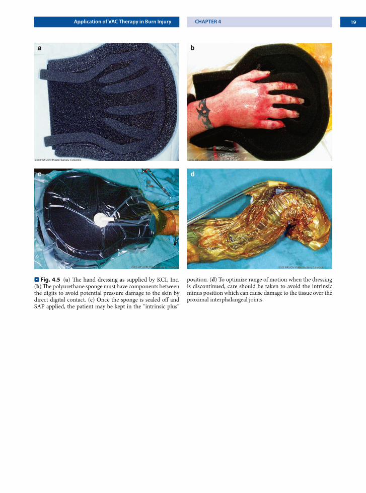
⊡⊡ Fig. 4.5 (a) The hand dressing as supplied by KCI, Inc. (b) The polyurethane sponge must have components between the digits to avoid potential pressure damage to the skin by direct digital contact. (c) Once the sponge is sealed off and SAP applied, the patient may be kept in the “intrinsic plus”
position. (d) To optimize range of motion when the dressing is discontinued, care should be taken to avoid the intrinsic minus position which can cause damage to the tissue over the proximal interphalangeal joints
20 CHAPTER 4 Application of VAC Therapy in Burn Injury
a b
c d
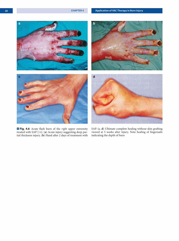
⊡⊡ Fig. 4.6 Acute flash burn of the right upper extremity treated with SAP [14]. (a) Acute injury suggesting deep par-tial thickness injury. (b) Hand after 2 days of treatment with
SAP. (c, d) Ultimate complete healing without skin grafting viewed at 5 weeks after injury. Note healing of fingernails indicating the depth of burn
21Application of VAC Therapy in Burn Injury CHAPTER 4
a b
c d
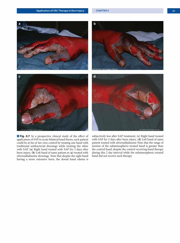
⊡⊡ Fig. 4.7 In a prospective clinical study of the effect of application of SAP to acute bilateral hand burns, each patient could be at his or her own control by treating one hand with traditional antibacterial dressings while treating the other with SAP. (a) Right hand treated with SAP for 2 days after burn injury. (b) Left hand of same patient as (a) treated with silversulfadiazine dressings. Note that despite the right hand having a more extensive burn, the dorsal hand edema is
subjectively less after SAP treatment. (c) Right hand treated with SAP for 2 days after burn injury. (d) Left hand of same patient treated with silversulfadiazine Note that the range of motion of the subatmospheric-treated hand is greater than the control hand, despite the control-receiving hand therapy during this 2 day interval while the subatmospheric-treated hand did not receive such therapy