Embed Size (px)
Citation preview
40-
BURROWING MECHANISM
The anguilliform fishes are capable of swimming
and burrowing. The simplest type A.bicolor swims about
freely and only occasionally hides in the bottom clay,
while the most advanced type A.fossorius usually·remains
in deep burrows most of the time and comes out only
oocasionally. S.bengalensis and P.boro occupies inter
mediate positions between these two extremes. The burrows
made by them are not so deep as those of A.fossorius. At
the same time, they do not swim about so freely as
A.bicolor. So in these four species the organs that are
concerned with locomotion, namely, the skeletal and mus
cular systems, show varying degrees of adaptive changes
depending on the extent of the burrowing habit.
The pattern of the body segments in fishes has
been observed by various authors, but only a few have
studied their functional mechanism. Owen (1866) des
cribed the lateral muscles of fishes as an aggregate
structure formed of a series of .transverse musc+es, but
failed to recognize their morphological importance.
Humphrey (1871) described the division of the lateral
muscles into dorsal and ventral moities by a horizontal
septum pas.s.ing inwards beneath the la,terel-line.
Chevrel(19a3) noted the conical structure of the myo
meres forming the lateral muscles. Greene and Greene
(1913) explained the mechanics of locomotion in fishes;
based on dissections of king salmon, Oncorhypchus
tachawytscha. A more complete study of the subject was
- 41 -
made by Roc~well, Evans and Pheasant (1938) and they
stressed the oblique nature of the attachment of the
myomeres to the verteb~al column. The pattern and dis
position of the muscles were also described by Shan
(1914) and Nishi (1938). The studies of Marey (1895),
Breeder (1926), Magnan (1930), Gray (1933) and Nvrsall
(1956) have enabled a clear understanding of the mecha
nics of locomotion of fishes in their natural aquatic
environment. But the question, how the body musculature
and the skeletal axis, which are normally designed for
swimming can also help the animal to push through mud
has not received much attention so far.
In a typical teleostean fish like the Scomber
or Mackerel, there is a thick sheet of muscle on each
side extending from the posterior region of the skull to
the hind, end of the vertebral column. This is dividedj
into as many muscle-segments or myotomes as there are
vertebrae. The myotomes are separated by the myocom-
mata (myosepta) and are innervated by the spinal serves
of the corresponding segments. A horizontal septum ex
tending the entire length of the body divides each myo
tome into a dorsal epaxial and a ventral hypaxial muscle.
The myocomma of the epaxial mu~cle is V-shaped with the
apex directed backwards. The two arms of the epaxial por
tion are distinguished into the mesio-dorsal and latero
dorsal portions. The hypaxial muscle of each segment is
a mirror image of the dorsal epaxial muscle and can be
similarly distinguished into latero-ventral and mesio-
I
- 42 -
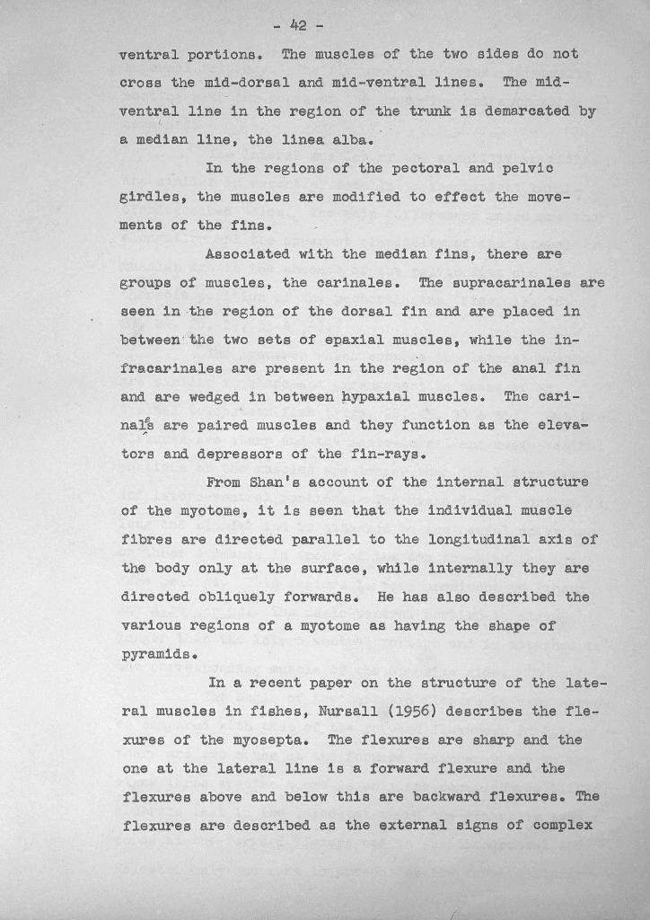
ventral portions. The muscles of the two sides do not
cross the mid-dorsal and mid-ventral lines. The mid-
ventral line in the region of the trunk is ~emarcated by~
a median line, the linea alba.
In the regions of the pectoral and pelvic
girdles, the muscles are modified to effect the move
ments of the fins.
Associated with the median fins, there are
groups of muscles, the carinales. The supracarinales are
seen in,the region of the dorsal fin and are placed in
between'the two sets of epaxial muscles, while the in
fracarinales are present in the region of the anal fin
and are wedged in between ~ypaxial muscles. The cari
na~B are paired muscles and they function as the eleva-r
tors and depressors of the fin-rays.
From Shan's account of the internal structure
of the myotome, it is seen that the individual muscle
fibres are directed parallel to the longitudinal axis of
the body only at the surface, while internally they are
directed obliquely forwards. He has also described the
various regions of a myotome as having the shape of
pyramids.
In a recent paper on the structure of the late
ral muscles in fishes, Nursall (1956) describes the f1e-
xures of the myosepta. The flexures are sharp and the
one at the lateral line is a forward flexure and the
flexures above and below this are backward flexures. The
flexures are described as the external signs of complex
f
- 43 -
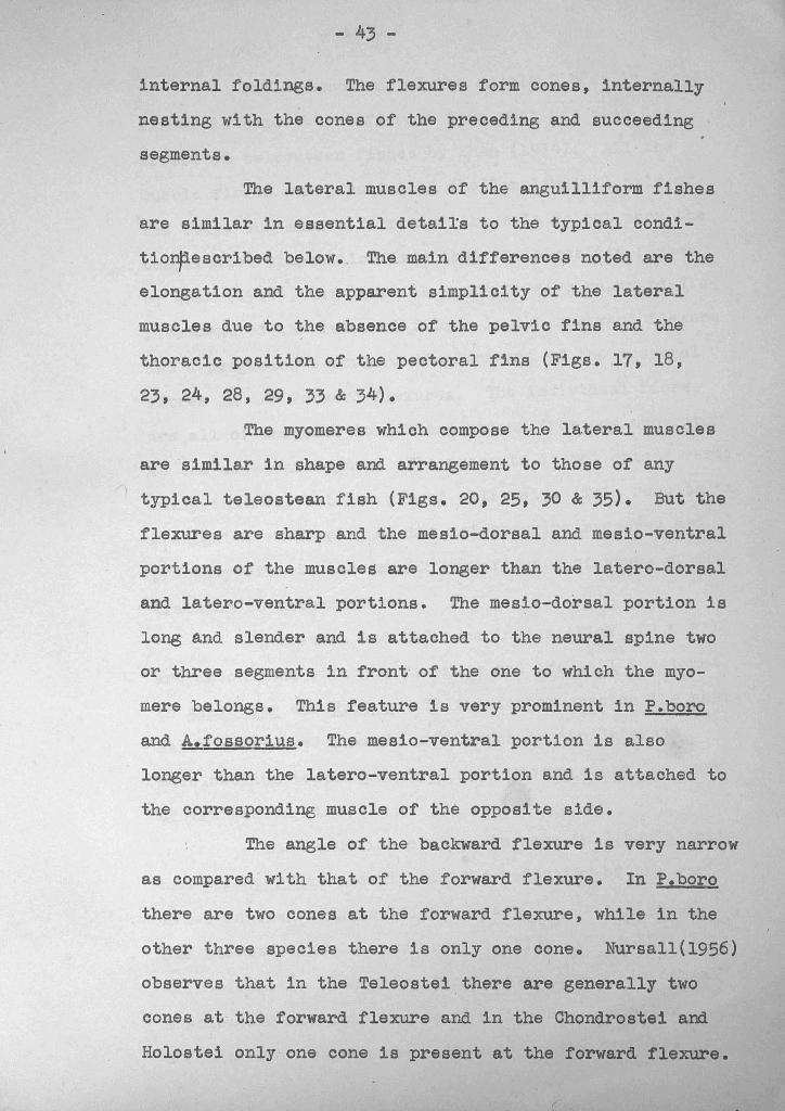
internal foldings. The flexures form cones, internally
nesting with the cones of the preceding and succeeding
segments.
The lateral muscles of the anguil1iform fishes
are similar in essential detai!s to the typical condi
tio~escribed below. The main differences noted are the
elongation and the apparent simplicity of the lateral
muscles due to the absence of the pelvic fins and the
thoracic position of the pectoral fins (Figs. 17, 18,
23, 24, 28, 29, 33 & 34).
The myomeres which compose the lateral muscles
are similar in shape and arrangement to those of any
typical te1eostean fish (Figs. 20, 25, 30 & 35). But the
flexures are sharp and the mesio-dorsal and mesio-ventral
portions of the muscles are longer than the 1atero-dorsal
and latero-ventral portions. The mesio-dorsal portion is
long and slender and is attached to the neural spine two
or three segments in front of the one to which the myo
mere belongs. This feature is very prominent in P.boro
and A.fossorius. The mesio-ventral portion is also
longer than the latero-ventral portion and is attached to
the corresponding muscle of the opposite side.
The angle of the backward flexure is very narrow
as compared with that of the forward flexure. In P.boro
there are two cones at the forward flexure, while in the
other three species there is only one cone. Nursal1(1956)I
observes that in the Te1eostei there are generally two
cones at· the forward flexure and in the Chondrostei and
Holostei only one cone is present at the forward flexure.
- 44 -
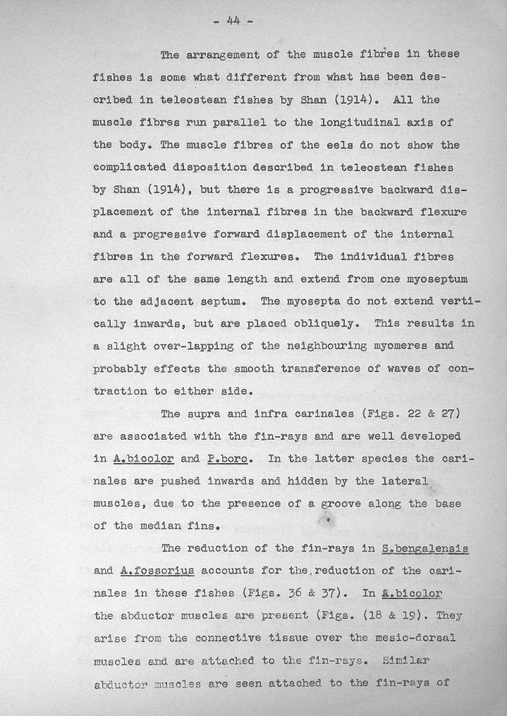
The arrangement of the muscle fibres in these
fishes is some what different from what has been des
cribed in teleostean fishes by Shan (1914). All the
muscle fibres run parallel to the longitudinal axis of
the body. The muscle fibres of the eels do not show the
complicated disposition described in teleostean fishes
by Shan (1914), but there is a progressive backward dis
placement of the internal fibres in the backward flexure
and a progressive forward displacement of the internal
fibres in the forward flexures. The individual fibres
are all of the same length and extend from one myoseptum
to the adjacent septum. The myosepta do not extend verti
cally inwards, but are placed obliquely. This results in
a slight over-lapping of the neighbouring myomeres and
probably effects the smooth transference of waves of con
traction to either side.
The supra and infra carinales (Figs. 22 & 27)
are associated with the fin-rays and are well developed
in A.bicolor and P.boro. In the latter species the cari
nales are pushed inwards and hidden by the lateral
muscles, due to the presence of a groove alo~g the base
of the median fins. v
The reduction of the fin-rays in Sobengalensis
and A.fossoriuB accounts for the. reduction of the cari
nales in these fishes (Figs. 36 & 37). In E.bicolor
the abductor muscles are present (Figs. (18 & 19). They
arise from the connective tissue over the mesio-dorsal
muscles and are attached to the fin-rays. Similar
abductor muscles are seen attached to the fin-rays of

- 45 -
. the anal fin also (Fig. 18).
An additional layer of muscle fibres running in
an oblique direction is seen in all these fishes except
P.boro. This may be termed the superficial oblique
muscle. This muscle is confined to the anterior region
of the trunk and extends obliquely from the mid-dorsal
line to the lateral line. The superficial oblique muscle
is devoid of ~~y indication of segmentation and forms a
thin investing layer over the lateral muscles of the
body wall.
The simplest condition of the' superficial
oblique muscle is found in A.bicolor (Fig. 17) where it
occupies the region between the cranial muscles and the
. base of the pectoral fin. The muscle fibres have an .
oblique course and extend from the mid-dorsal line to the
lateral line, immediately above the dorsal limit of the.
opercular chamber.
In S.benga1ensis the superficial oblique muscle
has a more elaborate arrangement (Fig. 28). It forms an
enveloping sheet ,of muscle over the dorsal moiety of about
twenty-five myomeres of the anterior region. A muscle
band from the anterior margin of the fourth myomere ex
tends forward on either side of the dorsal aspect of the
first three segments and is attached to the dorso-Iateral
region of the skull. These probably function as nec~
muscles. The dorsal. moiety of the lateral muscles of the
segments 4-14 is almost completely covered over by the
superficial oblique muscle. This continues further back-
- 46 -
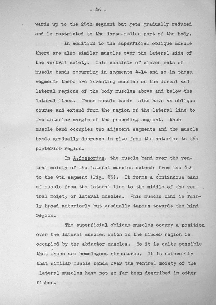
wards up to the 25th segment but gets gradually reduced
and is restricted to the dorso-median part of the body.
In addition to the superficial oblique muscle
there are also similar muscles over the lateral side of
the ventral moiety. This consists of eleven sets of
muscle bands occurring in segments 4-14 and so in these.
segments there are investing muscles on the dorsal and
lateral regions of the body muscles above and below the
lateral lines. These muec1e bands also have an oblique
course and extend from the region of the lateral line· to
the anterior margin of the preceding segment. Each
muscle band occupies two adjacent segments and the muscle
bands gradually decrease in size from the anterior to tlie
posterior region.
In A.fossorius, the muscle band over the ven
tral moiety of the lateral muscles extends from the 4th
to the 9th segment (Fig. ~3). It forms a continuous band
of muscle from the 1ateral·line to the middle of the ven- _
tra1 moiety of lateral muscles. This muscle band is fair
ly broad anteriorly but gradually tapers towards the hind
region.
The superficial oblique muscles occupy a position
over the lateral muscles which in the hinder region is
occupied by the abductor muscles. So it is quite possible
that these are homologous structures. It is noteworthy
that similar muscle bands over the ventral moiety, of the
lateral muscles have not so far been described in other
fishes.
- 47 -
The superficial oblique muscles are seen only in
the three species which burrow with the head and this
leads us to infer that these muscles assist in keeping the
anterior region straight and rigid in the act of burrowing.
The waves of contraction arise in the anterior region of
the body and proceed backwards and the head has to be kept
straight ~o enable it to push through mud. So the super
ficial oblique muscles may be regarded as a special
structure developed in response to the act of burrowing.
In support of this view, it may be pointed out that this
muscle is completely absent in P.boro, a fish which
burrows with its tailo
Locomotion in a typical flattened fish ~s
effected by the waves of contraction passing along the
sides of the body and by the strokes of the caudal fin.
The movement is initiated by the contraction of the first
few muscles of the anterior region on one side, with the
. result that the anterior region is thrown into a curve.
This curve is passed backwards in a series of waves, by
the alternate contractions and relaxations of the serially
arranged myomeres. Each successive muscle segment gives
an additional momentum to the wave and as soon as the
first wave has started backwards, a second one follows;
but on the oppq~ite side. This alternating sequence of
waves follows in regular suc~ession and the forward thrust
is attained by the pressure exerted by the body on the
water which fills the troughs in the wave series. All
these fishes can swim backwards with equal ease, by revers
ing the direction of wave motion.
- 48 -
The contraction of the individual myomere is
the result of the contraction of the muscle fibres that
compose the myomere. Since the muscle fibres are rather
short, the resulting contraction of ahy individual myo
mere is very insignificant. But when oontraction takes
place, simultaneously, a set of successive myomeres under-
go reduction in their total length and this results in
the curvature of a section of the body and when this
curve straightens, it exer s a force on the surrounding
medium, whether it be waterOD'mud and movement of the
fish is effected. The energy required to push the body
is the energy stored up in the curve, which in turn is
produced by the contraction of a set of successive myo
meres. So this can be taken as a case of bending and
can be treated as a physical phenomenon. Taking the
analogy of a bent bar, the bending moment is given by the
expression M = qAk:2 where M is the bending moment, q thefB.
young's modulus, A the area of cross-section of'the body,
k the radius of gyration and e the radius of curvature
of the bent bar.
The energy in the ben~ bar is given by the
expression E = t~Ak2L, where L is the length of the neu
tral axis represented by the vertabral column. It is
this energy that is stored up as a consequence of bending
that is released as kinetic energy resulting in motion.
In all these fishes, except in P.boro, the
hind part of the tail gets progressively flattened with
the result that the cross-sec~ion o~ this region becomes
elliptical. Owing to this change in. body-form, the
- 4 9 -
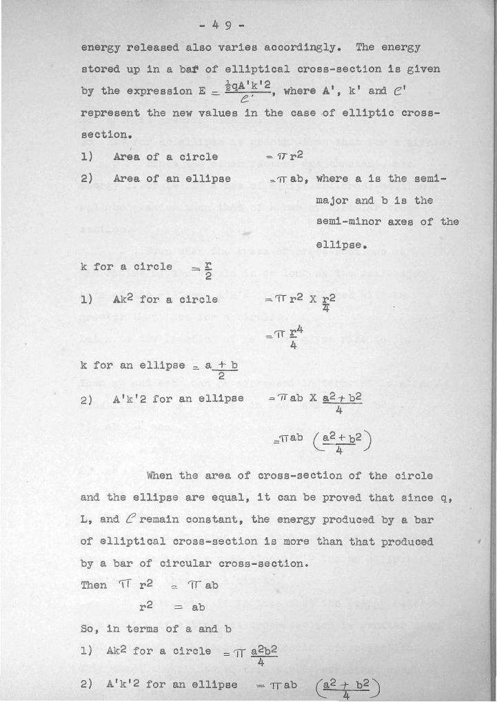
energy released also varies accordingly. The energy
stored up in a bar of elliptical cross-section is given1. Atkt2
by the expression E ::::. "2q e' ,where At, k t and e t
represent the new values in the case of elliptic'cross-
section.
1)
2)
I
Area of a circle
Area of an ellipse
= 'fTr2
:=-'iT ab, where a is the semi-
major and b is the
semi-minor axes of the
ellipse.
k for a circle =~2
1) Ak2 for a circle = 'iT r 2 X r 24
k for an ellipse - a + b- ~
A t k t 2 for an ellipse ='IT ab X a2 1- b24
When the area of cross-section of the circle
and the ellipse are equal, it can be proved that since q,
L, a.nd e remain constant, the energy produced by a bar
of elliptical cross-section is more than that produced '
by a bar of circular cross-section.
Then '1T r 2 =. 'IT ab
r 2 = ab
So, in terms of a and b
I) Ak2 for a circle =11 a2b24
2) At k t 2 for an ellipse
- 50 -
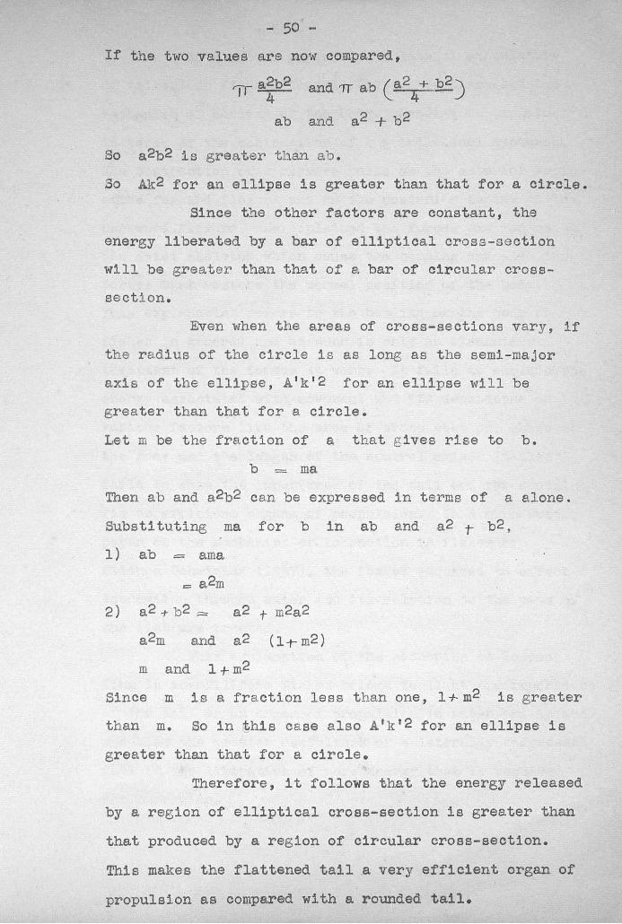
If the t·W'o values are now compared,
ab
and 'TT ab (a24+ b2 )
and a2 + b 2
So a 2b2 is greater than 13.1).
So Ak2 for an ellipse is greater than that for a circle.
Since the other factors are constant, the
energy liberated by a bar of elliptical cross-section
will be greater than that of a bar of circular cross-
section.
Even when the areas of cross-sections vary, if
the rae.ius of the circle is as long as the semi-major
axis of the ellipse, A'k'2 for an ellipse will be
greater than that for a circle.
Let m be the fraction of a that gives rise to b.
b = ma,
Then ab and a2b2 can be expressed in terms of a alone.
Substi tuting ma for b in ab and a2 T b2 ,
1) ab = ama
= a2m
2) a 2 -t- b 2 = a 2 t m2a 2
a2m and a 2 (Ii- m2 )
m and 1 +- m2
Since m is a fraction less than one, 1+m2 is greater
than m. So in ~hi8 case also A'k'2 for an ellipse is
greater than that for a circle.
Therefore, it follows that the energy released
by a region of elliptical croas-section is greater than
that produced by a region of circular croBs-section.
This makes the flattened tail a very efficient organ of
propulsion as compared with a rounded tail.

- 51 -In a recent paper on the lateral musculature
of teleostean fishes, Nursall (1956) has discussed the
mechanism of bending of the body. Bending 1s explained
in terms of the contraction of the individual myomeres.
The contraction of a myomere pulls on the anterior septum
o~he forward flexure and on the posterior septum of the
backward flexure. He explained the forces exerted on", '
the axial skeleton which cause the bending and also the
forces ~hkb restore the normal position of the body.
This explanation refers to the bending of the body of
fishes in general and as such is only an elementary
treatment of the forces at work. It fails to explain the
energy associated with movement and its dependance on
various factors like the area of cross section, shape of
the body and the length of the neutral axis. It also
fails to show the importance of the tail and the caudal
fin as efficient organs of propulsion. In a more recent
paper on the mechanics of locomotion in fishes by
Elienne Ochmichen (1957), the forces required to e~fect
locomotion through water and its relation to the mass of
the fish are treated.
This explanation of the mechanics of locomo
tion in anguilliform fishes brings to light the importance
of the tail as an organ of propulsion in water and in mud
and also the greater usefulness of a laterally compressed
tail in the liberation of more energy that is required
for burrowing.