Embed Size (px)
Citation preview

Pneumocystis carinii
By Sarah Weivoda & Kaleb Hale
This is a smear of P. Carinii isolated from human lung tissue.
Pathogen responsible for Causing interstitial plasma cell pneumonitis, which leads to Pneumocystis carinii pneumnia

Taxonomic Considerations Currently considered to be a fungus -Based on nucleic acid analysis Previously considered to be a protozoa
Domain: Eukaryota Kingdom: Fungi Phylum: Ascomycota Class: Pneumocystidomycetes Order: Pneumocystidales Family: Pneumocystidaceae Genus: Pneumocystis Species: carinii

History of P. CariniiFirst discovered in 1909 by Carlos ChagasFound to be associated with clinical pneumonia shortly
after World War 2 Until the 1980’s it occurred very rarely and only caused
pneumonia in people with congenital immunodeficiency's and patients immunocompromised by cancer.
From the 1980’s on the incidence of Pneumocystis Carinii associated pneumonia significantly increased due to the AIDS epidemic.
P. Carinii is now the leading cause of death by opportunistic infection in AIDS patients.

Geographic rangeAffects Humans and animals world wide
Infects a broad range of mammalian species including: -Humans -Mice -Rats -Cats -Dogs -Pigs-Most healthy children have been exposed to P. Carinii by age 3 to 4
-Primarily affects immunocompromised individuals
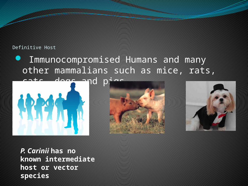
Definitive Host
Immunocompromised Humans and many other mammalians such as mice, rats, cats, dogs and pigs.
P. Carinii has no known intermediate host or vector species
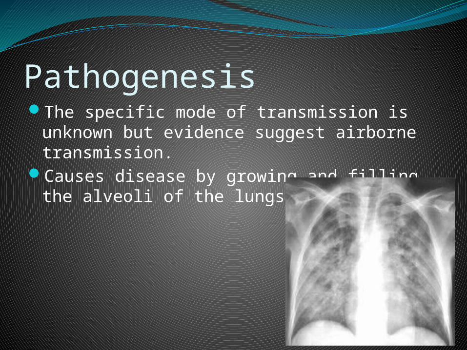
PathogenesisThe specific mode of transmission is unknown but
evidence suggest airborne transmission. Causes disease by growing and filling the alveoli of the
lungs

Clinical SignsPhysical Symptoms Initial
*Fever *Fatigue *Weight Loss Dyspnea Tachypnea Nonproductive cough Fevers Chills Sweats Progressive, profound fatigue Cyanosis around the mouth, hands,
feet, or mucous membranes
ProgressiveExtrapulmonary
manifestationsDeath

Laboratory DiagnosisTrophozoite Cyst

Morphology and Biology P. Carinii goes through 3 morphological stages:1. Trophozoite- pleomorphic in shape- Ranges from 1 to 5 µm in diameter- Has small filopodia 2. Precyst- Oval in shape - Has few filopodia - Has a cluster of mitochondria in its center3. Cyst- Spherical in shape- Has a thick membrane made of chitin- Contains 8 intracystic bodies
All morphological stages can be found within the lungs of the infected individual

Life Cycle

Treatment Drug of choice
Trimethoprim-sulfamethoxazoleRecommended others
PentamidineTrimethoprim plus daposoneAtovaquonePrimaquine plus clindamycin

References http://dpd.cdc.gov/dpdx/HTML/Pneumocystis.htmhttp://www.tulane.edu/~wiser/protozoology/notes/
aids.htmlhttp://www.aafp.org/afp/991015ap/1699.htmlhttp://emedicine.medscape.com/article/225976-over
viewhttp://www.nlm.nih.gov/medlineplus/ency/article/0
00671.htmRoberts, L. Janovy, J. Foundations of Parasitology, 8th
ed. New York: McGraw-Hill, 2009.