Embed Size (px)
Citation preview
JOURNAL OF SURGICAL RESEARCH 61, 323–329 (1996)ARTICLE NO. 0124
c-MYC Oncoprotein Production in ExperimentalVein Graft Intimal Hyperplasia1,2
JOSE A. RAMIREZ, M.D., LUIS A. SANCHEZ, M.D., MICHAEL L. MARIN, M.D.,3 ROSS T. LYON, M.D.,RICHARD E. PARSONS, M.D., WILLIAM D. SUGGS, M.D., AND FRANK J. VEITH, M.D.
The Division of Vascular Surgery, Department of Surgery, Montefiore Medical Center, The University Hospitalfor the Albert Einstein College of Medicine, New York, New York 10467
Submitted for publication October 18, 1994
MYC oncoprotein is expressed early after experimen-Purpose: The expression of c-MYC oncoprotein in tal vein grafting, with peak expression at 1 week. This
proliferating smooth muscle cells (SMCs) was ana- occurs during a period of maximal intimal thickening,lyzed in an experimental model of vein graft intimal SMC proliferation, and increased expression of PC10.thickening. Methods: Superficial epigastric vein grafts Expression of c-myc protooncogene may contribute towere inserted into the femoral arteries of male the induction and regulation of SMC proliferation,Sprague–Dawley rats. The vein grafts were harvested producing intimal hyperplasia. q 1996 Academic Press, Inc.
at 6 hr, 2 days, 1 week, 2 weeks, and 4 weeks aftergrafting and were rapidly frozen in liquid nitrogen.Immunohistochemical labeling and morphologic anal-
INTRODUCTIONysis of vein graft sections with a double staining tech-nique were used to identify c-MYC/alpha SMC actin
Smooth muscle cell (SMC) intimal hyperplasia is re-and proliferating cell nuclear antigen (PC10)/alphasponsible for 20–60% of clinical intermediate and lateSMC actin within intimal cells. c-MYC/alpha SMC actinvein graft failures [1, 2]. Although the exact etiology ofand PC10/alpha SMC actin positive cells were quanti-this lesion is unknown, several pathogenic mechanismstated in the perianastomotic area (R-1) and the bodyhave been proposed. Recurrent endothelial injury mayof the graft (R-2) for each time period. Total wall and
intimal thickness of perfusion fixed vein grafts were activate the migration and proliferation of SMCs withmeasured with a computer digitized system. Results: subsequent thickening of the intima and subintimalIntimal and total wall thickening in the R-1 region media [1, 3, 4]. Also implicated in the genesis of neointi-peaked at 1 week (27.4 and 579.4 mm, respectively) and mal hyperplasia is mechanical injury induced by com-were significantly thicker (P õ 0.01) than the same re- pliance mismatch and abnormal shear stresses whichgion at 6 hr after graft implantation (6.0 and 113.5 mm, may be related to aberrant flow patterns within therespectively). Staining for c-MYC and PC10 in R-1 was vessel [1]. Other possible etiological factors include im-also significantly higher (P õ 0.05) at 1 week (5.75 and munological activation of SMCs by interferon-g [5],7.00 positive cells/10 cells, respectively) compared with stimulation of cell proliferation by platelet-derivedthat at 6 hr (1.5 and 1.33, respectively). The R-1 region growth factor (PDGF) [6, 7], insulin-like growth factorstabilized and remodeled over the following 3 weeks, [8], transforming growth factor B1 [9], and active oxy-while c-MYC and PC10 staining progressively de- gen species [10] (i.e., O2, H2O2, and OH) generated dur-creased. In the R-2 region, intimal thickness signifi-
ing arterial wall injury. All have been shown to havecantly increased (P õ 0.05) from 6 hr (4.0 mm) to 1 weeka profound effect on SMCs and the vascular wall struc-(12.0 mm) and stabilized, while total wall thickness in-ture.creased throughout the first week and the difference
Injury events in the arterial wall have been linkedbecame significant at 2 weeks (P õ 0.05). Staining forto enhanced expression of protooncogenes which havec-MYC and PC10 paralleled the staining in R-1 with abeen demonstrated in SMCs of atheromatous plaquesignificant peak at 1 week (P õ 0.05). Conclusions: c-[11, 12]. One protooncogene—c-sis—codes for a pro-tein similar to the b-chain of PDGF, a potent stimulator1 Supported by grants from the U.S. Public Health Service (HLof SMC proliferation [13]. Protooncogenes, in general,02990-03), the James Hilton Manning and Emma Austin Manninghave the potential to induce and regulate a variety ofFoundation, The Anna S. Brown Trust, and the New York Institute
for Vascular Studies. nuclear functions. The c-myc protooncogene, a nuclear2 Presented at the 47th Scientific Meeting of The Society for Vascu- phosphoprotein, has been associated with the control
lar Surgery, Washington, DC, June 8–9, 1993. of cellular proliferation and differentiation [14]. Active3 To whom reprint requests should be addressed at The Division ofexpression of c-MYC has recently been demonstratedVascular Surgery, Montefiore Medical Center, 111 East 210th Street,
New York, NY 10467. in SMCs of human carotid plaque [15].
323 0022-4804/96 $18.00Copyright q 1996 by Academic Press, Inc.
All rights of reproduction in any form reserved.
AID JSR 4671 / m4999f$$69 01-29-96 10:55:54 srga AP: Surg Res

324 JOURNAL OF SURGICAL RESEARCH: VOL. 61, NO. 2, MARCH 1996
In rat arteries, the expression of c-myc has also beenidentified in SMCs below injured endothelium [16]. Invitro experimental studies have shown similar expres-sion of protooncogenes c-jun, c-fos, and c-myc withinendothelial and SMCs following various forms of stimu-lation [12].
The purpose of the present study was to analyze theproduction of c-MYC protooncoprotein in an experi-mental model of vein graft intimal thickening and tocorrelate it with SMC proliferation.
MATERIAL AND METHODS
Grafting Procedures
Thirty-two male Sprague–Dawley rats weighing between 325 and400 g were used in this study. All animals were treated in accor-dance with the ‘‘Principles of Laboratory Animal Care’’ (formulatedby the National Society for Medical Research) and the ‘‘Guide for theCare and Use of Laboratory Animals’’ (NIH Publication No. 86-23,revised 1985).
The rats were anesthetized with 40 mg/kg of sodium pentobarbitaladministered via intraperitoneal injection. Using a microsurgicaltechnique, the superficial epigastric vein was dissected free fromthe surrounding tissues, removed, and gently flushed with 2 ml ofheparinized saline solution. The common femoral artery was isolatedand 5 mm of artery were resected. The divided ends of the arterieswere irrigated with heparinized saline solution, and the previouslyharvested 5-mm-long segment of superficial epigastric vein was su-tured end-to-end to the artery as an interposition graft with eightinterrupted 10-O Dermalon microsutures (Davis & Geck, Danbury,CT) at each anastomosis. Arterial occlusion time ranged from 45 minto 1 hr. When the vascular clamps were removed, immediate bloodflow was restored in the vessel. Patency was ensured by the use ofstandard microsurgical techniques [17]. Bilateral interposition veingrafts were performed in all animals.
Four animals each were sacrificed at 6 hr, 2 days, 1 week, 2 weeks,and 4 weeks after vein grafting. Two grafts were harvested fromeach animal with a 1.5-mm segment of adjacent artery at both ends.One graft was divided in half longitudinally, placed in embeddingoil (Instrumedics, Teaneck, NJ), and snap frozen in liquid nitrogen.Frozen vein graft segments were made into blocks with OptimumCutting Temperature compound (Tissue-Tek, Elkhard, IN), cut into4-mm sections, and stored at 0707C for subsequent immunohisto-chemistry. The other graft was kept frozen for further studies.
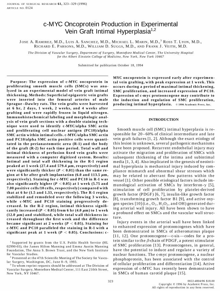
Three other animals were sacrificed at the same time intervalsand the vein grafts were harvested in the same manner. The graftswere perfusion fixed at 100 mmHg in 3% glutaraldehyde buffered to FIG. 1. Light micrograph (1400) of the body region (R-2) of su-pH 7.4 with sodium cacodylate. One was used for light microscopy perficial epigastric vein grafts from 6 hr (a) to 28 days (e) after graft-and the other was kept in glutaraldehyde for later studies. Control ing. (b) Two days after grafting, a layer of pseudointima composedfreshly harvested, superficial epigastric veins from the same animals of platelets and fibrin can be seen. (c) This pseudointima becomeswere processed in a similar fashion for immunohistochemistry and reorganized by 7 days with the initiation of a smooth muscle prolifer-light microscopy. ative response in the underlying intima. (d) At 14 days a thickened
hypercellular intima is recognized. (e) By 28 days reorganizationwithin the R-2 region of the graft results in a continuous neointimaImmunohistochemistry(1% azure II and methylene blue).
c-MYC and alpha smooth muscle cell actin. The 4-mm sections ofvascular tissue were thawed at room temperature and air dried for20 min. Sections were fixed in acetone and dried for 5 min prior to 1:250. Alpha SMC actin visualization was achieved by incubating
the tissue sections with Texas red avidin D (Vector Laboratories)antibody labeling. In order to identify c-MYC oncoprotein in alphaactin producing SMCs, a double staining sequential technique was diluted 1:20 in sterile water. All sections were examined using a
Nikon fluorescent microscope (Nikon, Melville, NY) equipped with aused [18].Tissue sections were incubated for 2 hr at 457C with mouse mono- super high-pressure mercury lamp power supply (Model HB-10101
AF) and a dual image quality emission filter FITC/Texas Red Dualclonal antibody to c-MYC (OM-11-906) (Cambridge Research Bio-chemicals, Wilmington, DE) diluted 1:250 with phosphate-buffered Band set (Omega Optical, Brattleboro, VT).
c-MYC oncoprotein was also identified using an immunoperoxidasesaline (PBS). Vein sections were then rinsed with PBS and immersedfor 30 min in fluorescein avidin D solution (Vector Laboratories, technique [19]. The 4-mm sections were incubated as described above
with mouse monoclonal antibody to c-MYC and rinsed with PBS.Burlingame, CA) diluted 1:20 with PBS. After rinsing again withPBS, SMCs were identified by incubating the sections for 30 min at The vein sections were incubated for 30 min at room temperature
with biotinylated secondary antibody solution (Vector Laboratories),457C with alpha SMC actin (A-2547) (Sigma, St. Louis, MO) diluted
AID JSR 4671 / m4999f$$69 01-29-96 10:55:54 srga AP: Surg Res

325RAMIREZ ET AL.: c-MYC ONCOPROTEIN IN RAT VEIN GRAFT
to visualize PC10. Double staining for alpha SMC actin was alsoperformed as previously described.
Immunohistochemical control slides were prepared using PBSwithout the primary antibodies to c-MYC, alpha SMC actin, andPC10. In addition, an absorption control was also performed usingthe c-MYC peptide OP-11-3069 (Cambridge Research Biochemicals,Wilmington, DE). These sections were incubated with c-MYC pri-mary antibody that had been preabsorbed for 30 min with c-MYCprotein at a concentration of 1 mg/ml of serum. All sections wereexamined as above for immunofluorescence.
Quantitation of Positive c-MYC and PC10 Cells
For each specimen studied, 10 cells from a representative area ofthe perianastomotic region (R-1) and 10 from the body of the graft(R-2) were counted within a single high-powered field (4001). Thenumber of c-MYC/alpha SMC actin and PC10/alpha SMC actin posi-tive cells in each group was calculated and pooled for analysis. Thehistologist evaluating the specimens was blinded to the region eachwas obtained from.
Light Microscopy and Morphometric Analysis
The glutaraldehyde-fixed vessels were postfixed in 1% osmium te-troxide and dehydrated in increasing concentrations of ethyl alcohol.Tissue samples were infiltrated with propylene oxide and flat embed-ded in Epon 812. After polymerization, 1-mm sections of each speci-men were cut longitudinally and stained with 1% azure II and meth-ylene blue.
Vein graft total wall and intimal thickness were measured in the1-mm sections and analyzed using a computer digitized morphometricsystem (Bioscan Optimas, Edmon, WA). Measurements were madein the perianastomotic region (R-1) and in the body of each graft (R-2) at a site 2 mm from the anastomosis.
Statistics
Statistical analysis of the data was performed using the Studentpaired t test. Statistical significance was assumed at the 95% confi-dence level (P õ 0.05). The data are represented as the mean plus/minus 1 standard deviation.
RESULTS
Graft Morphology and Morphometric Analysis
The control freshly harvested, superficial epigastricveins contained minimal elastic tissue which was notclearly organized into internal or external elastic lami-nae. A poorly defined intima was covered with a uniqueFIG. 2. Light micrographs (1100) of the perianastomotic region
(R-1) of superficial epigastric vein grafts (a) 6 hr, (b) 2 days, (c) 7 days, layer of endothelial cells. The medium was thin and(d) 14 days, and (e) 28 days after grafting. Progressive thickening andcellularity can be seen as the vein graft matures (a–e). Thickeningpeaks at 7 days (c) with presumed progression of vessel wall reorgani- TABLE 1zation occurring at a later time (d, e) (1% azure II and methyleneblue). Wall Thickness
Total wall thickness (mm) Intimal thickness (mm)Time period { standard deviation { standard deviationrinsed with PBS, and then incubated for 10 min at room temperature
with Vectastain Elite reagent (Vector Laboratories). The sectionsR-1 (6 hr) 113.5 { 7.7 6.0 { 1.0were rinsed with trizma solution and then incubated for 5 min atR-1 (2 days) 141.0 { 15.8 5.0 { 1.0room temperature with peroxidase substrate solution (Vector Labo-R-1 (7 days) 379.4 { 14.3 27.4 { 2.0ratories) and later rinsed with distilled water. Sections were counter-R-1 (14 days) 273.4 { 29.4 24.2 { 9.8stained with hematoxylin and eosin solution.R-1 (28 days) 317.8 { 40.3 21.7 { 10.4PC10 and alpha smooth muscle cell actin. The antiproliferating
cell nuclear antigen clone PC10 (1486 772) (Boehringer Mannheim, R-2 (6 hr) 24.0 { 7.2 4.0 { 1.0Indianapolis, IN) was used to identify proliferating cells. The 4-mm R-2 (2 days) 60.9 { 25.8 6.0 { 1.0frozen sections of vein grafts were thawed at room temperature, air R-2 (7 days) 136.6 { 59.8 12.0 { 2.1dried for 20 min, and then incubated at 47C overnight with PC10 R-2 (14 days) 211.5 { 61.4 12.1 { 1.1diluted 1:20 with PBS. The sections were then rinsed with PBS and R-2 (28 days) 159.3 { 77.2 10.4 { 8.4labeled with fluorescein avidin D solution as previously described
AID JSR 4671 / m4999f$$69 01-29-96 10:55:54 srga AP: Surg Res

326 JOURNAL OF SURGICAL RESEARCH: VOL. 61, NO. 2, MARCH 1996
FIG. 3. Double immunofluorescent labeling technique for c-MYC oncoprotein and alpha smooth muscle cell actin in a 1-week-old veingraft from the perianastomotic region (R-1). (a) c-MYC expression is seen in individual cells as bright yellow fluorescence (arrows). Alphasmooth muscle cell actin (red granular cytoplasmic material) aids in the identification of smooth muscle. (b) An absorption control for c-MYC and alpha smooth muscle cell actin shows no fluorescent staining (Texas red/FITC 1400).
FIG. 4. (a) Immunofluorescent colocalization of alpha smooth muscle cell actin and c-MYC oncoprotein in a vein graft 1 week aftergrafting in the R-2 (body) region of the graft. Bright yellow fluorescent staining documents the presence of the c-MYC oncoprotein (arrows).Granular red fluorescent staining throughout the cytoplasm identifies alpha smooth muscle cell actin. (b) An absorption control for c-MYCdemonstrates no staining (Texas red/FITC 1400).
FIG. 5. (a) Immunofluorescent double labeling of alpha smooth muscle cell actin and proliferating cell nuclear antigen (PC10). Brightgreen immunofluorescence is seen in the nucleus of cells that contain PCNA. Orange–yellow fluorescein is seen in those cells that containalpha smooth muscle cell actin. (b) Control specimens for PCNA demonstrate no staining (Texas red/FITC 1400).
composed of collagen and SMCs, approximately two to thelium and the rudimentary internal elastic laminaewas seen in R-1 (Fig. 2). There was intramural hemor-three layers thick, indistinguishable from the 6-hr vein
graft morphology (Fig. 1). rhage and the SMCs appeared spindle-shaped. Themean total wall thickness was 113.5 mm ({7.7) in R-Six hours after vein grafting, disruption of the endo-
AID JSR 4671 / m4999f$$69 01-29-96 10:55:54 srga AP: Surg Res

327RAMIREZ ET AL.: c-MYC ONCOPROTEIN IN RAT VEIN GRAFT
TABLE 2
c-myc/PC10 Staining
c-myc PC10
Mean No. Mean No.of positive of positive
No. of cells/10 Standard No. of cells/10 Standardspecimens cells deviation specimens cells deviation
R-1 (6 hr) 4 1.50 0.58 4 1.33 1.15R-1 (2 days) 4 3.25 0.96 4 2.67 1.53R-1 (7 days) 4 5.75 2.36 4 7.00 2.00R-1 (14 days) 4 3.25 1.50 4 3.33 1.15R-1 (28 days) 4 2.50 1.00 4 1.67 1.53
R-2 (6 hr) 4 0 0 4 0 0R-2 (2 days) 4 1.00 0.82 4 1.33 1.53R-2 (7 days) 4 3.25 0.96 4 6.33 1.15R-2 (14 days) 4 2.00 0.82 4 1.67 0.58R-2 (28 days) 4 2.75 1.26 4 2.67 2.08
1. At 2 days, inflammatory cells were adjacent to the 4a, and 4b). This was significantly increased comparedwith staining at 6 hr in R-1 (P Å 0.03) and R-2 (P Åendothelium and the matrix between the SMCs had
increased somewhat, with a mean total wall thickness 0.01). c-MYC expression then decreased at 2 and 4weeks in both the R-1 and the R-2 regions (Table 2).of 141.0 mm ({15.9) (Fig. 2b). Similar findings were
observed in R-2 at 2 days (Table 1, Fig. 1b). Staining for PC10 was also evaluated in conjunctionwith alpha SMC actin (Figs. 5a and 5b). At 6 hr 1.33The intimal and total wall thickness at 1 week after
graft implantation had significantly increased in R-1 { 1.15 PC10 positive cells/10 SMCs were noted in R-1, while no PC10 positive cells were noted in R-2. PC10from 6.0 { 1.0 mm at 6 hr to 27.4 { 2.0 mm (P Å 0.003)
and from 113.5 { 7.7 mm at 6 hr to 379.4 { 14.3 mm staining peaked at 7 days, with 7.00 { 2.00 PC10 posi-tive cells in R-1 and 6.33 { 1.15 PC10 positive cells in(P Å 0.002), respectively (Table 1). The matrix in the
media and intima increased, with a greater number R-2. This was significantly increased compared withstaining at 6 hr in R-1 (P Å 0.01) and R-2 (P Å 0.01).of spindle-shaped SMCs present (Fig. 2c). In R-2, the
intimal thickness increased significantly from 4.0{ 1.0 At 2 and 4 weeks, the staining in R-1 and R-2 progres-sively decreased (Table 2).mm at 6 hr to 12.0 { 2.1 mm at 1 week (Põ 0.05), while
the total wall thickness increased through the firstweek but the difference was not significant until Week DISCUSSION2 (P Å 0.04) (Fig. 1c). At 2 and 4 weeks, the intimal andtotal wall thickness in R-1 did not change significantly
Although neointimal hyperplasia is one of the majorcompared with those seen at 7 days, suggestive of reor-causes of vein and prosthetic graft failure [1, 3], theganization and remodeling (Figs. 2d and 2e; Table 1).biological events involved in this pathologic entity arepoorly understood. Smooth muscle cell proliferationImmunohistochemistryand migration are central events in this process whichis controlled, in part, by specific peptide growth factorsFour grafts for each time period were evaluated by
double immunohistochemical labeling with c-MYC/ [6–9]. Such factors have also been shown to be associ-ated with increased expression of specific early re-alpha SMC actin and PC10/alpha SMC actin antibod-
ies. The R-1 and R-2 areas of each graft were analyzed sponse genes such as c-myc, c-fos, and c-jun, and theseprotooncogenes may play an important role in regulat-separately. Staining for c-MYC localized within the nu-
clei in a granular fashion and staining for alpha SMC ing cellular proliferation and differentiation [6, 12, 16].The c-myc protooncogene is typically expressed in veryactin was principally distributed throughout the cyto-
plasm. limited amounts or may even be absent during quies-cent periods of the cell cycle [14, 20]. Brisk expressionFreshly harvested, superficial epigastric control
veins did not demonstrate c-MYC or PC10 in alpha of c-myc has been documented during periods of activecell division [16, 20].SMC actin positive cells. At 6 hr, limited c-MYC expres-
sion was seen in R-1 (1.5 { 0.58 c-MYC positive cells/ In the present study, a mouse monoclonal antibodyto c-MYC raised against the 45-kDa protein was used10 SMCs) and no expression was noted in R-2. At 2
days, 3.25 { 0.96 cells/10 SMCs in R-1 and 1.00 { 0.82 for identification. The proliferating capacity of cells ex-pressing c-MYC was identified with PC10, a proliferat-cells/10 SMCs in R-2 stained positive for c-MYC. One
week after grafting, c-MYC expression peaked at 5.75 ing antibody that recognizes a fixation and processingresistance epitope in proliferating tissues [21]. Control{ 2.36 in R-1 and 3.25 { 0.96 in R-2 (Figs. 3a, 3b,
AID JSR 4671 / m4999f$$69 01-29-96 10:55:54 srga AP: Surg Res

328 JOURNAL OF SURGICAL RESEARCH: VOL. 61, NO. 2, MARCH 1996
veins did not show staining for either of these antibod- The availability of an experimental model of intimalhyperplasia in vein grafts in which the expression of c-ies. This is consistent with the quiescent state and low
cell turnover in these vessels. Limited expressions of MYC oncoprotein can be controlled will permit a betterunderstanding of the etiologic role of this oncogene inc-MYC and PC10 were first noted 6 hr after grafting,
before any histologic changes could be detected. In this smooth muscle cell intimal hyperplasia.early period staining for both antibodies was localizedto the media of vein grafts at the perianastomotic site. ACKNOWLEDGMENTIn vitro studies performed by others have also reportedearly expression of c-MYC—within 2 hr after stimula- The authors thank Miguel Torres for his technical assistance with
this project.tion of vascular SMC cultures with angiotensin II [22].In our model, an almost fourfold increase and maxi-
mal staining for c-MYC at 1 week closely paralleled the REFERENCESincreased expression of PC10 during the same time
1. Bunt, T. J. Neointimal hyperplasia. In T. J. Bunt (Ed.), Iatro-period. In these vein grafts, c-MYC positive cells weregenic Vascular Injury: A Discourse on Surgical Technique. Mt.located predominantly in the perianastomotic areaKisco: Futura Publishing, 1990. Pp. 127–160.with some cells approaching the subendothelial region.
2. Imparato, A. M., Baumann, F. G., Pearson, J., et al. ElectronThe enhanced c-MYC production at 1 week also corre-microscopic studies of experimentally produced fibromuscular
lated with morphologically evident intimal thickening. arterial lesions. Surg. Gynecol. Obstet. 139: 497, 1974.The enhanced SMC proliferation and intimal thick- 3. Ross, R. Atherosclerosis: A question of endothelial cell integrityening during the period of maximal c-MYC oncoprotein and growth control of smooth muscle. Harvey Lect. 77: 1161,
1981–1982.production are consistent with but do not prove a regu-latory role for c-MYC in vein graft healing and SMC 4. Nikkari, S. T., Wight, T. N., and Clowes, A. W. Smooth muscle
cell (SMC) expression of extracellular matrix (ECM) genes afterhyperplasia.arterial injury. FASEB J. 7: A791, 1993. [Abstract]Marked SMC proliferative responses have also been
5. Jonasson, L., Holm, J., and Hansson, G. K. Smooth muscle cellsobserved in the arterial wall of balloon-injured vesselsexpress 1a antigens during arterial response to injury. Lab.[23, 24]. Zeymer et al. [25] observed a peak proliferative Invest. 58: 310, 1988.
response 1 week after injury, which is similar to results 6. Banskota, N. K., Taub, R., Zellner, K., and King, G. L. Insulin,observed in our study of vein graft SMCs. Protoonco- insulin-like growth factor I and platelet-derived growth factorgene expression has also been induced by balloon in- interact additively in the induction of the protooncogene c-myc
and cellular proliferation in cultured bovine aortic smooth mus-jury, and similar SMC growth regulating functionscle cells. Mol. Endocrinol. 3: 1183, 1989.have been postulated for c-fos and c-jun during postbal-
7. Williams, L. T. Signal transduction by the platelet-derivedlooning arterial wall healing [26]. Our results supportgrowth factor receptor. Science 243: 1564, 1989.a direct relationship between c-myc protooncogene ex-
8. Banskota, N. K., Taub, R., Zellner, K., Olsen, P., and King,pression and the proliferative state of the cells. How- G. L. Characterization of induction of protooncogene c-myc andever, the causative nature of this relationship remains cellular growth in human vascular smooth muscle cells by insu-to be documented. These data as well as the results lin and IGF-1. Diabetes 38: 123, 1989.reported by others [27] have led us to suspect that 9. Janat, M. F., and Liau, G. Transforming growth factor b1 is a
powerful modulator of platelet-derived growth factor action intransforming genes in vein graft SMCs may behave invascular smooth muscle cells. J. Cell. Physiol. 150: 232, 1992.a manner similar to the oncogenes in neoplastic cells.
10. Rao, G. N., and Berk, B. C. Active oxygen species stimulateThe high levels of c-MYC production in vein grafts arevascular smooth muscle cell growth and proto-oncogene expres-associated with proliferation and the diminished levels sion. Circ. Res. 70(3): 593, 1992.
of c-MYC production are associated with inhibition of11. Parkes, J. L., Cardell, R. R., Hubbard, F. C., Hubbard, D., Melt
differentiation and loss of proliferative capacity [20]. zer, A., and Penn, A. Cultured human atherosclerotic plaqueSeveral methods have been used to suppress c-myc smooth muscle cells retain transforming potential and display
enhanced expression of the myc protooncogene. Am. J. Pathol.expression and thereby control cellular proliferation. c-138: 765, 1991.myc Antisense oligonucleotides can effectively abolish
12. Hsieh, H.-J., Li, N.-Q., and Frangos, J. A. Pulsatile and steadyc-myc expression and may inhibit cellular proliferationflow induces c-fos expression in human endothelial cells. J. Cell.[28]. Heparin can suppress c-myc expression when ad-Physiol. 154: 143, 1993.
ministered in doses known to inhibit SMC proliferation13. Onraed-Dupriez, B. Atherosclerose et oncogenes. Pathol. Biol.
[29, 30]. Whether the decreased c-myc expression is 40: 56, 1992.the direct cause of the inhibition of SMC proliferation 14. Burck, K. B., Liu, E. T., and Larrick, J. W. (Eds.), Oncogenes.remains to be proven. An Introduction to the Concept of Cancer Genes. New York:
Springer-Verlag, 1988. Pp. 198–221.In conclusion, the expression of c-MYC in experimen-tal vein grafts directly correlates with SMC prolifera- 15. Marin, M. L., Gordon, R. E., Veith, F. J., Tulchin, N., and Pa-
netta, T. F. Distribution of c-myc oncoprotein in healthy andtion and the development of neointimal thickening. Itatherosclerotic human carotid arteries. J. Vasc. Surg. 18: 170,remains unclear as to whether c-MYC expression is the1993.principal regulatory component in the development of
16. Miano, J. M., Tota, R. R., Vlasic, N., Danishefsky, K. J., andintimal hyperplasia. These findings suggest that meth- Stemerman, M. B. Early protooncogene expression in rat aorticods to suppress c-myc expression may inhibit SMC pro- smooth muscle cells following endothelial removal. Am. J. Pa-
thol. 137: 761, 1990.liferation and intimal hyperplasia.
AID JSR 4671 / m4999f$$69 01-29-96 10:55:54 srga AP: Surg Res

329RAMIREZ ET AL.: c-MYC ONCOPROTEIN IN RAT VEIN GRAFT
17. Acland, R. Signs of patency in small vessel anastomosis. Sur- 24. Clowes, A. W., Reidy M. A., and Clowes, M. M. Mechanisms ofstenosis after arterial injury. Lab. Invest. 49: 208, 1983.gery 72: 744, 1972.
25. Zeymer, U., Fishbein, M. C., Forrester, J. S., and Cercek, B.18. Van der Loos, C. M., Becker, A. E., and Van den Oord, J. J.Proliferating cell nuclear antigen immunohistochemistry in ratPractical suggestions for successful immunoenzyme double-aorta after balloon denudation. Comparison with thymidine andstaining experiments. Histochem. J. 25: 1, 1993.bromodeoxyuridine labeling. Am. J. Pathol. 141: 685, 1992.
19. Hsu, S.-M., Raine, L., and Fanger, H. Use of avidin–biotin– 26. Miano, J. M., Vlasic, N., Tota, R. R, and Stemerman, M. B.peroxidase complex (ABC) in immunoperoxidase techniques: A Localization of fos and jun proteins in rat aortic smooth musclecomparison between ABC and unlabeled antibody (PAP) proce- cells after vascular injury. Am. J. Pathol. 142: 715, 1993.dures. J. Histochem. Cytochem. 29: 577, 1981. 27. Sadhu, D. N., Merchant, M., Safe, S. H., and Ramos, K. S.
20. Kato, G. J., and Dang, C. V. Function of the c-Myc oncoprotein. Modulation of protooncogene expression in rat aortic smoothFASEB J. 6: 3065, 1992. muscle cells by benzo[a]pyrene. Arch. Biochem. Biophys. 300:
124, 1993.21. Hall, P. A., Levison, D. A., Woods, A. L., Yu, C. C., Kellock, D.28. Ebbecke, M., Unterberg, C., Buchwald, A., Stohr, S., and Wie-B., Watkins, J. A., Barnes, D. M., et al. Proliferating cell nuclear
gand, V. Antiproliferative effects of a c-myc antisense oligonu-antigen (PCNA) immunolocalization in paraffin sections: Ancleotide on human arterial smooth muscle cells. Basic Res.index of cell proliferation with evidence of deregulated expres-Cardiol. 87: 585, 1992.sion in some neoplasms. J. Pathol. 162: 285, 1990.
29. Pukac, L. A., Castellot, J. J., Jr., Wright, T. C., Jr., Caleb, B. L.,22. Naftilan, A. J. The role of angiotensin II in vascular smooth and Karnovsky, M. J. Heparin inhibits c-fos and c-myc mRNA
muscle cell growth. J. Cardiovasc. Pharmacol. 20(Suppl 1): expression in vascular smooth muscle cells. Cell Regul. 1: 435,S37, 1992. 1990.
23. Clowes, A. W., Reidy, M. A., and Clowes, M. M. Kinetics of 30. Lindner, V., Olson, N. E., Clowes, A. W., and Reidy, M. A. Inhi-cellular proliferation after arterial injury. 1. Smooth muscle bition of smooth muscle cell proliferation in injured rat arteries.growth in the absence of endothelium. Lab. Invest. 49: 327, Interaction of heparin with basic fibroblast growth factor. J.
Clin. Invest. 90: 2044, 1992.1983.
AID JSR 4671 / m4999f$$69 01-29-96 10:55:54 srga AP: Surg Res