Embed Size (px)
Citation preview
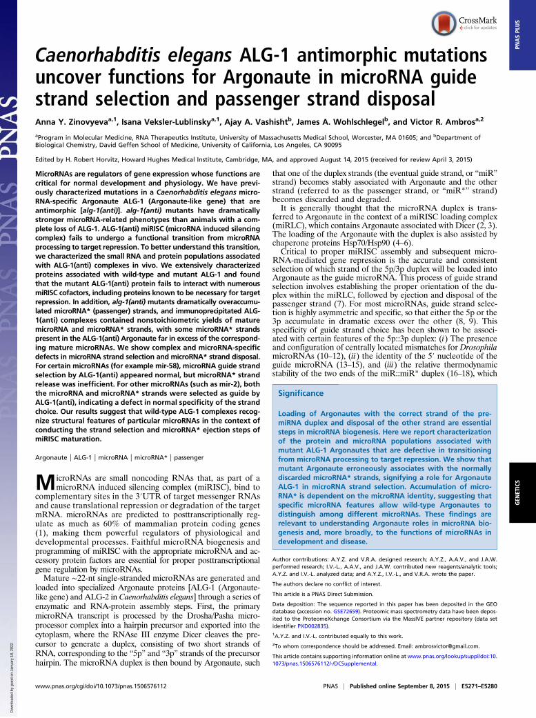
Caenorhabditis elegans ALG-1 antimorphic mutationsuncover functions for Argonaute in microRNA guidestrand selection and passenger strand disposalAnna Y. Zinovyevaa,1, Isana Veksler-Lublinskya,1, Ajay A. Vashishtb, James A. Wohlschlegelb, and Victor R. Ambrosa,2
aProgram in Molecular Medicine, RNA Therapeutics Institute, University of Massachusetts Medical School, Worcester, MA 01605; and bDepartment ofBiological Chemistry, David Geffen School of Medicine, University of California, Los Angeles, CA 90095
Edited by H. Robert Horvitz, Howard Hughes Medical Institute, Cambridge, MA, and approved August 14, 2015 (received for review April 3, 2015)
MicroRNAs are regulators of gene expression whose functions arecritical for normal development and physiology. We have previ-ously characterized mutations in a Caenorhabditis elegans micro-RNA-specific Argonaute ALG-1 (Argonaute-like gene) that areantimorphic [alg-1(anti)]. alg-1(anti) mutants have dramaticallystronger microRNA-related phenotypes than animals with a com-plete loss of ALG-1. ALG-1(anti) miRISC (microRNA induced silencingcomplex) fails to undergo a functional transition from microRNAprocessing to target repression. To better understand this transition,we characterized the small RNA and protein populations associatedwith ALG-1(anti) complexes in vivo. We extensively characterizedproteins associated with wild-type and mutant ALG-1 and foundthat the mutant ALG-1(anti) protein fails to interact with numerousmiRISC cofactors, including proteins known to be necessary for targetrepression. In addition, alg-1(anti) mutants dramatically overaccumu-lated microRNA* (passenger) strands, and immunoprecipitated ALG-1(anti) complexes contained nonstoichiometric yields of maturemicroRNA and microRNA* strands, with some microRNA* strandspresent in the ALG-1(anti) Argonaute far in excess of the correspond-ing mature microRNAs. We show complex and microRNA-specificdefects in microRNA strand selection and microRNA* strand disposal.For certain microRNAs (for example mir-58), microRNA guide strandselection by ALG-1(anti) appeared normal, but microRNA* strandrelease was inefficient. For other microRNAs (such as mir-2), boththe microRNA and microRNA* strands were selected as guide byALG-1(anti), indicating a defect in normal specificity of the strandchoice. Our results suggest that wild-type ALG-1 complexes recog-nize structural features of particular microRNAs in the context ofconducting the strand selection and microRNA* ejection steps ofmiRISC maturation.
Argonaute | ALG-1 | microRNA | microRNA* | passenger
MicroRNAs are small noncoding RNAs that, as part of amicroRNA induced silencing complex (miRISC), bind to
complementary sites in the 3′UTR of target messenger RNAsand cause translational repression or degradation of the targetmRNA. microRNAs are predicted to posttranscriptionally reg-ulate as much as 60% of mammalian protein coding genes(1), making them powerful regulators of physiological anddevelopmental processes. Faithful microRNA biogenesis andprogramming of miRISC with the appropriate microRNA and ac-cessory protein factors are essential for proper posttranscriptionalgene regulation by microRNAs.Mature ∼22-nt single-stranded microRNAs are generated and
loaded into specialized Argonaute proteins [ALG-1 (Argonaute-like gene) and ALG-2 in Caenorhabditis elegans] through a series ofenzymatic and RNA-protein assembly steps. First, the primarymicroRNA transcript is processed by the Drosha/Pasha micro-processor complex into a hairpin precursor and exported into thecytoplasm, where the RNAse III enzyme Dicer cleaves the pre-cursor to generate a duplex, consisting of two short strands ofRNA, corresponding to the “5p” and “3p” strands of the precursorhairpin. The microRNA duplex is then bound by Argonaute, such
that one of the duplex strands (the eventual guide strand, or “miR”strand) becomes stably associated with Argonaute and the otherstrand (referred to as the passenger strand, or “miR*” strand)becomes discarded and degraded.It is generally thought that the microRNA duplex is trans-
ferred to Argonaute in the context of a miRISC loading complex(miRLC), which contains Argonaute associated with Dicer (2, 3).The loading of the Argonaute with the duplex is also assisted bychaperone proteins Hsp70/Hsp90 (4–6).Critical to proper miRISC assembly and subsequent micro-
RNA-mediated gene repression is the accurate and consistentselection of which strand of the 5p/3p duplex will be loaded intoArgonaute as the guide microRNA. This process of guide strandselection involves establishing the proper orientation of the du-plex within the miRLC, followed by ejection and disposal of thepassenger strand (7). For most microRNAs, guide strand selec-tion is highly asymmetric and specific, so that either the 5p or the3p accumulate in dramatic excess over the other (8, 9). Thisspecificity of guide strand choice has been shown to be associ-ated with certain features of the 5p::3p duplex: (i) The presenceand configuration of centrally located mismatches for DrosophilamicroRNAs (10–12), (ii) the identity of the 5′ nucleotide of theguide microRNA (13–15), and (iii) the relative thermodynamicstability of the two ends of the miR::miR* duplex (16–18), which
Significance
Loading of Argonautes with the correct strand of the pre-miRNA duplex and disposal of the other strand are essentialsteps in microRNA biogenesis. Here we report characterizationof the protein and microRNA populations associated withmutant ALG-1 Argonautes that are defective in transitioningfrom microRNA processing to target repression. We show thatmutant Argonaute erroneously associates with the normallydiscarded microRNA* strands, signifying a role for ArgonauteALG-1 in microRNA strand selection. Accumulation of micro-RNA* is dependent on the microRNA identity, suggesting thatspecific microRNA features allow wild-type Argonautes todistinguish among different microRNAs. These findings arerelevant to understanding Argonaute roles in microRNA bio-genesis and, more broadly, to the functions of microRNAs indevelopment and disease.
Author contributions: A.Y.Z. and V.R.A. designed research; A.Y.Z., A.A.V., and J.A.W.performed research; I.V.-L., A.A.V., and J.A.W. contributed new reagents/analytic tools;A.Y.Z. and I.V.-L. analyzed data; and A.Y.Z., I.V.-L., and V.R.A. wrote the paper.
The authors declare no conflict of interest.
This article is a PNAS Direct Submission.
Data deposition: The sequence reported in this paper has been deposited in the GEOdatabase (accession no. GSE72659). Proteomic mass spectrometry data have been depos-ited to the ProteomeXchange Consortium via the MassIVE partner repository (data setidentifier PXD002835).1A.Y.Z. and I.V.-L. contributed equally to this work.2To whom correspondence should be addressed. Email: [email protected].
This article contains supporting information online at www.pnas.org/lookup/suppl/doi:10.1073/pnas.1506576112/-/DCSupplemental.
www.pnas.org/cgi/doi/10.1073/pnas.1506576112 PNAS | Published online September 8, 2015 | E5271–E5280
GEN
ETICS
PNASPL
US
Dow
nloa
ded
by g
uest
on
Janu
ary
18, 2
022
has a dramatic impact on strand choice in both flies and mammals.It should be noted that the relative thermodynamic stability of theduplex ends may be a primary determinant of strand selection forperfectly paired duplexes of siRNAs, but may be less important forthe mismatched duplexes of most microRNAs (10, 11, 16, 17).Additionally, in C. elegans, microRNAs that produce 5p guideswere reported to have similar thermodynamic stabilities of duplexends as microRNAs that produce 3p guides (9), suggesting that atleast in C. elegans other duplex features may be more importantfor strand selection (9). Features, such as microRNA duplex se-quence composition and position of mismatches, could instead bemajor contributors to strand selection (19–21).Although guide strand selection appears to be highly asym-
metric for most microRNAs, for a small subset of microRNAssubstantial quantities of both 5p and 3p strands can accumulate,and in at least some cases there is evidence for functional rolesfor both strands (22–25). It’s possible that the relative loading of5p and 3p strands as the guide could be regulated by upstreamsignals acting via Argonaute or other components of the miRLC.How C. elegans Dicer DCR-1 may contribute mechanistically tothe specificity of microRNA loading and guide strand selection isunknown. Mammalian Dicer-associated proteins TRBP/PACTcontribute to strand selection (26), and R2D2, a key componentof Drosophila miRLC, is dispensable for miR strand selection(27). Interestingly, mammalian Dicer itself may be dispensablefor asymmetric miRISC duplex loading, at least under certaincircumstances (28, 29). The Argonaute protein itself may havethe capacity to distinguish among specific microRNAs to de-termine the guide/miR vs. passenger/miR* fates of the 5p and 3pstrands. Indeed, a very recent report has suggested a direct rolefor mAGO2 in miR strand choice (30). All these considerationsemphasize the importance of acquiring a better understanding ofthe regulation of microRNA guide strand choice by Argonautecomplexes in vivo.We reported previously novel antimorphic alleles of the
C. elegans Argonaute gene alg-1 that broadly impair the functionsof many microRNAs, apparently by sequestering microRNAs intoimmature and ineffectual miRLC complexes (31). The mutantALG-1(anti) proteins exhibit an increased association with themiRLC component Dicer DCR-1 in vivo, and a decreased as-sociation with the miRISC effector AIN-1 (ALG-1 interactingprotein-1). We proposed that ALG-1(anti)–containing com-plexes associate with microRNAs, but fail to properly maturefrom the Dicer-containing miRLCs to the effector miRISCs,thereby sequestering microRNAs in immature (and nonfunc-tional) RISC complexes (31). To better understand both theprotein and the microRNA dynamics associated with this miRLC-to-miRISC maturation step, we further characterized the ALG-1(anti)–associated proteins and microRNAs using high-throughputproteomics and small RNAseq. Our results support the idea thatalg-1(anti) mutants are defective in transitioning from biogenesis(miRLC) to effector (miRISC) status and identify conservedArgonaute-interacting proteins that may be specific to eithermiRLC or miRISC complexes. Moreover, our small RNAseqanalysis shows that alg-1(anti) mutants accumulate miR* strands atlevels dramatically greater than the wild-type, indicating thatthe ALG-1(anti) mutant proteins are defective in microRNAstrand selection and miR* strand disposal. We show that thebiogenesis of different microRNAs can be impacted very differ-ently by ALG-1(anti). For example, in the case of mir-58, miR*strands are retained by the ALG-1(anti) Argonaute apparently aspart of the miR::miR* duplex, suggesting defective miR* strandrelease. In contrast, for mir-2, the ALG-1(anti) complexes con-tained far more miR* strands than miR, indicating that miR-2biogenesis suffers from defects in the strand selection step ofmiRISC maturation. These findings support a model whereinmicroRNA biogenesis and miRISC maturation involve criticalroles for Argonaute both in recognizing features that define spe-cific microRNAs and in exercising microRNA-specific programsof guide strand selection and miR* strand disposal.
Materials and MethodsC. elegans Culture and Genetics. C. elegans culture was performed usingstandard nematode growth conditions (32), except the HB101 Escherichiacoli strain was used as the food source. All strains were grown at 20 °C.Because of their strong heterochronic phenotypes, alg-1(anti) mutant ani-mals often burst through the vulva during the L4-adult molt (31). Therefore,alg-1(anti) mutations were maintained in a lin-31(n1053) genetic back-ground that impairs vulva development and thereby suppresses the burstingphenotype of alg-1 mutants, while leaving their heterochronic phenotypesintact. The adult-specific col-19::gfp reporter transgene is also present in allof the strains. Therefore, the alg-1(anti) strains used here contain lin-31;col-19::gfp, and in all experiments the wild-type controls were lin-31(n1053);col-19::gfp.
Northern Blotting. Total RNA was isolated from mixed population of animalsusing TRIzol reagent (Life Technologies), and Northern blots were performedas previously described (33, 34). For oligo probe sequences please see SIMaterials and Methods.
Firefly microRNA Quantifications. FirePlex microRNA assays were performed aspreviously described (31). For experiments involving 2′O-methyl oligonu-cleotide-mediated pull-down, equivalent fractions of input material andsupernatant were tested using the FirePlex assays to quantitatively assessmicroRNA abundance in the starting material and microRNA depletion fromthe supernatant by the 2′-O-methylated oligonucleotide.
Small RNA Library Preparation and High-Throughput Sequencing. Small RNAcDNA libraries were prepared as previously described (35) and sequenced onthe Illumina GAIIx instrument or the NextSeq500 (for L2 staged samples)using standard manufacturer’s protocols.
Computational Analysis of cDNA Library Sequence Data. Detailed description ofcDNA library sequence data analysis can be found in SI Materials and Methods.
ALG-1 Immunoprecipitation and Western Blot Analysis. Detailed descriptionof antibodies, lysate preparation, immunoprecipitation, and Western blotanalysis can be found in SI Materials and Methods.
2′O-Methyl Oligo Pull-Downs. The 2′O-methyl oligo pull-downs from extracts ofwhole worms were performed as described previously (36). For mass spec-trometry, each sample contained 20 mg of total protein. Sequences of the2′-O-methylated, biotinylated oligonucleotides can be found in SI Materialsand Methods.
Mass Spectrometry Analysis of ALG-1 Immunopurified Complexes and 2′-O-MethylOligonucleotide Pulldown Complexes and Computational Analysis of ProteomicData. For a detailed description of mass spectrometry analysis and computa-tional analysis of ALG-1 associated proteomes, as well as data analysis of miR-58and miR-58* associated proteins, see SI Materials and Methods.
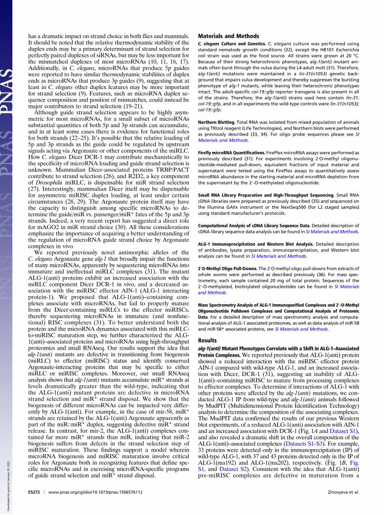
Resultsalg-1(anti)Mutant Phenotypes Correlate with a Shift in ALG-1–AssociatedProtein Complexes.We reported previously that ALG-1(anti) proteinshowed a reduced interaction with the miRISC effector proteinAIN-1 compared with wild-type ALG-1, and an increased associa-tion with Dicer, DCR-1 (31), suggesting an inability of ALG-1(anti)–containing miRISC to mature from processing complexesto effector complexes. To determine if interactions of ALG-1 withother proteins were affected by the alg-1(anti) mutations, we con-ducted ALG-1 IP from wild-type and alg-1(anti) animals followedby MudPIT (Multidimensional Protein Identification Technology)analysis to determine the composition of the associating complexes.The MudPIT data confirmed the results of our previous Westernblot experiments, of a reduced ALG-1(anti) association with AIN-1and an increased association with DCR-1 (Fig. 1A and Dataset S1),and also revealed a dramatic shift in the overall composition of theALG-1(anti)–associated complexes (Datasets S1–S3). For example,33 proteins were detected only in the immunoprecipitation (IP) ofwild-type ALG-1, with 37 and 43 proteins detected only in the IP ofALG-1(ma192) and ALG-1(ma202), respectively. (Fig. 1B, Fig.S1, and Dataset S2). Consistent with the idea that ALG-1(anti)pre-miRISC complexes are defective in maturation from a
E5272 | www.pnas.org/cgi/doi/10.1073/pnas.1506576112 Zinovyeva et al.
Dow
nloa
ded
by g
uest
on
Janu
ary
18, 2
022
miRISC biogenesis stage to the mature effector miRISC complex,we observed that the ALG-1(anti) IP recovered an increased yield,compared with wild-type, of certain hsp-60 and hsp-70 familyproteins and other putative chaperones (Datasets S1–S3), and areduced yield of several known miRISC effector components,including the poly-A binding proteins PAB-1 and PAB-2 (DatasetS1), which have been shown to play critical roles in target re-pression (37 and 38; reviewed in ref. 39). This trend was seenacross the two alg-1(anti) mutants examined: alg-1(ma192) andalg-1(ma202) (Fig. 1 and Dataset S1), which affect the PIWI andMID domains of the protein, respectively (31). ALG-1(ma192)and ALG-1(ma202) coimmunoprecipitated with overlapping yetnonidentical sets of proteins (Fig. 1, Fig. S1, and Dataset S1),consistent with the hypothesis that the two mutations may nothave identical effects on the ability of ALG-1 to interact withvarious partners.It should be noted that the proteins coimmunoprecipitated
with ALG-1 in these experiments could include factors that di-rectly interact with ALG-1 or that indirectly associate withmiRISC by binding to target mRNAs, because our IP experi-ments were performed in the absence of RNase treatment.
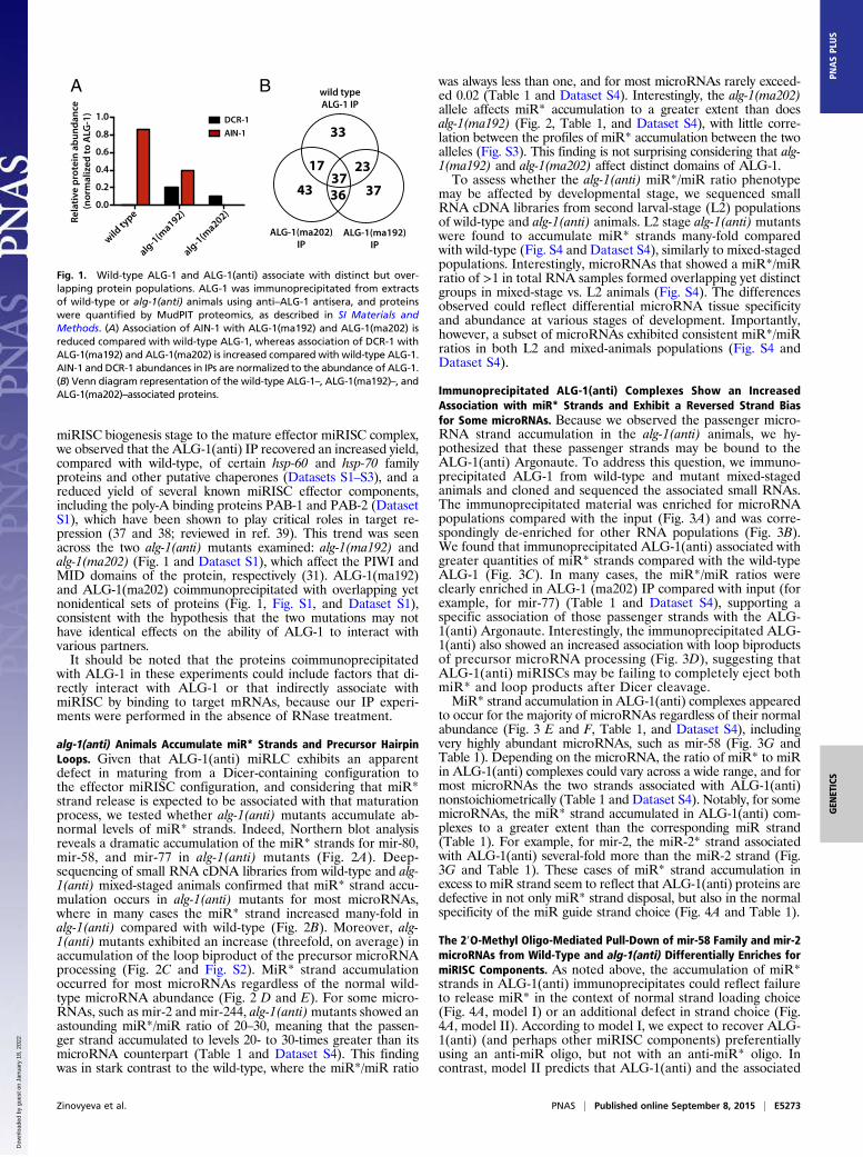
alg-1(anti) Animals Accumulate miR* Strands and Precursor HairpinLoops. Given that ALG-1(anti) miRLC exhibits an apparentdefect in maturing from a Dicer-containing configuration tothe effector miRISC configuration, and considering that miR*strand release is expected to be associated with that maturationprocess, we tested whether alg-1(anti) mutants accumulate ab-normal levels of miR* strands. Indeed, Northern blot analysisreveals a dramatic accumulation of the miR* strands for mir-80,mir-58, and mir-77 in alg-1(anti) mutants (Fig. 2A). Deep-sequencing of small RNA cDNA libraries from wild-type and alg-1(anti) mixed-staged animals confirmed that miR* strand accu-mulation occurs in alg-1(anti) mutants for most microRNAs,where in many cases the miR* strand increased many-fold inalg-1(anti) compared with wild-type (Fig. 2B). Moreover, alg-1(anti) mutants exhibited an increase (threefold, on average) inaccumulation of the loop biproduct of the precursor microRNAprocessing (Fig. 2C and Fig. S2). MiR* strand accumulationoccurred for most microRNAs regardless of the normal wild-type microRNA abundance (Fig. 2 D and E). For some micro-RNAs, such as mir-2 and mir-244, alg-1(anti)mutants showed anastounding miR*/miR ratio of 20–30, meaning that the passen-ger strand accumulated to levels 20- to 30-times greater than itsmicroRNA counterpart (Table 1 and Dataset S4). This findingwas in stark contrast to the wild-type, where the miR*/miR ratio
was always less than one, and for most microRNAs rarely exceed-ed 0.02 (Table 1 and Dataset S4). Interestingly, the alg-1(ma202)allele affects miR* accumulation to a greater extent than doesalg-1(ma192) (Fig. 2, Table 1, and Dataset S4), with little corre-lation between the profiles of miR* accumulation between the twoalleles (Fig. S3). This finding is not surprising considering that alg-1(ma192) and alg-1(ma202) affect distinct domains of ALG-1.To assess whether the alg-1(anti) miR*/miR ratio phenotype
may be affected by developmental stage, we sequenced smallRNA cDNA libraries from second larval-stage (L2) populationsof wild-type and alg-1(anti) animals. L2 stage alg-1(anti) mutantswere found to accumulate miR* strands many-fold comparedwith wild-type (Fig. S4 and Dataset S4), similarly to mixed-stagedpopulations. Interestingly, microRNAs that showed a miR*/miRratio of >1 in total RNA samples formed overlapping yet distinctgroups in mixed-stage vs. L2 animals (Fig. S4). The differencesobserved could reflect differential microRNA tissue specificityand abundance at various stages of development. Importantly,however, a subset of microRNAs exhibited consistent miR*/miRratios in both L2 and mixed-animals populations (Fig. S4 andDataset S4).
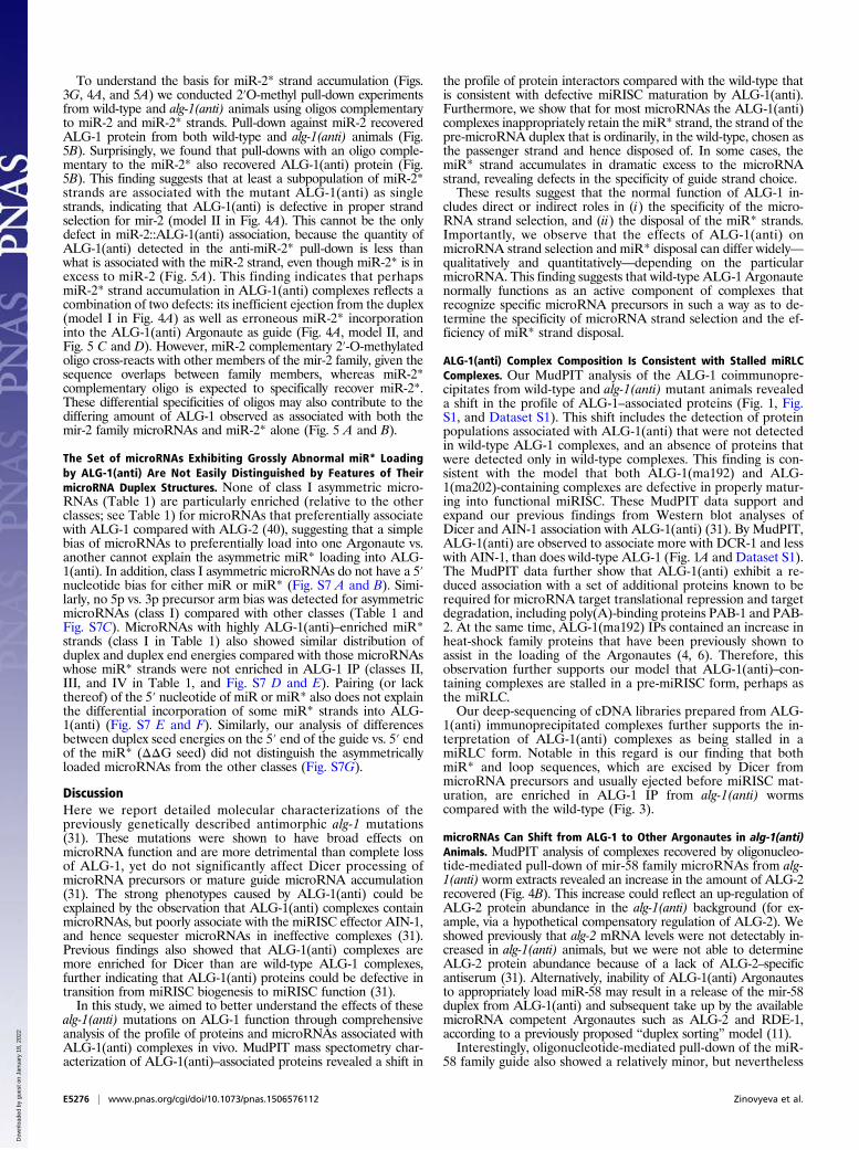
Immunoprecipitated ALG-1(anti) Complexes Show an IncreasedAssociation with miR* Strands and Exhibit a Reversed Strand Biasfor Some microRNAs. Because we observed the passenger micro-RNA strand accumulation in the alg-1(anti) animals, we hy-pothesized that these passenger strands may be bound to theALG-1(anti) Argonaute. To address this question, we immuno-precipitated ALG-1 from wild-type and mutant mixed-stagedanimals and cloned and sequenced the associated small RNAs.The immunoprecipitated material was enriched for microRNApopulations compared with the input (Fig. 3A) and was corre-spondingly de-enriched for other RNA populations (Fig. 3B).We found that immunoprecipitated ALG-1(anti) associated withgreater quantities of miR* strands compared with the wild-typeALG-1 (Fig. 3C). In many cases, the miR*/miR ratios wereclearly enriched in ALG-1 (ma202) IP compared with input (forexample, for mir-77) (Table 1 and Dataset S4), supporting aspecific association of those passenger strands with the ALG-1(anti) Argonaute. Interestingly, the immunoprecipitated ALG-1(anti) also showed an increased association with loop biproductsof precursor microRNA processing (Fig. 3D), suggesting thatALG-1(anti) miRISCs may be failing to completely eject bothmiR* and loop products after Dicer cleavage.MiR* strand accumulation in ALG-1(anti) complexes appeared
to occur for the majority of microRNAs regardless of their normalabundance (Fig. 3 E and F, Table 1, and Dataset S4), includingvery highly abundant microRNAs, such as mir-58 (Fig. 3G andTable 1). Depending on the microRNA, the ratio of miR* to miRin ALG-1(anti) complexes could vary across a wide range, and formost microRNAs the two strands associated with ALG-1(anti)nonstoichiometrically (Table 1 and Dataset S4). Notably, for somemicroRNAs, the miR* strand accumulated in ALG-1(anti) com-plexes to a greater extent than the corresponding miR strand(Table 1). For example, for mir-2, the miR-2* strand associatedwith ALG-1(anti) several-fold more than the miR-2 strand (Fig.3G and Table 1). These cases of miR* strand accumulation inexcess to miR strand seem to reflect that ALG-1(anti) proteins aredefective in not only miR* strand disposal, but also in the normalspecificity of the miR guide strand choice (Fig. 4A and Table 1).
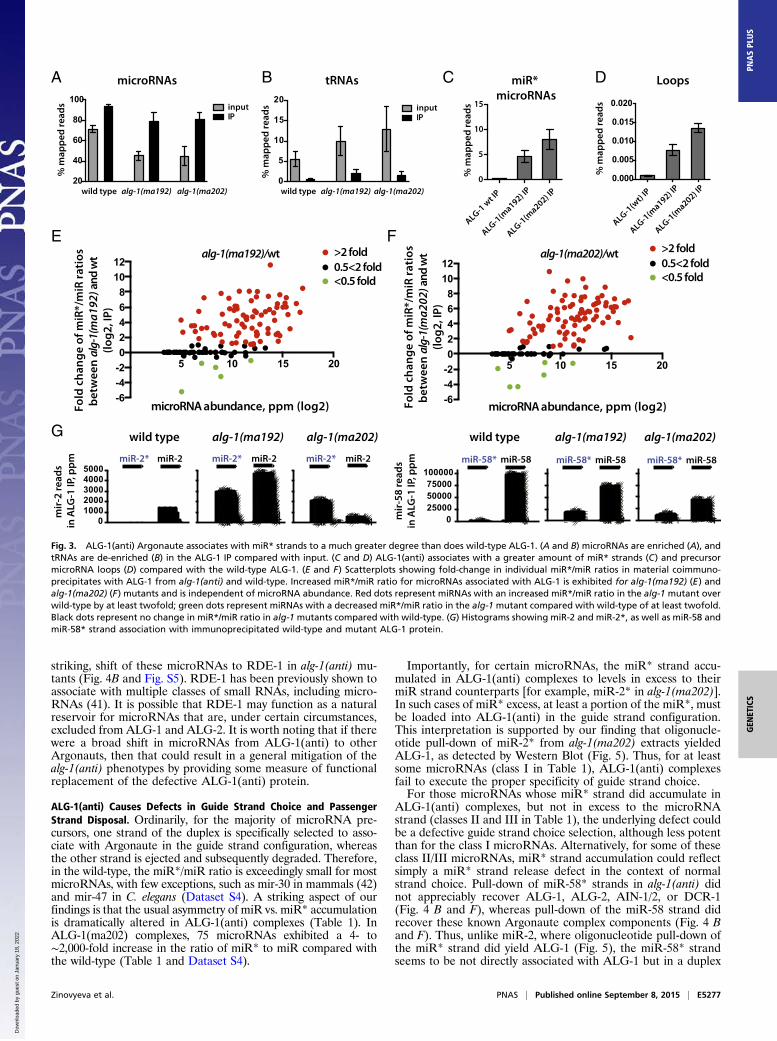
The 2′O-Methyl Oligo-Mediated Pull-Down of mir-58 Family and mir-2microRNAs from Wild-Type and alg-1(anti) Differentially Enriches formiRISC Components. As noted above, the accumulation of miR*strands in ALG-1(anti) immunoprecipitates could reflect failureto release miR* in the context of normal strand loading choice(Fig. 4A, model I) or an additional defect in strand choice (Fig.4A, model II). According to model I, we expect to recover ALG-1(anti) (and perhaps other miRISC components) preferentiallyusing an anti-miR oligo, but not with an anti-miR* oligo. Incontrast, model II predicts that ALG-1(anti) and the associated
BARe
lativ
e pr
otei
n ab
unda
nce
(nor
mal
ized
to A
LG-1
)
wild ty
pe
alg-1(ma192)
alg-1(ma202)
0.0
0.2
0.4
0.6
0.8
1.0 DCR-1AIN-1
3643
wild typeALG-1 IP
ALG-1(ma192)IP
33
3737
2317
ALG-1(ma202)IP
Fig. 1. Wild-type ALG-1 and ALG-1(anti) associate with distinct but over-lapping protein populations. ALG-1 was immunoprecipitated from extractsof wild-type or alg-1(anti) animals using anti–ALG-1 antisera, and proteinswere quantified by MudPIT proteomics, as described in SI Materials andMethods. (A) Association of AIN-1 with ALG-1(ma192) and ALG-1(ma202) isreduced compared with wild-type ALG-1, whereas association of DCR-1 withALG-1(ma192) and ALG-1(ma202) is increased compared with wild-type ALG-1.AIN-1 and DCR-1 abundances in IPs are normalized to the abundance of ALG-1.(B) Venn diagram representation of the wild-type ALG-1–, ALG-1(ma192)–, andALG-1(ma202)–associated proteins.
Zinovyeva et al. PNAS | Published online September 8, 2015 | E5273
GEN
ETICS
PNASPL
US
Dow
nloa
ded
by g
uest
on
Janu
ary
18, 2
022
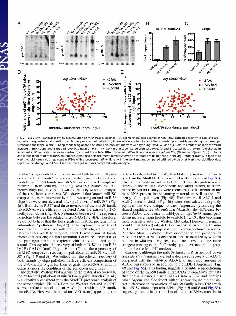
miRISC components should be recovered both by anti-miR pull-down and by anti-miR* pull-down. To distinguish between thesemodels for mir-58 family microRNAs, we examined complexesrecovered from wild-type and alg-1(ma192) lysates by 2′O-methyl oligo-mediated pull-down followed by MudPIT analysisof the associated complexes. We observed that known miRISCcomponents were recovered by pull-down using an anti–miR-58oligo but were not detected after pull-down of miR-58* (Fig.4B). Both the miR-58* and three members of the mir-58 familymicroRNAs were efficiently depleted from the extract by 2′O-methyl pull-down (Fig. 4C), presumably because of the sequencehomology between the related microRNAs (Fig. 4D). Therefore,we do not believe that the low signals for miRISC proteins in theanti–miR-58* pull-down (Fig. 4B) can be explained by inefficientbase pairing of passenger with anti–miR-58* oligo. Rather, weinterpret this result to support model I, where mir-58 familymicroRNA passenger strand accumulation reflects retention ofthe passenger strand in duplexes with an ALG-loaded guidestrand. This explains the recovery of both miR-58* and miR-58by IP of ALG-1(anti) (Fig. 4 E and G) and the asymmetry ofmiRISC component recovery in pull-down of miR-58 vs. miR-58* (Fig. 4 B and H). We believe that the efficient recovery ofboth strands by oligo pull-down reflects efficient competition ofthe 2′-O-methyl oligos for their cognate microRNA strands inextracts under the condition of the pull-down experiment.Incidentally, Western blot analysis of the material recovered by
the 2′O-methyl pull-down of mir-58 family guide strands (Fig. 4F)is qualitatively consistent with the MudPIT proteomic analysis ofthe same samples (Fig. 4B). Both the Western blot and MudPITshowed reduced association of ALG-1(anti) with mir-58 familymicroRNAs. However, the signal for ALG-1(anti) appeared more
reduced as detected by the Western blot compared with the wild-type than the MudPIT data indicate (Fig. 4 B and F and Fig. S5).This finding could in part reflect the fact that the protein abun-dances of the miRISC components and other factors, as deter-mined by MudPIT analysis, were normalized to the amount of themicroRNA present in the starting material, as well as the effi-ciency of the pull-down (Fig. 4B). Furthermore, if ALG-1 andALG-2 protein yields (Fig. 4B) were recalculated using onlypeptides that were unique to each Argonaute (discarding theshared peptides; see Materials and Methods), the difference be-tween ALG-1 abundance in wild-type vs. alg-1(anti) animal pull-downs increases from twofold to ∼sixfold (Fig. 4B), thus becomingmore consistent with the Western blot data. However, it remainspossible that ALG-1(anti) detection on aWestern blot by the anti–ALG-1 antibody is hampered for unknown technical reasons.Another MudPIT/Western blot discrepancy, the presence ofALG-1 in the miR-58*-associated material as detected byWesternblotting in wild-type (Fig. 4F), could be a result of the morestringent washing of the 2′-O-methyl pull-down material in prep-aration for the MudPIT analysis.Curiously, although the miR-58 family miR strand pull-down
from alg-1(anti) animals yielded a decreased recovery of ALG-1compared with the wild-type ALG-1, an increased amount ofALG-2 was recovered, in addition to the RDE-1 Argonaute (Fig.4B and Fig. S5). This finding suggests a possible reapportioningof some of the mir-58 family microRNAs in alg-1(anti) mutantsthat normally associate with ALG-1 into ALG-2 and perhapsother Argonautes. Consistent with this scenario, we did not de-tect a decrease in association of mir-58 family microRNAs withthe miRISC effector protein AIN-1 (Fig. 4 B and F and Fig. S5),suggesting that at least a portion of the microRNAs must be in
wild ty
pe
alg-1(ma192)
alg-1(ma202)
0
2
4
6
8
10wt alg-1(ma192)
alg-1(ma202)
alg-1(ma195)
alg-1(ma203)
alg-1(0)
miR
-80
U6
wt alg-1(ma192)
alg-1(ma202)
alg-1(ma195)
alg-1(ma203)
alg-1(0)
U6m
iR-5
8
miR
-77
U6
wt alg-1(ma192)
alg-1(ma202)
alg-1(ma195)
alg-1(ma203)
alg-1(0)
int
wild ty
pe
alg-1(ma192)
alg-1(ma202)
0.00
0.01
0.02
0.03
0.04
A B C
ED
% o
f map
ped
read
s
% o
f map
ped
read
s
miR* microRNAs Loops
5 10 15 20
-4-2024681012
microRNA abundance, ppm (log2)
Fold
cha
nge
of m
iR*/
mic
roRN
A ra
tios
betw
een
alg-
1(m
a192
) and
wt
(log2
, inp
ut)
alg-1(ma192)/wt >2 fold0.5<2 fold<0.5 fold
5 10 15 20
-4-2024681012
microRNA abundance, ppm (log2) Fo
ld c
hang
e of
miR
*/m
icro
RNA
ratio
s be
twee
n al
g-1(
ma2
02) a
nd w
t (lo
g2, i
nput
)
>2 fold0.5<2 fold<0.5 fold
alg-1(ma202)/wt
pre pre pre
int
Fig. 2. alg-1(anti) mutants show an accumulation of miR* strands in total RNA. (A) Northern blot analysis of total RNA extracted from wild-type and alg-1mutants using probes against miR* strands (pre, precursor microRNA; int, intermediate species of microRNA processing presumably containing the passengerstrand and the loop). (B and C) Deep-sequencing analysis of small RNA populations from wild-type, alg-1(ma192) and alg-1(ma202) mutant animals shows anincrease in miR* populations (B) and loop accumulation (C) in the alg-1 mutants compared with wild-type. (D and E) Scatterplots showing fold-change inindividual miR*/miR ratios between alg-1(anti) and wild-type total RNA. Increased miR*/miR ratio is seen in alg-1(ma192) (D) and alg-1(ma202) (E) mutantsand is independent of microRNA abundance (ppm). Red dots represent microRNAs with an increased miR*/miR ratio in the alg-1 mutant over wild-type of atleast twofold; green dots represent miRNAs with a decreased miR*/miR ratio in the alg-1 mutant compared with wild-type of at least twofold. Black dotsrepresent no change in miR*/miR ratio in the alg-1 mutants compared with wild-type.
E5274 | www.pnas.org/cgi/doi/10.1073/pnas.1506576112 Zinovyeva et al.
Dow
nloa
ded
by g
uest
on
Janu
ary
18, 2
022
miRISCs that contain AIN-1. The observed AIN-1 and DCR-1signal (Fig. 4F) is likely to at least in part reflect complexes ofmiR-58 with ALG-2. Similar observations were made for themir-52 family microRNA pull-downs (Fig. S6). Interestingly, mir-58
family microRNA association with AIN-2 was not observed inalg-1(ma192) mutant animals (Fig. 4B), suggesting that AIN-2may differ from its homolog AIN-1 with respect to its interactionwith Argonautes ALG-1 and ALG-2.
Table 1. microRNAs classified according to their miR*/miR ratio in input or ALG-1 IPs from alg-1(anti) mutants and wild-type
Class mir
Wild-type alg-1(ma192) alg-1(ma202) Fold-changein miR*/miR
ratiosbetween
ALG-1(ma202)IP and
ALG-1(wt) IP
AveragemicroRNAabundance
(ppm)
AveragemiR*/miR
ratio
AveragemicroRNAabundance
(ppm)
AveragemiR*/miR
ratio
AveragemicroRNAabundance
(ppm)
AveragemiR*/miR
ratio
Input IP Input IP Input IP Input IP Input IP Input IP
Class I:Asymmetric(IP miR*/miR > 1)
mir-244 335 336 0.149 0.068 1,644 467 0.762 0.937 132 52 21.024 21.700 321mir-87 297 327 0.102 0.005 258 181 2.400 1.081 158 94 13.566 9.205 1,942mir-2 518 966 0.091 0.020 1,308 3,748 3.520 0.484 140 442 30.950 4.261 212mir-86 3,622 4,019 0.006 0.002 3,073 6,598 0.216 0.112 1,206 1,407 5.593 2.496 1,005mir-252 310 1,187 0.006 0.002 129 1,618 4.769 0.526 240 1,024 3.349 1.565 818mir-241 2,460 2,454 0.016 0.007 746 470 0.460 0.491 524 262 1.206 1.559 218mir-45 2,269 1,868 0.014 0.006 6,359 2,322 0.010 0.014 10,737 6,100 1.298 1.127 183
Class II:Asymmetric(inputmiR*/miR > 1)
mir-235 998 5,019 0.022 0.003 821 6,086 2.879 0.416 453 2,528 11.603 0.487 192mir-90 2,323 13,937 0.054 0.002 4,580 11,900 0.316 0.022 1,572 8,970 3.301 0.256 105mir-54 20,260 26,474 0.012 0.003 11,576 30,654 0.140 0.017 4,804 14,626 1.818 0.330 112mir-786 329 314 0.139 0.051 2,067 2,586 0.149 0.037 167 118 1.188 0.373 7mir-79 3,644 2,092 0.011 0.012 15,606 12,644 0.238 0.159 8,892 6,286 1.024 0.607 51
Class III:Accumulated
mir-793 843 710 0.001 0.000 688 439 0.104 0.040 613 447 0.350 0.214 492mir-77 34,022 14,471 0.002 0.001 19,458 18,383 0.114 0.063 53,246 27,746 0.190 0.467 463mir-67 4,987 4,783 0.002 0.001 3,705 4,158 0.197 0.197 3,536 5,558 0.533 0.336 457mir-82 23,791 24,879 0.001 0.000 7,637 5,013 0.065 0.010 18,774 19,543 0.133 0.057 213mir-64 16,189 18,788 0.000 0.000 3,522 8,989 0.077 0.008 13,263 16,721 0.087 0.027 170mir-1 68,107 82,018 0.002 0.000 13,608 6,031 0.183 0.065 29,357 26,087 0.202 0.068 160mir-43 1,365 580 0.012 0.006 3,638 791 0.232 0.291 5,604 1,702 0.716 0.833 136mir-50 9,314 10,552 0.004 0.001 2,792 1,126 0.151 0.017 698 659 0.564 0.087 134mir-52 61,157 45,748 0.001 0.000 112,055 286,661 0.131 0.017 35,070 49,065 0.312 0.057 131mir-792 217 186 0.003 0.002 111 119 0.071 0.082 53 50 0.286 0.237 125lin-4 28,721 34,049 0.000 0.000 12,529 3,248 0.438 0.083 9,432 5,610 0.051 0.003 114
mir-248 267 314 0.001 0.000 122 84 0.104 0.033 61 49 0.066 0.028 113mir-58 99,377 86,512 0.003 0.002 186,421 106,646 0.066 0.162 75,670 48,452 0.119 0.205 112mir-38 4,656 2,615 0.007 0.001 12,010 1,343 0.025 0.014 6,628 1,940 0.142 0.129 95
mir-1829c 458 385 0.007 0.001 568 718 0.011 0.017 138 113 0.024 0.067 92mir-84 7,428 8,996 0.004 0.001 5,116 3,399 0.097 0.029 11,952 14,754 0.184 0.063 92mir-81 24,466 27,301 0.001 0.000 7,395 8,512 0.190 0.013 15,353 28,579 0.142 0.012 84mir-74 3,845 2,478 0.001 0.000 6,947 2,253 0.005 0.002 2,180 1,301 0.043 0.009 79mir-71 54,306 58,135 0.009 0.003 34,269 18,007 0.354 0.206 29,816 28,479 0.688 0.248 78mir-55 39,420 39,856 0.001 0.000 37,619 77,519 0.047 0.002 19,635 35,531 0.059 0.006 75
Class IV:Unaffected
mir-259 1,321 1,470 0.005 0.004 1,013 2,108 0.130 0.020 544 816 0.072 0.018 4mir-250 6,847 1,897 0.013 0.002 10,518 2,811 0.007 0.005 24,762 9,243 0.010 0.009 4
mir-1829a 71 136 0.284 0.030 49 92 1.107 0.108 43 74 0.506 0.096 3mir-356b 53 96 0.047 0.021 24 60 0.431 0.141 45 114 0.291 0.064 3mir-59 105 53 0.010 0.016 564 182 0.028 0.032 529 151 0.010 0.047 3mir-63 2,915 2,920 0.034 0.017 1,520 999 0.035 0.013 3,177 4,966 0.105 0.041 2mir-61 1,652 848 0.003 0.002 2,693 1,119 0.008 0.005 1,637 1,117 0.013 0.005 2mir-36 1,492 468 0.105 0.027 9,510 2,694 0.047 0.027 13,420 6,710 0.056 0.057 2mir-230 355 285 0.202 0.093 409 328 0.195 0.089 466 762 0.612 0.190 2mir-2214 189 84 0.021 0.112 403 148 0.056 0.202 617 151 0.050 0.220 2mir-4816 97 57 0.073 0.021 124 54 0.210 0.038 133 112 0.202 0.037 2mir-75 3,038 2,426 0.044 0.026 4,394 5,836 0.107 0.072 29,063 89,083 0.117 0.044 2mir-65 30,192 20,108 0.000 0.000 5,417 4,792 0.005 0.001 24,775 43,527 0.001 0.000 2mir-62 486 485 0.244 0.018 1,397 478 0.123 0.027 1,070 928 0.075 0.021 1mir-791 47 364 0.797 0.117 37 422 2.076 0.076 45 221 0.694 0.053 0mir-34 3,106 3,459 0.084 0.054 2,950 1,253 0.233 0.096 4,709 1,753 0.149 0.023 0
Class I: Asymmetric. MicroRNAs that had a miR*/miR ratio >1 in ALG-1(ma202) IP. Class II: Asymmetric in input. MicroRNAs that had a miR*/miR ratio >1 ininput for the ALG-1(ma202) IP. Class III: Accumulated. 20 microRNAs representative of the microRNAs whose miR*/miR ratio was significantly increased inALG-1(ma202) IP compared with wild-type ALG-1 IP. See Dataset S4 for a complete list. Class IV: Unaffected. MicroRNAs whose miR*/miR ratio changed ≤4between wild-type ALG-1 IP and ALG-1(ma202) IP. ppm, parts per million.
Zinovyeva et al. PNAS | Published online September 8, 2015 | E5275
GEN
ETICS
PNASPL
US
Dow
nloa
ded
by g
uest
on
Janu
ary
18, 2
022
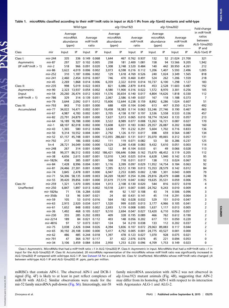
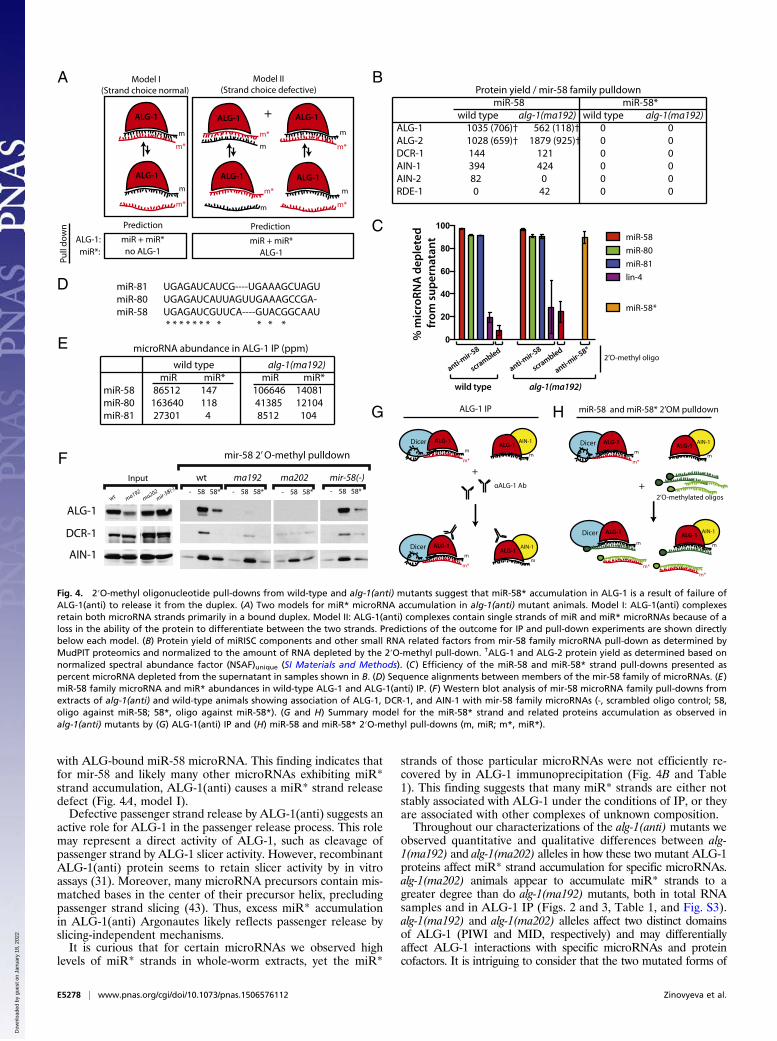
To understand the basis for miR-2* strand accumulation (Figs.3G, 4A, and 5A) we conducted 2′O-methyl pull-down experimentsfrom wild-type and alg-1(anti) animals using oligos complementaryto miR-2 and miR-2* strands. Pull-down against miR-2 recoveredALG-1 protein from both wild-type and alg-1(anti) animals (Fig.5B). Surprisingly, we found that pull-downs with an oligo comple-mentary to the miR-2* also recovered ALG-1(anti) protein (Fig.5B). This finding suggests that at least a subpopulation of miR-2*strands are associated with the mutant ALG-1(anti) as singlestrands, indicating that ALG-1(anti) is defective in proper strandselection for mir-2 (model II in Fig. 4A). This cannot be the onlydefect in miR-2::ALG-1(anti) association, because the quantity ofALG-1(anti) detected in the anti-miR-2* pull-down is less thanwhat is associated with the miR-2 strand, even though miR-2* is inexcess to miR-2 (Fig. 5A). This finding indicates that perhapsmiR-2* strand accumulation in ALG-1(anti) complexes reflects acombination of two defects: its inefficient ejection from the duplex(model I in Fig. 4A) as well as erroneous miR-2* incorporationinto the ALG-1(anti) Argonaute as guide (Fig. 4A, model II, andFig. 5 C and D). However, miR-2 complementary 2′-O-methylatedoligo cross-reacts with other members of the mir-2 family, given thesequence overlaps between family members, whereas miR-2*complementary oligo is expected to specifically recover miR-2*.These differential specificities of oligos may also contribute to thediffering amount of ALG-1 observed as associated with both themir-2 family microRNAs and miR-2* alone (Fig. 5 A and B).
The Set of microRNAs Exhibiting Grossly Abnormal miR* Loadingby ALG-1(anti) Are Not Easily Distinguished by Features of TheirmicroRNA Duplex Structures. None of class I asymmetric micro-RNAs (Table 1) are particularly enriched (relative to the otherclasses; see Table 1) for microRNAs that preferentially associatewith ALG-1 compared with ALG-2 (40), suggesting that a simplebias of microRNAs to preferentially load into one Argonaute vs.another cannot explain the asymmetric miR* loading into ALG-1(anti). In addition, class I asymmetric microRNAs do not have a 5′nucleotide bias for either miR or miR* (Fig. S7 A and B). Simi-larly, no 5p vs. 3p precursor arm bias was detected for asymmetricmicroRNAs (class I) compared with other classes (Table 1 andFig. S7C). MicroRNAs with highly ALG-1(anti)–enriched miR*strands (class I in Table 1) also showed similar distribution ofduplex and duplex end energies compared with those microRNAswhose miR* strands were not enriched in ALG-1 IP (classes II,III, and IV in Table 1, and Fig. S7 D and E). Pairing (or lackthereof) of the 5′ nucleotide of miR or miR* also does not explainthe differential incorporation of some miR* strands into ALG-1(anti) (Fig. S7 E and F). Similarly, our analysis of differencesbetween duplex seed energies on the 5′ end of the guide vs. 5′ endof the miR* (ΔΔG seed) did not distinguish the asymmetricallyloaded microRNAs from the other classes (Fig. S7G).
DiscussionHere we report detailed molecular characterizations of thepreviously genetically described antimorphic alg-1 mutations(31). These mutations were shown to have broad effects onmicroRNA function and are more detrimental than complete lossof ALG-1, yet do not significantly affect Dicer processing ofmicroRNA precursors or mature guide microRNA accumulation(31). The strong phenotypes caused by ALG-1(anti) could beexplained by the observation that ALG-1(anti) complexes containmicroRNAs, but poorly associate with the miRISC effector AIN-1,and hence sequester microRNAs in ineffective complexes (31).Previous findings also showed that ALG-1(anti) complexes aremore enriched for Dicer than are wild-type ALG-1 complexes,further indicating that ALG-1(anti) proteins could be defective intransition from miRISC biogenesis to miRISC function (31).In this study, we aimed to better understand the effects of these
alg-1(anti) mutations on ALG-1 function through comprehensiveanalysis of the profile of proteins and microRNAs associated withALG-1(anti) complexes in vivo. MudPIT mass spectometry char-acterization of ALG-1(anti)–associated proteins revealed a shift in
the profile of protein interactors compared with the wild-type thatis consistent with defective miRISC maturation by ALG-1(anti).Furthermore, we show that for most microRNAs the ALG-1(anti)complexes inappropriately retain the miR* strand, the strand of thepre-microRNA duplex that is ordinarily, in the wild-type, chosen asthe passenger strand and hence disposed of. In some cases, themiR* strand accumulates in dramatic excess to the microRNAstrand, revealing defects in the specificity of guide strand choice.These results suggest that the normal function of ALG-1 in-
cludes direct or indirect roles in (i) the specificity of the micro-RNA strand selection, and (ii) the disposal of the miR* strands.Importantly, we observe that the effects of ALG-1(anti) onmicroRNA strand selection and miR* disposal can differ widely—qualitatively and quantitatively—depending on the particularmicroRNA. This finding suggests that wild-type ALG-1 Argonautenormally functions as an active component of complexes thatrecognize specific microRNA precursors in such a way as to de-termine the specificity of microRNA strand selection and the ef-ficiency of miR* strand disposal.
ALG-1(anti) Complex Composition Is Consistent with Stalled miRLCComplexes. Our MudPIT analysis of the ALG-1 coimmunopre-cipitates from wild-type and alg-1(anti) mutant animals revealeda shift in the profile of ALG-1–associated proteins (Fig. 1, Fig.S1, and Dataset S1). This shift includes the detection of proteinpopulations associated with ALG-1(anti) that were not detectedin wild-type ALG-1 complexes, and an absence of proteins thatwere detected only in wild-type complexes. This finding is con-sistent with the model that both ALG-1(ma192) and ALG-1(ma202)-containing complexes are defective in properly matur-ing into functional miRISC. These MudPIT data support andexpand our previous findings from Western blot analyses ofDicer and AIN-1 association with ALG-1(anti) (31). By MudPIT,ALG-1(anti) are observed to associate more with DCR-1 and lesswith AIN-1, than does wild-type ALG-1 (Fig. 1A and Dataset S1).The MudPIT data further show that ALG-1(anti) exhibit a re-duced association with a set of additional proteins known to berequired for microRNA target translational repression and targetdegradation, including poly(A)-binding proteins PAB-1 and PAB-2. At the same time, ALG-1(ma192) IPs contained an increase inheat-shock family proteins that have been previously shown toassist in the loading of the Argonautes (4, 6). Therefore, thisobservation further supports our model that ALG-1(anti)–con-taining complexes are stalled in a pre-miRISC form, perhaps asthe miRLC.Our deep-sequencing of cDNA libraries prepared from ALG-
1(anti) immunoprecipitated complexes further supports the in-terpretation of ALG-1(anti) complexes as being stalled in amiRLC form. Notable in this regard is our finding that bothmiR* and loop sequences, which are excised by Dicer frommicroRNA precursors and usually ejected before miRISC mat-uration, are enriched in ALG-1 IP from alg-1(anti) wormscompared with the wild-type (Fig. 3).
microRNAs Can Shift from ALG-1 to Other Argonautes in alg-1(anti)Animals. MudPIT analysis of complexes recovered by oligonucleo-tide-mediated pull-down of mir-58 family microRNAs from alg-1(anti) worm extracts revealed an increase in the amount of ALG-2recovered (Fig. 4B). This increase could reflect an up-regulation ofALG-2 protein abundance in the alg-1(anti) background (for ex-ample, via a hypothetical compensatory regulation of ALG-2). Weshowed previously that alg-2 mRNA levels were not detectably in-creased in alg-1(anti) animals, but we were not able to determineALG-2 protein abundance because of a lack of ALG-2–specificantiserum (31). Alternatively, inability of ALG-1(anti) Argonautesto appropriately load miR-58 may result in a release of the mir-58duplex from ALG-1(anti) and subsequent take up by the availablemicroRNA competent Argonautes such as ALG-2 and RDE-1,according to a previously proposed “duplex sorting” model (11).Interestingly, oligonucleotide-mediated pull-down of the miR-
58 family guide also showed a relatively minor, but nevertheless
E5276 | www.pnas.org/cgi/doi/10.1073/pnas.1506576112 Zinovyeva et al.
Dow
nloa
ded
by g
uest
on
Janu
ary
18, 2
022
striking, shift of these microRNAs to RDE-1 in alg-1(anti) mu-tants (Fig. 4B and Fig. S5). RDE-1 has been previously shown toassociate with multiple classes of small RNAs, including micro-RNAs (41). It is possible that RDE-1 may function as a naturalreservoir for microRNAs that are, under certain circumstances,excluded from ALG-1 and ALG-2. It is worth noting that if therewere a broad shift in microRNAs from ALG-1(anti) to otherArgonauts, then that could result in a general mitigation of thealg-1(anti) phenotypes by providing some measure of functionalreplacement of the defective ALG-1(anti) protein.
ALG-1(anti) Causes Defects in Guide Strand Choice and PassengerStrand Disposal. Ordinarily, for the majority of microRNA pre-cursors, one strand of the duplex is specifically selected to asso-ciate with Argonaute in the guide strand configuration, whereasthe other strand is ejected and subsequently degraded. Therefore,in the wild-type, the miR*/miR ratio is exceedingly small for mostmicroRNAs, with few exceptions, such as mir-30 in mammals (42)and mir-47 in C. elegans (Dataset S4). A striking aspect of ourfindings is that the usual asymmetry of miR vs. miR* accumulationis dramatically altered in ALG-1(anti) complexes (Table 1). InALG-1(ma202) complexes, 75 microRNAs exhibited a 4- to∼2,000-fold increase in the ratio of miR* to miR compared withthe wild-type (Table 1 and Dataset S4).
Importantly, for certain microRNAs, the miR* strand accu-mulated in ALG-1(anti) complexes to levels in excess to theirmiR strand counterparts [for example, miR-2* in alg-1(ma202)].In such cases of miR* excess, at least a portion of the miR*, mustbe loaded into ALG-1(anti) in the guide strand configuration.This interpretation is supported by our finding that oligonucle-otide pull-down of miR-2* from alg-1(ma202) extracts yieldedALG-1, as detected by Western Blot (Fig. 5). Thus, for at leastsome microRNAs (class I in Table 1), ALG-1(anti) complexesfail to execute the proper specificity of guide strand choice.For those microRNAs whose miR* strand did accumulate in
ALG-1(anti) complexes, but not in excess to the microRNAstrand (classes II and III in Table 1), the underlying defect couldbe a defective guide strand choice selection, although less potentthan for the class I microRNAs. Alternatively, for some of theseclass II/III microRNAs, miR* strand accumulation could reflectsimply a miR* strand release defect in the context of normalstrand choice. Pull-down of miR-58* strands in alg-1(anti) didnot appreciably recover ALG-1, ALG-2, AIN-1/2, or DCR-1(Fig. 4 B and F), whereas pull-down of the miR-58 strand didrecover these known Argonaute complex components (Fig. 4 Band F). Thus, unlike miR-2, where oligonucleotide pull-down ofthe miR* strand did yield ALG-1 (Fig. 5), the miR-58* strandseems to be not directly associated with ALG-1 but in a duplex
BA sANRtsANRorcim
G
% m
appe
d re
ads
0
5
10
15
20
alg-1(ma202)wild type alg-1(ma192)
inputIP
alg-1(ma202)
% m
appe
d re
ads input
IP
wild type alg-1(ma192)20
40
60
80
100
5 10 15 20
-6-4-2024681012
microRNA abundance, ppm (log2)
E
Fol
d ch
ange
of m
iR*/
miR
rati
os
betw
een
alg-
1(m
a192
) and
wt
(log2
, IP)
C
ALG-1 w
t IP
ALG-1(ma192) IP
ALG-1(ma202) IP
0
5
10
15
ALG-1(w
t) IP
ALG-1(ma192) IP
ALG-1(ma202) IP
0.000
0.005
0.010
0.015
0.020
D
% m
appe
d re
ads
% m
appe
d re
ads
miR* microRNAs
Loops
Falg-1(ma192)/wt
wild type alg-1(ma192) alg-1(ma202)
mir
-2 re
ads
in A
LG-1
IP, p
pm
mir
-58
read
sin
ALG
-1 IP
, ppm
010002000300040005000
miR-2miR-2* miR-58miR-58*miR-2miR-2* miR-2miR-2*
0250005000075000100000
miR-58miR-58* miR-58miR-58*
>2 fold0.5<2 fold<0.5 fold
5 10 15 20
-6-4-2024681012
microRNA abundance, ppm (log2)
alg-1(ma202)/wt >2 fold0.5<2 fold<0.5 fold
Fol
d ch
ange
of m
iR*/
miR
rati
os
betw
een
alg-
1(m
a202
) and
wt
(log2
, IP)
wild type alg-1(ma192) alg-1(ma202)
Fig. 3. ALG-1(anti) Argonaute associates with miR* strands to a much greater degree than does wild-type ALG-1. (A and B) microRNAs are enriched (A), andtRNAs are de-enriched (B) in the ALG-1 IP compared with input. (C and D) ALG-1(anti) associates with a greater amount of miR* strands (C) and precursormicroRNA loops (D) compared with the wild-type ALG-1. (E and F) Scatterplots showing fold-change in individual miR*/miR ratios in material coimmuno-precipitates with ALG-1 from alg-1(anti) and wild-type. Increased miR*/miR ratio for microRNAs associated with ALG-1 is exhibited for alg-1(ma192) (E) andalg-1(ma202) (F) mutants and is independent of microRNA abundance. Red dots represent miRNAs with an increased miR*/miR ratio in the alg-1 mutant overwild-type by at least twofold; green dots represent miRNAs with a decreased miR*/miR ratio in the alg-1 mutant compared with wild-type of at least twofold.Black dots represent no change in miR*/miR ratio in alg-1 mutants compared with wild-type. (G) Histograms showing miR-2 and miR-2*, as well as miR-58 andmiR-58* strand association with immunoprecipitated wild-type and mutant ALG-1 protein.
Zinovyeva et al. PNAS | Published online September 8, 2015 | E5277
GEN
ETICS
PNASPL
US
Dow
nloa
ded
by g
uest
on
Janu
ary
18, 2
022
with ALG-bound miR-58 microRNA. This finding indicates thatfor mir-58 and likely many other microRNAs exhibiting miR*strand accumulation, ALG-1(anti) causes a miR* strand releasedefect (Fig. 4A, model I).Defective passenger strand release by ALG-1(anti) suggests an
active role for ALG-1 in the passenger release process. This rolemay represent a direct activity of ALG-1, such as cleavage ofpassenger strand by ALG-1 slicer activity. However, recombinantALG-1(anti) protein seems to retain slicer activity by in vitroassays (31). Moreover, many microRNA precursors contain mis-matched bases in the center of their precursor helix, precludingpassenger strand slicing (43). Thus, excess miR* accumulationin ALG-1(anti) Argonautes likely reflects passenger release byslicing-independent mechanisms.It is curious that for certain microRNAs we observed high
levels of miR* strands in whole-worm extracts, yet the miR*
strands of those particular microRNAs were not efficiently re-covered by in ALG-1 immunoprecipitation (Fig. 4B and Table1). This finding suggests that many miR* strands are either notstably associated with ALG-1 under the conditions of IP, or theyare associated with other complexes of unknown composition.Throughout our characterizations of the alg-1(anti) mutants we
observed quantitative and qualitative differences between alg-1(ma192) and alg-1(ma202) alleles in how these two mutant ALG-1proteins affect miR* strand accumulation for specific microRNAs.alg-1(ma202) animals appear to accumulate miR* strands to agreater degree than do alg-1(ma192) mutants, both in total RNAsamples and in ALG-1 IP (Figs. 2 and 3, Table 1, and Fig. S3).alg-1(ma192) and alg-1(ma202) alleles affect two distinct domainsof ALG-1 (PIWI and MID, respectively) and may differentiallyaffect ALG-1 interactions with specific microRNAs and proteincofactors. It is intriguing to consider that the two mutated forms of
C
G H
BA
D
E % m
icro
RNA
dep
lete
d fr
om s
uper
nata
nt
miR-58miR-80miR-81lin-4
miR-58*
wild type alg-1(ma192)
anti-mir-5
8 0
20
40
60
80
100
scrambled
anti-mir-5
8
scrambled
anti-mir-5
8* 2’O-methyl oligo
Protein yield / mir-58 family pulldown miR-58 miR-58* wild type alg-1(ma192) wild type alg-1(ma192) ALG-1 1035 (706)† 562 (118)† 0 0 ALG-2 1028 (659)† 1879 (925)† 0 0 DCR-1 144 121 0 0 AIN-1 394 424 0 0 AIN-2 82 0 0 0 RDE-1 0 42 0 0
F
miR-81 UGAGAUCAUCG----UGAAAGCUAGUmiR-80 UGAGAUCAUUAGUUGAAAGCCGA-miR-58 UGAGAUCGUUCA----GUACGGCAAU * * * * * * * * * * *
ma192- 58 58* - 58 58*
ALG-1
DCR-1
AIN-1
Input wt ma202 mir-58(-)
wt ma202mir-5
8(-)
ma192 - 58 58* - 58 58*
mir-58 2’ O-methyl pulldown
ALG-1 IP
AIN-1ALG-1
m
Dicer ALG-1m
m*
Dicer ALG-1m
m*
AIN-1ALG-1
m
+αALG-1 Ab
wild type alg-1(ma192) miR miR* miR miR* miR-58 86512 147 106646 14081 miR-80 163640 118 41385 12104 miR-81 27301 4 8512 104
microRNA abundance in ALG-1 IP (ppm)
Dicer ALG-1m
m*
AIN-1ALG-1
m
miR-58 and miR-58* 2’OM pulldown
Dicer ALG-1
m
+2’O-methylated oligos
AIN-1ALG-1
m
m*
m*
ALG-1:miR*:
Pull
dow
n
m
m*
Prediction
miR + miR* miR + miR*ALG-1no ALG-1
Prediction
m*
m
ALG-1m
m*
ALG-1
m*
m*
Model II(Strand choice defective)
Model I(Strand choice normal)
ALG-1ALG-1
m
m*
ALG-1
m
ALG-1
+ ALG-1
ALG-1m
Fig. 4. 2′O-methyl oligonucleotide pull-downs from wild-type and alg-1(anti) mutants suggest that miR-58* accumulation in ALG-1 is a result of failure ofALG-1(anti) to release it from the duplex. (A) Two models for miR* microRNA accumulation in alg-1(anti) mutant animals. Model I: ALG-1(anti) complexesretain both microRNA strands primarily in a bound duplex. Model II: ALG-1(anti) complexes contain single strands of miR and miR* microRNAs because of aloss in the ability of the protein to differentiate between the two strands. Predictions of the outcome for IP and pull-down experiments are shown directlybelow each model. (B) Protein yield of miRISC components and other small RNA related factors from mir-58 family microRNA pull-down as determined byMudPIT proteomics and normalized to the amount of RNA depleted by the 2′O-methyl pull-down. †ALG-1 and ALG-2 protein yield as determined based onnormalized spectral abundance factor (NSAF)unique (SI Materials and Methods). (C) Efficiency of the miR-58 and miR-58* strand pull-downs presented aspercent microRNA depleted from the supernatant in samples shown in B. (D) Sequence alignments between members of the mir-58 family of microRNAs. (E)miR-58 family microRNA and miR* abundances in wild-type ALG-1 and ALG-1(anti) IP. (F) Western blot analysis of mir-58 microRNA family pull-downs fromextracts of alg-1(anti) and wild-type animals showing association of ALG-1, DCR-1, and AIN-1 with mir-58 family microRNAs (-, scrambled oligo control; 58,oligo against miR-58; 58*, oligo against miR-58*). (G and H) Summary model for the miR-58* strand and related proteins accumulation as observed inalg-1(anti) mutants by (G) ALG-1(anti) IP and (H) miR-58 and miR-58* 2′O-methyl pull-downs (m, miR; m*, miR*).
E5278 | www.pnas.org/cgi/doi/10.1073/pnas.1506576112 Zinovyeva et al.
Dow
nloa
ded
by g
uest
on
Janu
ary
18, 2
022
ALG-1 Argonaute both disrupt miRLC to miRISC maturation butimpose distinct effects on the ability of ALG-1 to interact withspecific microRNA and protein cofactors.
Distinct Effects of ALG-1(anti) on DifferentmicroRNAs.Our data indicateat least two distinct effects of ALG-1(anti) on microRNA bio-genesis that are exhibited to varying degrees by different micro-RNAs: loss of specificity of guide strand selection (for example,mir-2) (Table 1), and defective passenger strand release (for ex-ample: mir-58) (Table 1). At this point we cannot estimate howmany microRNAs in classes I and II/III could be defective in onlypassenger strand release (such as mir-58), or only guide strand se-lection, or could be affected by compound defects in both processes.We speculate that differing effects of ALG-1(anti) on miR
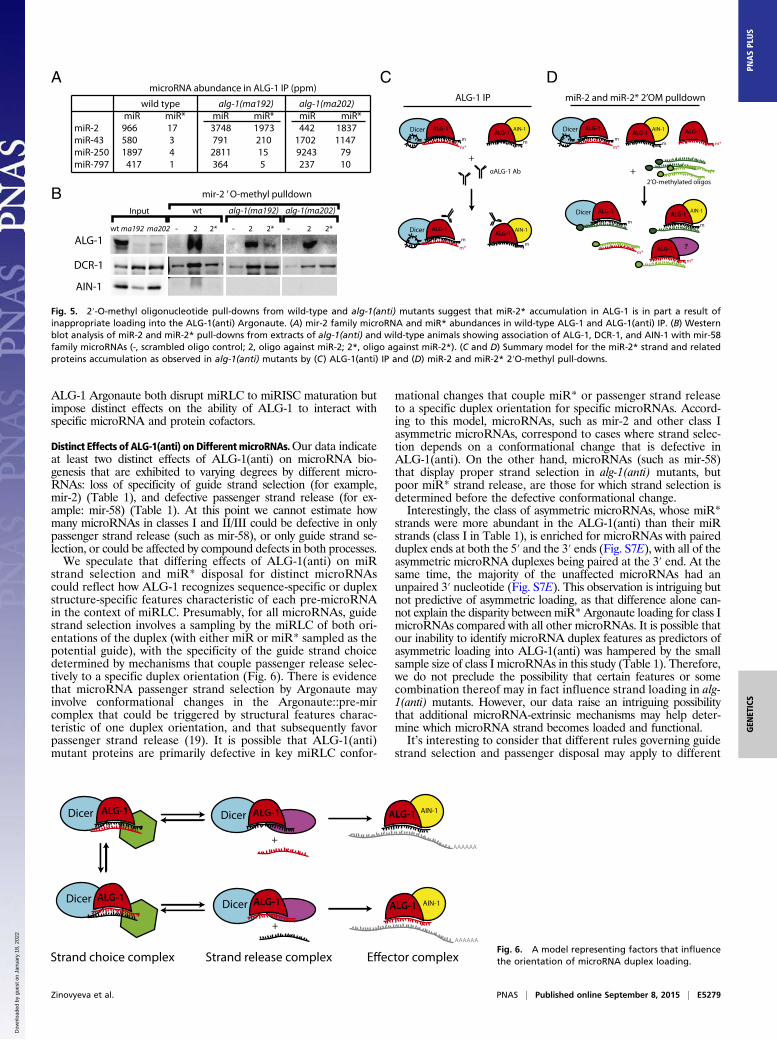
strand selection and miR* disposal for distinct microRNAscould reflect how ALG-1 recognizes sequence-specific or duplexstructure-specific features characteristic of each pre-microRNAin the context of miRLC. Presumably, for all microRNAs, guidestrand selection involves a sampling by the miRLC of both ori-entations of the duplex (with either miR or miR* sampled as thepotential guide), with the specificity of the guide strand choicedetermined by mechanisms that couple passenger release selec-tively to a specific duplex orientation (Fig. 6). There is evidencethat microRNA passenger strand selection by Argonaute mayinvolve conformational changes in the Argonaute::pre-mircomplex that could be triggered by structural features charac-teristic of one duplex orientation, and that subsequently favorpassenger strand release (19). It is possible that ALG-1(anti)mutant proteins are primarily defective in key miRLC confor-
mational changes that couple miR* or passenger strand releaseto a specific duplex orientation for specific microRNAs. Accord-ing to this model, microRNAs, such as mir-2 and other class Iasymmetric microRNAs, correspond to cases where strand selec-tion depends on a conformational change that is defective inALG-1(anti). On the other hand, microRNAs (such as mir-58)that display proper strand selection in alg-1(anti) mutants, butpoor miR* strand release, are those for which strand selection isdetermined before the defective conformational change.Interestingly, the class of asymmetric microRNAs, whose miR*
strands were more abundant in the ALG-1(anti) than their miRstrands (class I in Table 1), is enriched for microRNAs with pairedduplex ends at both the 5′ and the 3′ ends (Fig. S7E), with all of theasymmetric microRNA duplexes being paired at the 3′ end. At thesame time, the majority of the unaffected microRNAs had anunpaired 3′ nucleotide (Fig. S7E). This observation is intriguing butnot predictive of asymmetric loading, as that difference alone can-not explain the disparity between miR* Argonaute loading for class ImicroRNAs compared with all other microRNAs. It is possible thatour inability to identify microRNA duplex features as predictors ofasymmetric loading into ALG-1(anti) was hampered by the smallsample size of class I microRNAs in this study (Table 1). Therefore,we do not preclude the possibility that certain features or somecombination thereof may in fact influence strand loading in alg-1(anti) mutants. However, our data raise an intriguing possibilitythat additional microRNA-extrinsic mechanisms may help deter-mine which microRNA strand becomes loaded and functional.It’s interesting to consider that different rules governing guide
strand selection and passenger disposal may apply to different
DCR-1
AIN-1
ALG-1 wt ma192 ma202
Input
mir-2 ’ O-methyl pulldown
- 2 2* - 2 2* - 2 2*
wt alg-1(ma192) alg-1(ma202)
A
B
wild type alg-1(ma192) alg-1(ma202) miR miR* miR miR* miR miR* miR-2 966 17 3748 1973 442 1837 miR-43 580 3 791 210 1702 1147 miR-250 1897 4 2811 15 9243 79 miR-797 417 1 364 5 237 10
microRNA abundance in ALG-1 IP (ppm)
?
CmiR-2 and miR-2* 2’OM pulldown
Dicer ALG-1
m
m*
AIN-1ALG-1
m
Dicer ALG-1
m
+2’O-methylated oligos
AIN-1ALG-1
m
m*
ALG-1
m*
ALG-1
m*
ALG-1 IP
AIN-1ALG-1
m
Dicer ALG-1
m
m*
Dicer ALG-1
m
m*
AIN-1ALG-1
m
+αALG-1 Ab
D
Fig. 5. 2′-O-methyl oligonucleotide pull-downs from wild-type and alg-1(anti) mutants suggest that miR-2* accumulation in ALG-1 is in part a result ofinappropriate loading into the ALG-1(anti) Argonaute. (A) mir-2 family microRNA and miR* abundances in wild-type ALG-1 and ALG-1(anti) IP. (B) Westernblot analysis of miR-2 and miR-2* pull-downs from extracts of alg-1(anti) and wild-type animals showing association of ALG-1, DCR-1, and AIN-1 with mir-58family microRNAs (-, scrambled oligo control; 2, oligo against miR-2; 2*, oligo against miR-2*). (C and D) Summary model for the miR-2* strand and relatedproteins accumulation as observed in alg-1(anti) mutants by (C) ALG-1(anti) IP and (D) miR-2 and miR-2* 2′O-methyl pull-downs.
AIN-1Dicer ALG-1
Dicer ALG-1AIN-1
Dicer ALG-1
Dicer ALG-1
Strand choice complex Strand release complex
ALG-1
ALG-1
+
+
AAAAAA
AAAAAA
Fig. 6. A model representing factors that influencethe orientation of microRNA duplex loading.
Zinovyeva et al. PNAS | Published online September 8, 2015 | E5279
GEN
ETICS
PNASPL
US
Dow
nloa
ded
by g
uest
on
Janu
ary
18, 2
022
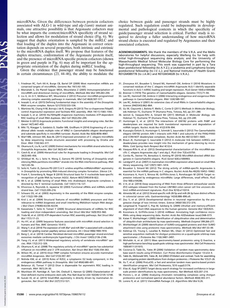
microRNAs. Given the differences between protein cofactorsassociated with ALG-1 in wild-type and alg-1(anti) mutant ani-mals, one attractive possibility is that Argonaute cofactors maybe what imparts the context/microRNA specificity of strand se-lection and allows for modulation of strand choice (Fig. 6). Wesuggest that duplex orientation is sampled by the miRLC andthat loading of the duplex into the Argonaute in correct orien-tation depends on several properties, both intrinsic and extrinsicto the microRNA duplex itself. We propose that features of theduplex structure, conformation of the Argonaute protein itself,and the presence of microRNA-specific protein cofactors (shownin green and purple in Fig. 6) may all be important for the ap-propriate orientation of the duplex during miRLC loading.Given the evidence that passenger strand may be functional
in certain circumstances (23, 44–46), the ability to modulate the
choice between guide and passenger strands must be highlyregulated. Such regulation could be indispensable in develop-mental and pathological contexts in which the specificity ofguide/passenger strand selection is critical. Further study is re-quired to develop a fuller understanding of how microRNAstrand choice is executed and regulated by Argonautes and theirassociated cofactors.
ACKNOWLEDGMENTS. We thank the members of the V.R.A. and the Mellolaboratories for helpful discussions, especially Weifeng Gu for help withinitial high-throughput sequencing data analysis; and the University ofMassachusetts Medical School Molecular Biology Core for performing thehigh-throughput sequencing. This work was supported in part by a TaraBean postdoctoral fellowship (to A.Y.Z.); a Leukemia and Lymphoma Societypostdoctoral fellowship (to I.V.-L.); and National Institutes of Health GrantsR01GM089778 (to J.A.W.) and R01GM34028 (to V.R.A.).
1. Friedman RC, Farh KK-H, Burge CB, Bartel DP (2009) Most mammalian mRNAs areconserved targets of microRNAs. Genome Res 19(1):92–105.
2. Maniataki E, De Planell Saguer MDA, Mourelatos Z (2005) Immunoprecipitation ofmicroRNPs and directional cloning of microRNAs. Methods Mol Biol 309:283–294.
3. Liu X, Jin D-Y, McManus MT, Mourelatos Z (2012) Precursor microRNA-programmedsilencing complex assembly pathways in mammals. Mol Cell 46(4):507–517.
4. Iwasaki S, et al. (2015) Defining fundamental steps in the assembly of the DrosophilaRNAi enzyme complex. Nature 521(7553):533–536.
5. Martinez NJ, Chang H-M, Borrajo J de R, Gregory RI (2013) The co-chaperones Fkbp4/5control Argonaute2 expression and facilitate RISC assembly. RNA 19(11):1583–1593.
6. Iwasaki S, et al. (2010) Hsc70/Hsp90 chaperone machinery mediates ATP-dependentRISC loading of small RNA duplexes. Mol Cell 39(2):292–299.
7. Meister G (2013) Argonaute proteins: Functional insights and emerging roles. Nat RevGenet 14(7):447–459.
8. Miki TS, Rüegger S, Gaidatzis D, Stadler MB, Großhans H (2014) Engineering of a con-ditional allele reveals multiple roles of XRN2 in Caenorhabditis elegans developmentand substrate specificity in microRNA turnover. Nucleic Acids Res 42(6):4056–4067.
9. Warf MB, Johnson WE, Bass BL (2011) Improved annotation of C. elegans microRNAsby deep sequencing reveals structures associated with processing by Drosha andDicer. RNA 17(4):563–577.
10. Okamura K, Liu N, Lai EC (2009) Distinct mechanisms for microRNA strand selection byDrosophila Argonautes. Mol Cell 36(3):431–444.
11. Czech B, et al. (2009) Hierarchical rules for Argonaute loading in Drosophila. Mol Cell36(3):445–456.
12. Ghildiyal M, Xu J, Seitz H, Weng Z, Zamore PD (2010) Sorting of Drosophila smallsilencing RNAs partitions microRNA* strands into the RNA interference pathway. RNA16(1):43–56.
13. Seitz H, Tushir JS, Zamore PD (2011) A 5′-uridine amplifies miRNA/miRNA* asymmetryin Drosophila by promoting RNA-induced silencing complex formation. Silence 2:4.
14. Frank F, Sonenberg N, Nagar B (2010) Structural basis for 5′-nucleotide base-specificrecognition of guide RNA by human AGO2. Nature 465(7299):818–822.
15. Mi S, et al. (2008) Sorting of small RNAs into Arabidopsis Argonaute complexes isdirected by the 5′ terminal nucleotide. Cell 133(1):116–127.
16. Khvorova A, Reynolds A, Jayasena SD (2003) Functional siRNAs and miRNAs exhibitstrand bias. Cell 115(2):209–216.
17. Schwarz DS, et al. (2003) Asymmetry in the assembly of the RNAi enzyme complex.Cell 115(2):199–208.
18. Krol J, et al. (2004) Structural features of microRNA (miRNA) precursors and theirrelevance to miRNA biogenesis and small interfering RNA/short hairpin RNA design.J Biol Chem 279(40):42230–42239.
19. Kawamata T, Seitz H, Tomari Y (2009) Structural determinants of miRNAs for RISCloading and slicer-independent unwinding. Nat Struct Mol Biol 16(9):953–960.
20. Yoda M, et al. (2010) ATP-dependent human RISC assembly pathways. Nat Struct MolBiol 17(1):17–23.
21. Hu HY, et al. (2009) Sequence features associated with microRNA strand selection inhumans and flies. BMC Genomics 10:413.
22. Wang Y, et al. (2014) The expression ofmiR-30a* andmiR-30e* is associatedwith a dualisticmodel for grading ovarian papillary serious carcinoma. Int J Oncol 44(6):1904–1914.
23. Bang C, et al. (2014) Cardiac fibroblast-derived microRNA passenger strand-enrichedexosomes mediate cardiomyocyte hypertrophy. J Clin Invest 124(5):2136–2146.
24. Yang J-S, et al. (2011) Widespread regulatory activity of vertebrate microRNA* spe-cies. RNA 17(2):312–326.
25. Okamura K, et al. (2008) The regulatory activity of microRNA* species has substantialinfluence on microRNA and 3′ UTR evolution. Nat Struct Mol Biol 15(4):354–363.
26. Wilson RC, et al. (2015) Dicer-TRBP complex formation ensures accurate mammalianmicroRNA biogenesis. Mol Cell 57(3):397–407.
27. Nishida KM, et al. (2013) Roles of R2D2, a cytoplasmic D2 body component, in theendogenous siRNA pathway in Drosophila. Mol Cell 49(4):680–691.
28. Betancur JG, Tomari Y (2012) Dicer is dispensable for asymmetric RISC loading inmammals. RNA 18(1):24–30.
29. Murchison EP, Partridge JF, Tam OH, Cheloufi S, Hannon GJ (2005) Characterization ofDicer-deficient murine embryonic stem cells. Proc Natl Acad Sci USA 102(34):12135–12140.
30. Suzuki HI, et al. (2015) Small-RNA asymmetry is directly driven by mammalian Ar-gonautes. Nat Struct Mol Biol 22(7):512–521.
31. Zinovyeva AY, Bouasker S, Simard MJ, Hammell CM, Ambros V (2014) Mutations inconserved residues of the C. elegans microRNA Argonaute ALG-1 identify separablefunctions in ALG-1 miRISC loading and target repression. PLoS Genet 10(4):e1004286.
32. Brenner S (1974) The genetics of Caenorhabditis elegans. Genetics 77(1):71–94.33. Lee RC, Hammell CM, Ambros V (2006) Interacting endogenous and exogenous RNAi
pathways in Caenorhabditis elegans. RNA 12(4):589–597.34. Lee RC, Ambros V (2001) An extensive class of small RNAs in Caenorhabditis elegans.
Science 294(5543):862–864.35. Gu W, Claycomb J, Batista P, Mello C, Conte D (2011) Methods in Molecular Biology,
eds Hobman TC, Duchaine TF (Humana Press, Totowa, NJ), pp 251–280.36. Jannot G, Vasquez-Rifo A, Simard MJ (2011) Methods in Molecular Biology, eds
Hobman TC, Duchaine TF (Humana Press, Totowa, NJ), pp 233–249.37. Huntzinger E, et al. (2013) The interactions of GW182 proteins with PABP and
deadenylases are required for both translational repression and degradation ofmiRNA targets. Nucleic Acids Res 41(2):978–994.
38. Kuzuoglu-Öztürk D, Huntzinger E, Schmidt S, Izaurralde E (2012) The Caenorhabditiselegans GW182 protein AIN-1 interacts with PAB-1 and subunits of the PAN2-PAN3and CCR4-NOT deadenylase complexes. Nucleic Acids Res 40(12):5651–5665.
39. Braun JE, Huntzinger E, Izaurralde E (2012) A molecular link between miRISCs anddeadenylases provides new insight into the mechanism of gene silencing by micro-RNAs. Cold Spring Harb Perspect Biol 4(12):4.
40. Vasquez-Rifo A, et al. (2012) Developmental characterization of the microRNA-spe-cific C. elegans Argonautes alg-1 and alg-2. PLoS One 7(3):e33750.
41. Corrêa RL, Steiner FA, Berezikov E, Ketting RF (2010) MicroRNA-directed siRNA bio-genesis in Caenorhabditis elegans. PLoS Genet 6(4):e1000903.
42. Landgraf P, et al. (2007) A mammalian microRNA expression atlas based on small RNAlibrary sequencing. Cell 129(7):1401–1414.
43. Bouasker S, Simard MJ (2012) The slicing activity of miRNA-specific Argonautes isessential for the miRNA pathway in C. elegans. Nucleic Acids Res 40(20):10452–10462.
44. Kozomara A, Hunt S, Ninova M, Griffiths-Jones S, Ronshaugen M (2014) Target re-pression induced by endogenous microRNAs: Large differences, small effects. PLoSOne 9(8):e104286.
45. Ji H, et al. (2014) Deep sequencing of RNA from three different extracellular vesicle(EV) subtypes released from the human LIM1863 colon cancer cell line uncovers dis-tinct miRNA-enrichment signatures. PLoS One 9(10):e110314.
46. Almeida MI, et al. (2012) Strand-specific miR-28-5p and miR-28-3p have distinct effectsin colorectal cancer cells. Gastroenterology 142(4):886–896.e9.
47. Zou Y, et al. (2013) Developmental decline in neuronal regeneration by the pro-gressive change of two intrinsic timers. Science 340(6130):372–376.
48. Langmead B, Trapnell C, Pop M, Salzberg SL (2009) Ultrafast and memory-efficientalignment of short DNA sequences to the human genome. Genome Biol 10(3):R25.
49. Kozomara A, Griffiths-Jones S (2014) miRBase: Annotating high confidence micro-RNAs using deep sequencing data. Nucleic Acids Res 42(Database issue):D68–D73.
50. Kaiser P, Wohlschlegel J (2005) Identification of ubiquitination sites and determinationof ubiquitin-chain architectures by mass spectrometry. Methods Enzymol 399:266–277.
51. Wohlschlegel JA (2009) Identification of SUMO-conjugated proteins and their SUMOattachment sites using proteomic mass spectrometry. Methods Mol Biol 497:33–49.
52. Kelstrup CD, Young C, Lavallee R, Nielsen ML, Olsen JV (2012) Optimized fast andsensitive acquisition methods for shotgun proteomics on a quadrupole orbitrap massspectrometer. J Proteome Res 11(6):3487–3497.
53. Michalski A, et al. (2011) Mass spectrometry-based proteomics using Q Exactive, ahigh-performance benchtop quadrupole orbitrap mass spectrometer.Mol Cell Proteomics10(9):M111.011015.
54. Cociorva D, Tabb DL, Yates JR (2006) Validation of tandem mass spectrometry data-base search results using DTASelect. Curr Protoc Bioinformatics Chapter 13:Unit 4.
55. Tabb DL, McDonald WH, Yates JR, 3rd (2002) DTASelect and contrast: Tools for assemblingand comparing protein identifications from shotgun proteomics. J Proteome Res 1(1):21–26.
56. Xu T, et al. (2006) ProLuCID, a fast and sensitive tandem mass spectra-based proteinidentification program. Mol Cell Proteomics, 5(10 suppl):S174.
57. Elias JE, Gygi SP (2007) Target-decoy search strategy for increased confidence in large-scale protein identifications by mass spectrometry. Nat Methods 4(3):207–214.
58. Florens L, et al. (2006) Analyzing chromatin remodeling complexes using shotgunproteomics and normalized spectral abundance factors. Methods 40(4):303–311.
59. Lorenz R, et al. (2011) ViennaRNA Package 2.0. Algorithms Mol Biol 6:26.
E5280 | www.pnas.org/cgi/doi/10.1073/pnas.1506576112 Zinovyeva et al.
Dow
nloa
ded
by g
uest
on
Janu
ary
18, 2
022