Embed Size (px)
Citation preview
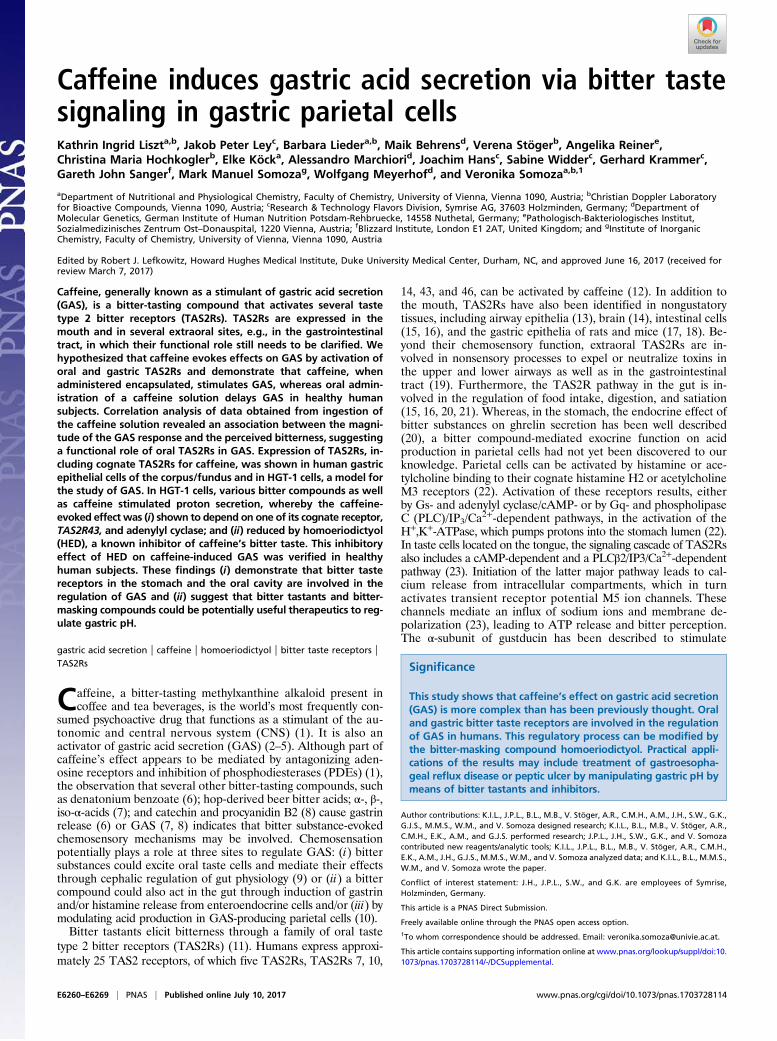
Caffeine induces gastric acid secretion via bitter tastesignaling in gastric parietal cellsKathrin Ingrid Liszta,b, Jakob Peter Leyc, Barbara Liedera,b, Maik Behrensd, Verena Stögerb, Angelika Reinere,Christina Maria Hochkoglerb, Elke Köcka, Alessandro Marchiorid, Joachim Hansc, Sabine Widderc, Gerhard Krammerc,Gareth John Sangerf, Mark Manuel Somozag, Wolfgang Meyerhofd, and Veronika Somozaa,b,1
aDepartment of Nutritional and Physiological Chemistry, Faculty of Chemistry, University of Vienna, Vienna 1090, Austria; bChristian Doppler Laboratoryfor Bioactive Compounds, Vienna 1090, Austria; cResearch & Technology Flavors Division, Symrise AG, 37603 Holzminden, Germany; dDepartment ofMolecular Genetics, German Institute of Human Nutrition Potsdam-Rehbruecke, 14558 Nuthetal, Germany; ePathologisch-Bakteriologisches Institut,Sozialmedizinisches Zentrum Ost–Donauspital, 1220 Vienna, Austria; fBlizzard Institute, London E1 2AT, United Kingdom; and gInstitute of InorganicChemistry, Faculty of Chemistry, University of Vienna, Vienna 1090, Austria
Edited by Robert J. Lefkowitz, Howard Hughes Medical Institute, Duke University Medical Center, Durham, NC, and approved June 16, 2017 (received forreview March 7, 2017)
Caffeine, generally known as a stimulant of gastric acid secretion(GAS), is a bitter-tasting compound that activates several tastetype 2 bitter receptors (TAS2Rs). TAS2Rs are expressed in themouth and in several extraoral sites, e.g., in the gastrointestinaltract, in which their functional role still needs to be clarified. Wehypothesized that caffeine evokes effects on GAS by activation oforal and gastric TAS2Rs and demonstrate that caffeine, whenadministered encapsulated, stimulates GAS, whereas oral admin-istration of a caffeine solution delays GAS in healthy humansubjects. Correlation analysis of data obtained from ingestion ofthe caffeine solution revealed an association between the magni-tude of the GAS response and the perceived bitterness, suggestinga functional role of oral TAS2Rs in GAS. Expression of TAS2Rs, in-cluding cognate TAS2Rs for caffeine, was shown in human gastricepithelial cells of the corpus/fundus and in HGT-1 cells, a model forthe study of GAS. In HGT-1 cells, various bitter compounds as wellas caffeine stimulated proton secretion, whereby the caffeine-evoked effect was (i) shown to depend on one of its cognate receptor,TAS2R43, and adenylyl cyclase; and (ii) reduced by homoeriodictyol(HED), a known inhibitor of caffeine’s bitter taste. This inhibitoryeffect of HED on caffeine-induced GAS was verified in healthyhuman subjects. These findings (i) demonstrate that bitter tastereceptors in the stomach and the oral cavity are involved in theregulation of GAS and (ii) suggest that bitter tastants and bitter-masking compounds could be potentially useful therapeutics to reg-ulate gastric pH.
gastric acid secretion | caffeine | homoeriodictyol | bitter taste receptors |TAS2Rs
Caffeine, a bitter-tasting methylxanthine alkaloid present incoffee and tea beverages, is the world’s most frequently con-
sumed psychoactive drug that functions as a stimulant of the au-tonomic and central nervous system (CNS) (1). It is also anactivator of gastric acid secretion (GAS) (2–5). Although part ofcaffeine’s effect appears to be mediated by antagonizing aden-osine receptors and inhibition of phosphodiesterases (PDEs) (1),the observation that several other bitter-tasting compounds, suchas denatonium benzoate (6); hop-derived beer bitter acids; α-, β-,iso-α-acids (7); and catechin and procyanidin B2 (8) cause gastrinrelease (6) or GAS (7, 8) indicates that bitter substance-evokedchemosensory mechanisms may be involved. Chemosensationpotentially plays a role at three sites to regulate GAS: (i) bittersubstances could excite oral taste cells and mediate their effectsthrough cephalic regulation of gut physiology (9) or (ii) a bittercompound could also act in the gut through induction of gastrinand/or histamine release from enteroendocrine cells and/or (iii) bymodulating acid production in GAS-producing parietal cells (10).Bitter tastants elicit bitterness through a family of oral taste
type 2 bitter receptors (TAS2Rs) (11). Humans express approxi-mately 25 TAS2 receptors, of which five TAS2Rs, TAS2Rs 7, 10,
14, 43, and 46, can be activated by caffeine (12). In addition tothe mouth, TAS2Rs have also been identified in nongustatorytissues, including airway epithelia (13), brain (14), intestinal cells(15, 16), and the gastric epithelia of rats and mice (17, 18). Be-yond their chemosensory function, extraoral TAS2Rs are in-volved in nonsensory processes to expel or neutralize toxins inthe upper and lower airways as well as in the gastrointestinaltract (19). Furthermore, the TAS2R pathway in the gut is in-volved in the regulation of food intake, digestion, and satiation(15, 16, 20, 21). Whereas, in the stomach, the endocrine effect ofbitter substances on ghrelin secretion has been well described(20), a bitter compound-mediated exocrine function on acidproduction in parietal cells had not yet been discovered to ourknowledge. Parietal cells can be activated by histamine or ace-tylcholine binding to their cognate histamine H2 or acetylcholineM3 receptors (22). Activation of these receptors results, eitherby Gs- and adenylyl cyclase/cAMP- or by Gq- and phospholipaseC (PLC)/IP3/Ca
2+-dependent pathways, in the activation of theH+,K+-ATPase, which pumps protons into the stomach lumen (22).In taste cells located on the tongue, the signaling cascade of TAS2Rsalso includes a cAMP-dependent and a PLCβ2/IP3/Ca2+-dependentpathway (23). Initiation of the latter major pathway leads to cal-cium release from intracellular compartments, which in turnactivates transient receptor potential M5 ion channels. Thesechannels mediate an influx of sodium ions and membrane de-polarization (23), leading to ATP release and bitter perception.The α-subunit of gustducin has been described to stimulate
Significance
This study shows that caffeine’s effect on gastric acid secretion(GAS) is more complex than has been previously thought. Oraland gastric bitter taste receptors are involved in the regulationof GAS in humans. This regulatory process can be modified bythe bitter-masking compound homoeriodictyol. Practical appli-cations of the results may include treatment of gastroesopha-geal reflux disease or peptic ulcer by manipulating gastric pH bymeans of bitter tastants and inhibitors.
Author contributions: K.I.L., J.P.L., B.L., M.B., V. Stöger, A.R., C.M.H., A.M., J.H., S.W., G.K.,G.J.S., M.M.S., W.M., and V. Somoza designed research; K.I.L., B.L., M.B., V. Stöger, A.R.,C.M.H., E.K., A.M., and G.J.S. performed research; J.P.L., J.H., S.W., G.K., and V. Somozacontributed new reagents/analytic tools; K.I.L., J.P.L., B.L., M.B., V. Stöger, A.R., C.M.H.,E.K., A.M., J.H., G.J.S., M.M.S., W.M., and V. Somoza analyzed data; and K.I.L., B.L., M.M.S.,W.M., and V. Somoza wrote the paper.
Conflict of interest statement: J.H., J.P.L., S.W., and G.K. are employees of Symrise,Holzminden, Germany.
This article is a PNAS Direct Submission.
Freely available online through the PNAS open access option.1To whom correspondence should be addressed. Email: [email protected].
This article contains supporting information online at www.pnas.org/lookup/suppl/doi:10.1073/pnas.1703728114/-/DCSupplemental.
E6260–E6269 | PNAS | Published online July 10, 2017 www.pnas.org/cgi/doi/10.1073/pnas.1703728114

PDEs, resulting in low cAMP levels and PKA activities, whichkeep the IP3 type 3 receptor hypophosphorylated and sensitized(24). Therefore, the tonic activity of α-gustducin regulates tastecell responsivity. Transducin, a similar G protein also present intaste cells, can replace the function of α-gustducin (25, 26).TAS2R-expressing cells in the gastrointestinal tract have beenreported to coexpress the downstream taste signaling components,suggesting that similar signal transduction pathways could alsomediate gastrointestinal physiology (27). However, the detailedsignal transduction pathways in extraoral chemosensitive cellsare yet unknown.This study investigated whether gastric and oral TAS2Rs
contribute to the regulation of caffeine-induced mechanisms ofGAS in humans. To study this hypothesis, the effect of caffeineon GAS was investigated in a human intervention trial, takinginto account taste receptor activation in the mouth and the stom-ach. The underlying gastric mechanisms were studied by TAS2Rexpression analysis and by means of the validated HGT-1 cell cul-ture model, which maintains the relevant characteristics of humanparietal cells (28, 29).
ResultsOral Bitter Perception Reduces GAS in Human Subjects. Real-timegastric pH measurements were performed after caffeine adminis-tration in human subjects by means of Heidelberg pH diagnosticcapsules (29–32). Heidelberg pH capsules are used to determinegastric acid secretory ability under conditions simulating the in-gestion of food or beverages by means of radiotelemetry. For themeasurements, overnight-fasted subjects swallow the pH capsule,followed by a saturated sodium bicarbonate solution. Ingestion ofthe bicarbonate solution triggers an increase in stomach pH anda subsequent attempt by the parietal cells to reestablish acidity.The impact of foods or beverages on the reacidification time canbe analyzed by administration of the test material before or afterthe pH challenge. In this study, subjects swallowed a caffeinesolution with or without a bitter-masking compound, homoer-iodictyol (HED) (33, 34) 5 min after or 25 min before the bi-carbonate challenge. Reacidification time was measured forthree distinct delivery protocols (1–3), each of which assessesdifferent sites of TAS2R activation (Fig. 1A). The subjects un-derwent the following interventions in 11 consecutive study sitevisits (Fig. 1B): For the first 8 visits, test compounds were ad-ministered 5 min after the bicarbonate solution. In deliveryprotocol (1), subjects drank 125 mL water, a caffeine solution(37.5, 75, or 150 mg caffeine in 125 mL water) with or without30 mg HED, or an HED solution (30 mg HED in 125 mL water),thereby stimulating oral and gastric TAS2Rs (Fig. 1B). For de-livery protocols 2 and 3, a dose of 150 mg caffeine was adminis-tered along with 125 mL water, either encapsulated to selectivelystimulate gastric TAS2Rs, or as a sip-and-spit solution to activateonly oral TAS2Rs, respectively (Fig. 1B).During the final three visits, subjects were asked to drink
125 mL water or 150 mg caffeine with or without 30 mg HED in125 mL water (delivery protocol 2) 25 min before the bicarbonatechallenge to evaluate the effect of administration time. The in-tervention time of 25 min was chosen according to previouspublications that demonstrated that caffeine starts to stimulategastric acid after 30 min (2, 5).Drinking the volume water control solution 5 min after the
bicarbonate challenge resulted in a mean reacidification time of23 ± 1 min (individual representative gastrogram shown in Fig.1C). Oral application of caffeine by sip-and-spit or drinking ledto prolongations (P < 0.05) of reacidification time by deltareacidification time values (reacidification timetest compound −reacidification timewater) of 20 ± 6 min and 8 ± 2 min, re-spectively, compared with administration of a volume watercontrol solution, indicating a delay of GAS (Fig. 1D). Stimula-tion of gastric sites only by encapsulated caffeine resulted in a
shorter delta reacidification time of 5 ± 3 min relative to sip-and-spit administration (P < 0.05; Fig. 1 C and D). Individual gas-trograms were quantified by determining the slope after theonset of reacidification. A higher slope indicates that, whenreacidification has started, the gastric pH returns to its initial pHfaster. The slope of the gastrogram (relative to water control)obtained after administration of encapsulated caffeine washigher (0.20 ± 0.16 pH units per min) compared with the slopecalculated after drinking (−0.20 ± 0.10 pH units per min) andsip-and-spit intervention (−0.39 ± 0.05 pH units per min),whereby stimulation of oral receptors occurred (Fig. 1E). Toextend the time period over which the effect of caffeine on GAScould be measured, we repeated the experiments with encapsu-lated caffeine administered with 125 mL water 25 min before thealkaline challenge. This intervention allows gastric pH changesto be recorded over a time period of 25 min to approximately85 min after caffeine administration and revealed a stimulationof GAS, indicated by a reduced delta reacidification time of−23 ± 4 min by caffeine (Fig. 2B) compared with control treat-ment (empty capsule plus 125 mL water reacidification time,41 ± 4 min; P < 0.01).
HED Reduces the Caffeine-Evoked Effects on GAS in Human Subjects.To determine if TAS2R bitter-taste receptors mediate the effectof caffeine on GAS, 125 mL water containing 150 mg caffeineand/or 30 mg of the bitter-masking compound HED (33, 34)were swallowed 5 min after the alkaline challenge (deliveryprotocol 1). Administration of HED alone resulted in a reac-idification time of 21 ± 2 min, comparable to that of water (24 ±1 min) as volume control.Unexpectedly, concomitant administration of HED and caf-
feine resulted in accelerated gastric emptying in 4 of 10 subjects,as indicated by passing of the Heidelberg capsule into the duo-denum before complete reacidification. The same effect wasobserved in 2 of 10 subjects after drinking a solution of 30 mgHED dissolved in 125 mL water. When HED and caffeine wereadministered encapsulated (delivery protocol 2), reacidificationtimes could be analyzed in only six subjects, as four subjectsdemonstrated accelerated gastric emptying as seen after oral andgastric delivery (protocol 1). These results raised the questionwhether the bitter-masking compound HED promotes gastricmotility by stimulating gastric relaxation. Experiments usingstrips of dissections of human stomach biopsy specimensrevealed that treatment with 1 mM HED in an organ bathinduced a maximum relaxation after 40 min, with mean tensionvalues of 45.4 ± 6.7%, compared with water control values of107 ± 5.7% (Fig. S1 A and B).In those subjects who were subjected to delivery protocol
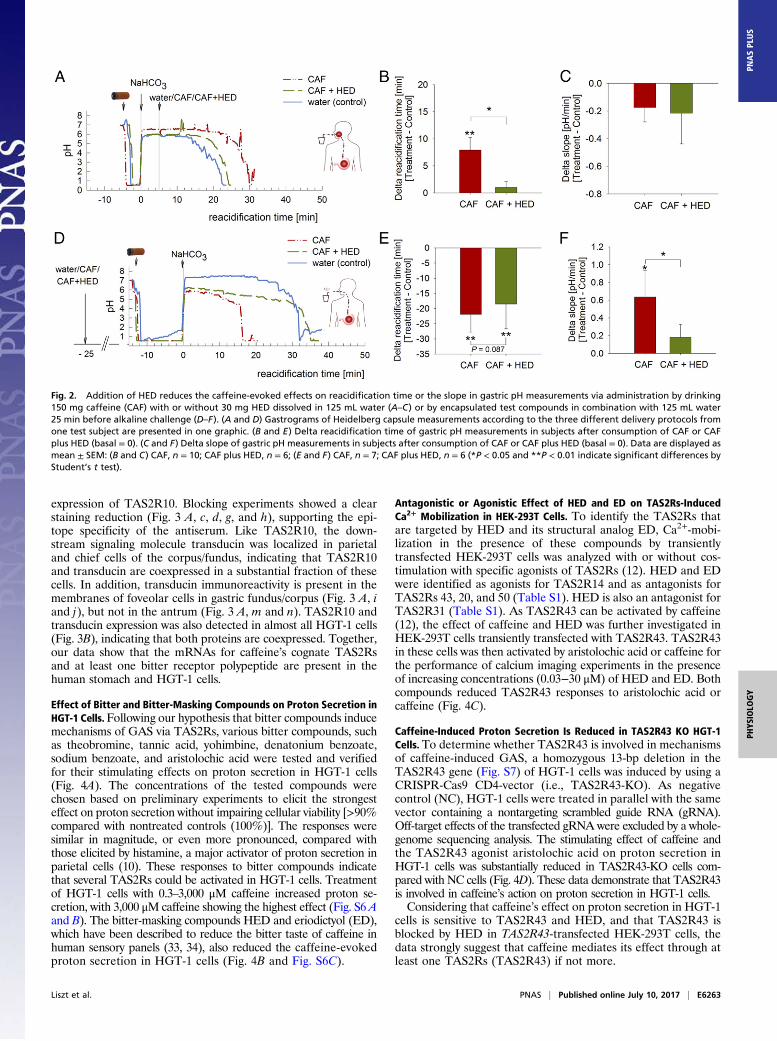
1 and did not respond with accelerated gastric emptying, HEDlargely reversed the effects of caffeine on reacidifcation time:whereas drinking of the caffeine solution 5 min after alkalinechallenge resulted in a delta reacidification time of 8 ± 2 min,concomitant caffeine and HED administration revealed a meanvalue of 1 ± 1 min (Fig. 2 A and B), but showed no effect on theslope of the gastrogram (Fig. 2C). In contrast, gastric adminis-tration of encapsulated caffeine 25 min before alkaline challenge(delivery protocol 2) induced GAS compared with administrationof water, resulting in a delta reacidification time of −23 ± 4 min(Fig. 2 D and E). Although the reversing effect of HED on thecaffeine-mediated reacidification shown in Fig. 2D did not reachstatistical significance in terms of reacidification time (P = 0.087;Fig. 2E), concomitant application of HED and caffeine reducedthe slope of the gastrogram compared with caffeine administra-tion, with mean respective values of 0.18 ± 0.13 pH units per minand 0.64 ± 0.26 pH units per min (P < 0.05; Fig. 2F).The potent attenuation of caffeine’s effects on GAS by the
bitter-masking agent HED suggests that TAS2Rs are criticallyinvolved in caffeine’s action in the mouth and the stomach.
Liszt et al. PNAS | Published online July 10, 2017 | E6261
PHYS
IOLO
GY
PNASPL
US

Sensory Evaluation. To verify that the subjects were capable ofsensing caffeine bitterness, the bitter recognition threshold of thesame subjects who underwent the gastric pH measurements wasdetermined by means of a threshold test, which yielded a resultof 117 ± 44 mg/L for caffeine. In addition, the subjects rated thebitterness of 1,200 mg/L caffeine in the absence or presence of240 mg/L HED in a blinded duo sensory test and confirmed thebitter-masking effect of HED reported by Ley et al. (33):Whereas the mean bitterness rating (±SD) for the caffeine so-lution was 7.5 ± 1.7, ratings for caffeine plus HED revealedmean values of 5.8 ± 1.9, corresponding to a −20 ± 8% reductionof caffeine-mediated bitterness by HED (Fig. S2A). The subjects’caffeine bitterness scores correlated with reacidification time(correlation coefficient, 0.66; P = 0.03; n = 10; Fig. S2 B and C)after caffeine administration by drinking (delivery protocol 1,5 min after alkaline challenge), as well as with reacidificationtime after caffeine plus HED administered by drinking (corre-lation coefficient, 0.89; P < 0.05; n = 6; 5 min after alkalinechallenge). No statistically significant correlation between bitterintensity rating and reacidification time was calculated after ad-ministration of encapsulated caffeine (delivery protocol 1; P > 0.05).
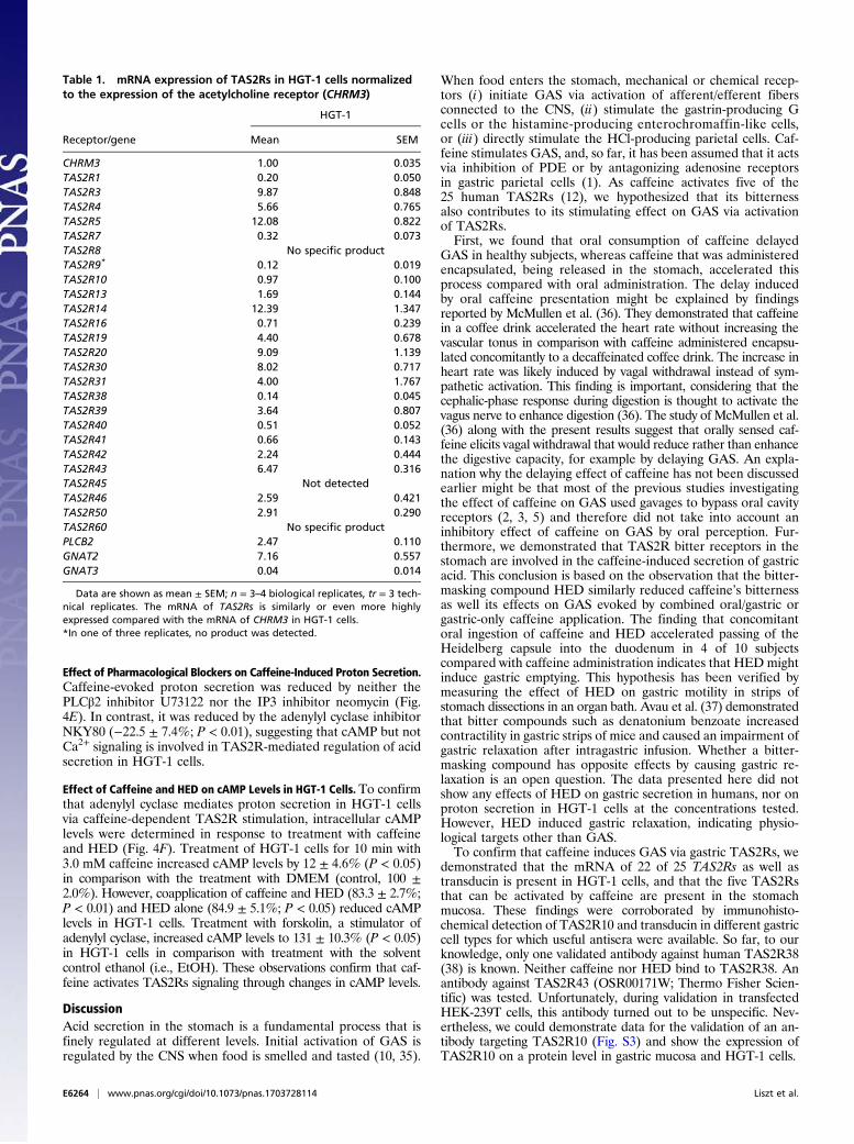
TAS2R Expression in HGT-1 Cells and Human Gastric Tissue. ThemRNA expression of 25 human TAS2Rs in the HGT-1 cell linewas investigated by quantitative RT-PCR (RT-qPCR) studies.The genes for the five TAS2Rs known to be activated by caf-feine, TAS2Rs 7, 10, 14, 43, and 46 (12), as well as several otherTAS2R genes, are expressed at similar or even higher levels thanthe M3 acetylcholine receptor CHRM3 gene, a major regulatorof GAS (Table 1). Although TAS2R5 and TAS2R14 are the mosthighly expressed TAS2Rs, TAS2R8, 45, and 60 mRNAs were notfound in HGT-1 cells. HGT-1 cells also express mRNAs forTAS2R downstream signaling proteins PLCβ2, transducin(GNAT2), and α-gustducin (GNAT3) (11, 23) (Table 1). Likethe parietal cell line HGT-1, the human gastric epitheliumcontains transcripts for the five cognate caffeine bitter receptorsTAS2R7, TAS2R10, TAS2R14, TAS2R43 and TAS2R46 atlevels similar to those of the M3 acetylcholine receptor, with ratiosrelative to that receptor of 0.76 ± 0.039, 0.97 ± 0.190, 1.16 ± 0.025,0.62 ± 0.017, and 0.83 ± 0.071, respectively.The presence of the broadly tuned, caffeine-sensitive TAS2R10
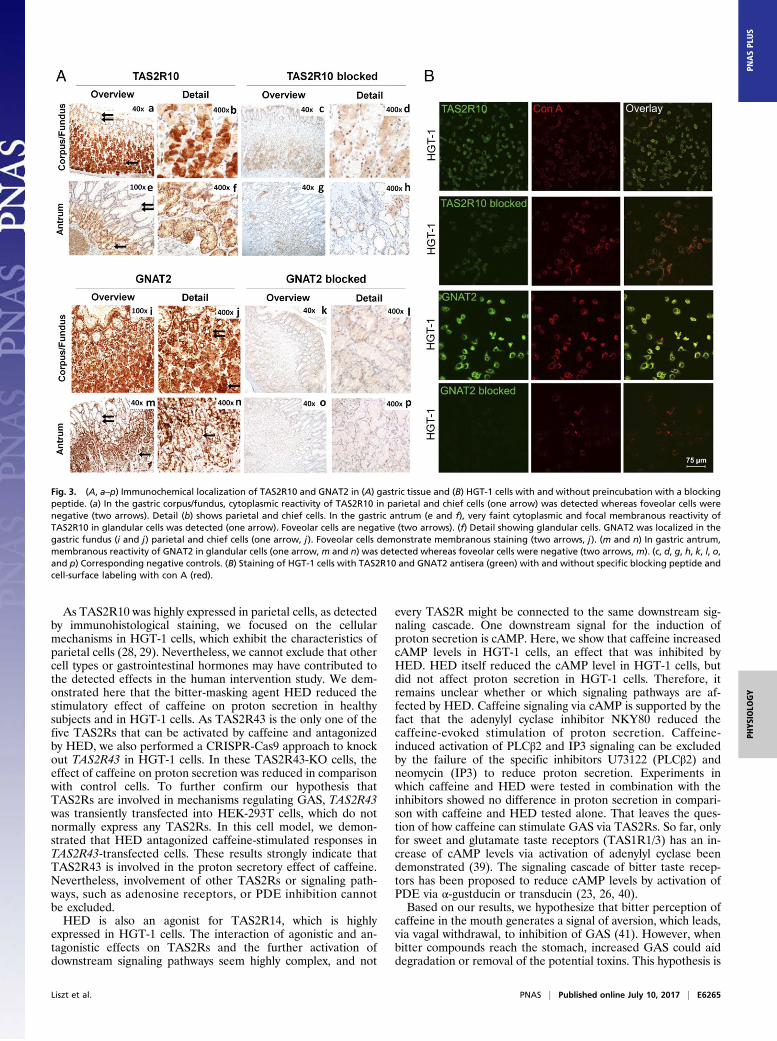
receptor (12) in the gastric epithelium was confirmed by immu-nohistochemical staining of stomach surgical specimens from theantrum and fundus/corpus region. The specificity of the TAS2R10antibody was verified in transiently transfected HEK-293T cells(Fig. S3). In gastric mucosa, cell types were identified by H&Estaining (Figs. S4 and S5). Parietal cells are localized in the glandsof gastric fundus and body, and are scattered in the middle and, toa lesser extent, in the bottom part of the mucosa (Fig. S4). Theyare characterized by broad pink cytoplasms. Chief cells stain withbasophilic cytoplasm and are mainly located in the bottom partsof the mucosa (Fig. 3A and Fig. S4). Localization of TAS2R10staining was confined to parietal cells and to gastric chief cells inthe fundus/corpus, showing strong cytoplasmic granular re-activity (Fig. 3 A, a and b). Staining of glandular cells in thegastric antrum was faint, consisting of very weak cytoplasmic andfocal intermediate membranous reaction (Fig. 3 A, e and f). Incontrast, mucus-producing foveolar cells in the fundus/corpus(Fig. 3 A, a and b) and antrum (Fig. 3 A, e and f) did not show
Fig. 1. Results of the gastric pH measurements demonstrate that the effectof caffeine (CAF) on reacidification time is influenced by the type of ad-ministration. (A) Overview of the different administration types in the hu-man intervention trial. (B) Overview of the study procedure. (C) Gastrogramsof different Heidelberg capsule measurements from one test subject com-bined in one graphic show that 150 mg caffeine diluted or administeredwith 125 mL water (blue line) administered by sip and spit (3) prolongs thereacidification time (i.e., time until the original pH is reached again) morethan administration via drinking (2) or in encapsulated form (1). (D) Deltareacidification time of gastrograms show that sip-and-spit administration
resulted in the highest prolongation of reacidification time compared withgastric and gastric plus oral administration. (E) Delta slope of the gastro-grams indicate that encapsulated administration (gastric delivery) stronglystimulate GAS when reacidification has started. Data are displayed asmean ± SEM, n = 5–10; one-way ANOVA with Holm–�Sídák post hoc test;significant (P < 0.05) differences are indicated by distinct letters [*P < 0.05,significant vs. water control (basal = 0) tested with paired Student t test].
E6262 | www.pnas.org/cgi/doi/10.1073/pnas.1703728114 Liszt et al.
expression of TAS2R10. Blocking experiments showed a clearstaining reduction (Fig. 3 A, c, d, g, and h), supporting the epi-tope specificity of the antiserum. Like TAS2R10, the down-stream signaling molecule transducin was localized in parietaland chief cells of the corpus/fundus, indicating that TAS2R10and transducin are coexpressed in a substantial fraction of thesecells. In addition, transducin immunoreactivity is present in themembranes of foveolar cells in gastric fundus/corpus (Fig. 3 A, iand j), but not in the antrum (Fig. 3 A, m and n). TAS2R10 andtransducin expression was also detected in almost all HGT-1 cells(Fig. 3B), indicating that both proteins are coexpressed. Together,our data show that the mRNAs for caffeine’s cognate TAS2Rsand at least one bitter receptor polypeptide are present in thehuman stomach and HGT-1 cells.
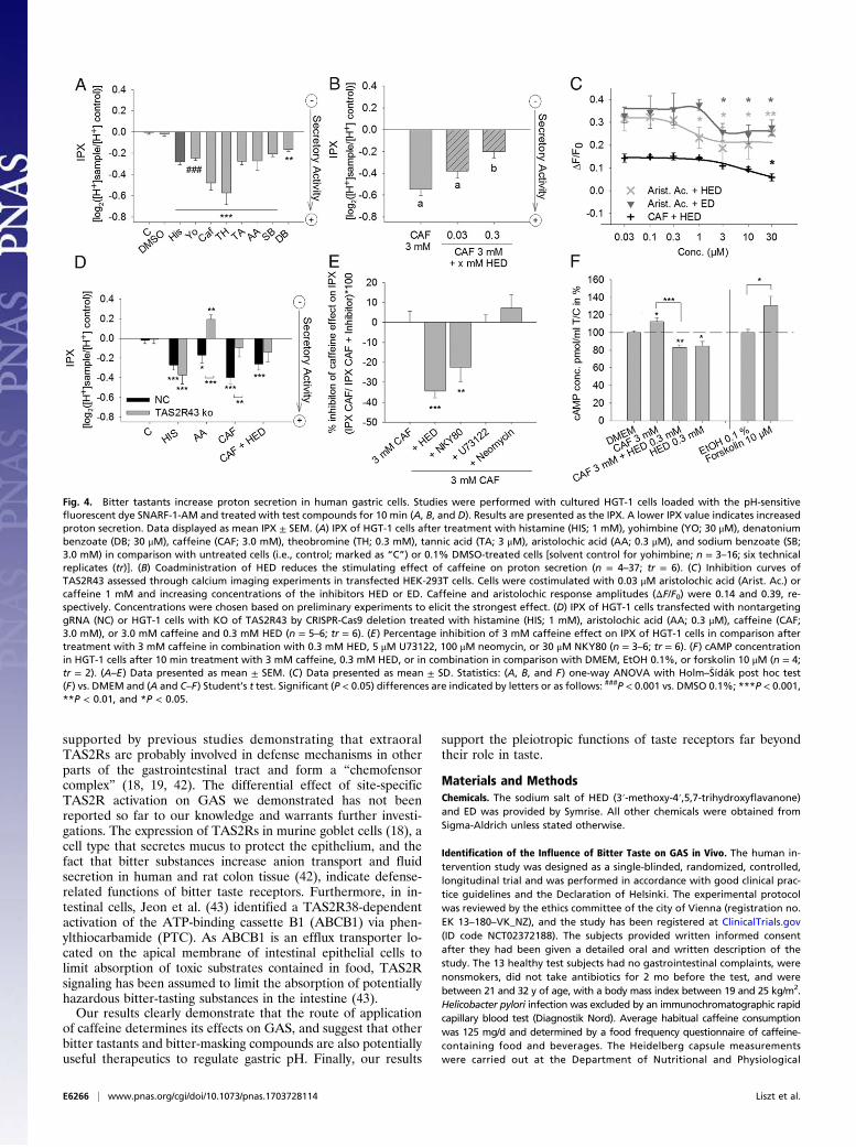
Effect of Bitter and Bitter-Masking Compounds on Proton Secretion inHGT-1 Cells.Following our hypothesis that bitter compounds inducemechanisms of GAS via TAS2Rs, various bitter compounds, suchas theobromine, tannic acid, yohimbine, denatonium benzoate,sodium benzoate, and aristolochic acid were tested and verifiedfor their stimulating effects on proton secretion in HGT-1 cells(Fig. 4A). The concentrations of the tested compounds werechosen based on preliminary experiments to elicit the strongesteffect on proton secretion without impairing cellular viability [>90%compared with nontreated controls (100%)]. The responses weresimilar in magnitude, or even more pronounced, compared withthose elicited by histamine, a major activator of proton secretion inparietal cells (10). These responses to bitter compounds indicatethat several TAS2Rs could be activated in HGT-1 cells. Treatmentof HGT-1 cells with 0.3–3,000 μM caffeine increased proton se-cretion, with 3,000 μM caffeine showing the highest effect (Fig. S6 Aand B). The bitter-masking compounds HED and eriodictyol (ED),which have been described to reduce the bitter taste of caffeine inhuman sensory panels (33, 34), also reduced the caffeine-evokedproton secretion in HGT-1 cells (Fig. 4B and Fig. S6C).
Antagonistic or Agonistic Effect of HED and ED on TAS2Rs-InducedCa2+ Mobilization in HEK-293T Cells. To identify the TAS2Rs thatare targeted by HED and its structural analog ED, Ca2+-mobi-lization in the presence of these compounds by transientlytransfected HEK-293T cells was analyzed with or without cos-timulation with specific agonists of TAS2Rs (12). HED and EDwere identified as agonists for TAS2R14 and as antagonists forTAS2Rs 43, 20, and 50 (Table S1). HED is also an antagonist forTAS2R31 (Table S1). As TAS2R43 can be activated by caffeine(12), the effect of caffeine and HED was further investigated inHEK-293T cells transiently transfected with TAS2R43. TAS2R43in these cells was then activated by aristolochic acid or caffeine forthe performance of calcium imaging experiments in the presenceof increasing concentrations (0.03−30 μM) of HED and ED. Bothcompounds reduced TAS2R43 responses to aristolochic acid orcaffeine (Fig. 4C).
Caffeine-Induced Proton Secretion Is Reduced in TAS2R43 KO HGT-1Cells. To determine whether TAS2R43 is involved in mechanismsof caffeine-induced GAS, a homozygous 13-bp deletion in theTAS2R43 gene (Fig. S7) of HGT-1 cells was induced by using aCRISPR-Cas9 CD4-vector (i.e., TAS2R43-KO). As negativecontrol (NC), HGT-1 cells were treated in parallel with the samevector containing a nontargeting scrambled guide RNA (gRNA).Off-target effects of the transfected gRNA were excluded by a whole-genome sequencing analysis. The stimulating effect of caffeine andthe TAS2R43 agonist aristolochic acid on proton secretion inHGT-1 cells was substantially reduced in TAS2R43-KO cells com-pared with NC cells (Fig. 4D). These data demonstrate that TAS2R43is involved in caffeine’s action on proton secretion in HGT-1 cells.Considering that caffeine’s effect on proton secretion in HGT-1
cells is sensitive to TAS2R43 and HED, and that TAS2R43 isblocked by HED in TAS2R43-transfected HEK-293T cells, thedata strongly suggest that caffeine mediates its effect through atleast one TAS2Rs (TAS2R43) if not more.
Fig. 2. Addition of HED reduces the caffeine-evoked effects on reacidification time or the slope in gastric pH measurements via administration by drinking150 mg caffeine (CAF) with or without 30 mg HED dissolved in 125 mL water (A–C) or by encapsulated test compounds in combination with 125 mL water25 min before alkaline challenge (D–F). (A and D) Gastrograms of Heidelberg capsule measurements according to the three different delivery protocols fromone test subject are presented in one graphic. (B and E) Delta reacidification time of gastric pH measurements in subjects after consumption of CAF or CAFplus HED (basal = 0). (C and F) Delta slope of gastric pH measurements in subjects after consumption of CAF or CAF plus HED (basal = 0). Data are displayed asmean ± SEM: (B and C) CAF, n = 10; CAF plus HED, n = 6; (E and F) CAF, n = 7; CAF plus HED, n = 6 (*P < 0.05 and **P < 0.01 indicate significant differences byStudent’s t test).
Liszt et al. PNAS | Published online July 10, 2017 | E6263
PHYS
IOLO
GY
PNASPL
US
Effect of Pharmacological Blockers on Caffeine-Induced Proton Secretion.Caffeine-evoked proton secretion was reduced by neither thePLCβ2 inhibitor U73122 nor the IP3 inhibitor neomycin (Fig.4E). In contrast, it was reduced by the adenylyl cyclase inhibitorNKY80 (−22.5 ± 7.4%; P < 0.01), suggesting that cAMP but notCa2+ signaling is involved in TAS2R-mediated regulation of acidsecretion in HGT-1 cells.
Effect of Caffeine and HED on cAMP Levels in HGT-1 Cells. To confirmthat adenylyl cyclase mediates proton secretion in HGT-1 cellsvia caffeine-dependent TAS2R stimulation, intracellular cAMPlevels were determined in response to treatment with caffeineand HED (Fig. 4F). Treatment of HGT-1 cells for 10 min with3.0 mM caffeine increased cAMP levels by 12 ± 4.6% (P < 0.05)in comparison with the treatment with DMEM (control, 100 ±2.0%). However, coapplication of caffeine and HED (83.3 ± 2.7%;P < 0.01) and HED alone (84.9 ± 5.1%; P < 0.05) reduced cAMPlevels in HGT-1 cells. Treatment with forskolin, a stimulator ofadenylyl cyclase, increased cAMP levels to 131 ± 10.3% (P < 0.05)in HGT-1 cells in comparison with treatment with the solventcontrol ethanol (i.e., EtOH). These observations confirm that caf-feine activates TAS2Rs signaling through changes in cAMP levels.
DiscussionAcid secretion in the stomach is a fundamental process that isfinely regulated at different levels. Initial activation of GAS isregulated by the CNS when food is smelled and tasted (10, 35).
When food enters the stomach, mechanical or chemical recep-tors (i) initiate GAS via activation of afferent/efferent fibersconnected to the CNS, (ii) stimulate the gastrin-producing Gcells or the histamine-producing enterochromaffin-like cells,or (iii) directly stimulate the HCl-producing parietal cells. Caf-feine stimulates GAS, and, so far, it has been assumed that it actsvia inhibition of PDE or by antagonizing adenosine receptorsin gastric parietal cells (1). As caffeine activates five of the25 human TAS2Rs (12), we hypothesized that its bitternessalso contributes to its stimulating effect on GAS via activationof TAS2Rs.First, we found that oral consumption of caffeine delayed
GAS in healthy subjects, whereas caffeine that was administeredencapsulated, being released in the stomach, accelerated thisprocess compared with oral administration. The delay inducedby oral caffeine presentation might be explained by findingsreported by McMullen et al. (36). They demonstrated that caffeinein a coffee drink accelerated the heart rate without increasing thevascular tonus in comparison with caffeine administered encapsu-lated concomitantly to a decaffeinated coffee drink. The increase inheart rate was likely induced by vagal withdrawal instead of sym-pathetic activation. This finding is important, considering that thecephalic-phase response during digestion is thought to activate thevagus nerve to enhance digestion (36). The study of McMullen et al.(36) along with the present results suggest that orally sensed caf-feine elicits vagal withdrawal that would reduce rather than enhancethe digestive capacity, for example by delaying GAS. An expla-nation why the delaying effect of caffeine has not been discussedearlier might be that most of the previous studies investigatingthe effect of caffeine on GAS used gavages to bypass oral cavityreceptors (2, 3, 5) and therefore did not take into account aninhibitory effect of caffeine on GAS by oral perception. Fur-thermore, we demonstrated that TAS2R bitter receptors in thestomach are involved in the caffeine-induced secretion of gastricacid. This conclusion is based on the observation that the bitter-masking compound HED similarly reduced caffeine’s bitternessas well its effects on GAS evoked by combined oral/gastric orgastric-only caffeine application. The finding that concomitantoral ingestion of caffeine and HED accelerated passing of theHeidelberg capsule into the duodenum in 4 of 10 subjectscompared with caffeine administration indicates that HED mightinduce gastric emptying. This hypothesis has been verified bymeasuring the effect of HED on gastric motility in strips ofstomach dissections in an organ bath. Avau et al. (37) demonstratedthat bitter compounds such as denatonium benzoate increasedcontractility in gastric strips of mice and caused an impairment ofgastric relaxation after intragastric infusion. Whether a bitter-masking compound has opposite effects by causing gastric re-laxation is an open question. The data presented here did notshow any effects of HED on gastric secretion in humans, nor onproton secretion in HGT-1 cells at the concentrations tested.However, HED induced gastric relaxation, indicating physio-logical targets other than GAS.To confirm that caffeine induces GAS via gastric TAS2Rs, we
demonstrated that the mRNA of 22 of 25 TAS2Rs as well astransducin is present in HGT-1 cells, and that the five TAS2Rsthat can be activated by caffeine are present in the stomachmucosa. These findings were corroborated by immunohisto-chemical detection of TAS2R10 and transducin in different gastriccell types for which useful antisera were available. So far, to ourknowledge, only one validated antibody against human TAS2R38(38) is known. Neither caffeine nor HED bind to TAS2R38. Anantibody against TAS2R43 (OSR00171W; Thermo Fisher Scien-tific) was tested. Unfortunately, during validation in transfectedHEK-239T cells, this antibody turned out to be unspecific. Nev-ertheless, we could demonstrate data for the validation of an an-tibody targeting TAS2R10 (Fig. S3) and show the expression ofTAS2R10 on a protein level in gastric mucosa and HGT-1 cells.
Table 1. mRNA expression of TAS2Rs in HGT-1 cells normalizedto the expression of the acetylcholine receptor (CHRM3)
Receptor/gene
HGT-1
Mean SEM
CHRM3 1.00 0.035TAS2R1 0.20 0.050TAS2R3 9.87 0.848TAS2R4 5.66 0.765TAS2R5 12.08 0.822TAS2R7 0.32 0.073TAS2R8 No specific productTAS2R9* 0.12 0.019TAS2R10 0.97 0.100TAS2R13 1.69 0.144TAS2R14 12.39 1.347TAS2R16 0.71 0.239TAS2R19 4.40 0.678TAS2R20 9.09 1.139TAS2R30 8.02 0.717TAS2R31 4.00 1.767TAS2R38 0.14 0.045TAS2R39 3.64 0.807TAS2R40 0.51 0.052TAS2R41 0.66 0.143TAS2R42 2.24 0.444TAS2R43 6.47 0.316TAS2R45 Not detectedTAS2R46 2.59 0.421TAS2R50 2.91 0.290TAS2R60 No specific productPLCB2 2.47 0.110GNAT2 7.16 0.557GNAT3 0.04 0.014
Data are shown as mean ± SEM; n = 3–4 biological replicates, tr = 3 tech-nical replicates. The mRNA of TAS2Rs is similarly or even more highlyexpressed compared with the mRNA of CHRM3 in HGT-1 cells.*In one of three replicates, no product was detected.
E6264 | www.pnas.org/cgi/doi/10.1073/pnas.1703728114 Liszt et al.
As TAS2R10 was highly expressed in parietal cells, as detectedby immunohistological staining, we focused on the cellularmechanisms in HGT-1 cells, which exhibit the characteristics ofparietal cells (28, 29). Nevertheless, we cannot exclude that othercell types or gastrointestinal hormones may have contributed tothe detected effects in the human intervention study. We dem-onstrated here that the bitter-masking agent HED reduced thestimulatory effect of caffeine on proton secretion in healthysubjects and in HGT-1 cells. As TAS2R43 is the only one of thefive TAS2Rs that can be activated by caffeine and antagonizedby HED, we also performed a CRISPR-Cas9 approach to knockout TAS2R43 in HGT-1 cells. In these TAS2R43-KO cells, theeffect of caffeine on proton secretion was reduced in comparisonwith control cells. To further confirm our hypothesis thatTAS2Rs are involved in mechanisms regulating GAS, TAS2R43was transiently transfected into HEK-293T cells, which do notnormally express any TAS2Rs. In this cell model, we demon-strated that HED antagonized caffeine-stimulated responses inTAS2R43-transfected cells. These results strongly indicate thatTAS2R43 is involved in the proton secretory effect of caffeine.Nevertheless, involvement of other TAS2Rs or signaling path-ways, such as adenosine receptors, or PDE inhibition cannotbe excluded.HED is also an agonist for TAS2R14, which is highly
expressed in HGT-1 cells. The interaction of agonistic and an-tagonistic effects on TAS2Rs and the further activation ofdownstream signaling pathways seem highly complex, and not
every TAS2R might be connected to the same downstream sig-naling cascade. One downstream signal for the induction ofproton secretion is cAMP. Here, we show that caffeine increasedcAMP levels in HGT-1 cells, an effect that was inhibited byHED. HED itself reduced the cAMP level in HGT-1 cells, butdid not affect proton secretion in HGT-1 cells. Therefore, itremains unclear whether or which signaling pathways are af-fected by HED. Caffeine signaling via cAMP is supported by thefact that the adenylyl cyclase inhibitor NKY80 reduced thecaffeine-evoked stimulation of proton secretion. Caffeine-induced activation of PLCβ2 and IP3 signaling can be excludedby the failure of the specific inhibitors U73122 (PLCβ2) andneomycin (IP3) to reduce proton secretion. Experiments inwhich caffeine and HED were tested in combination with theinhibitors showed no difference in proton secretion in compari-son with caffeine and HED tested alone. That leaves the ques-tion of how caffeine can stimulate GAS via TAS2Rs. So far, onlyfor sweet and glutamate taste receptors (TAS1R1/3) has an in-crease of cAMP levels via activation of adenylyl cyclase beendemonstrated (39). The signaling cascade of bitter taste recep-tors has been proposed to reduce cAMP levels by activation ofPDE via α-gustducin or transducin (23, 26, 40).Based on our results, we hypothesize that bitter perception of
caffeine in the mouth generates a signal of aversion, which leads,via vagal withdrawal, to inhibition of GAS (41). However, whenbitter compounds reach the stomach, increased GAS could aiddegradation or removal of the potential toxins. This hypothesis is
Fig. 3. (A, a–p) Immunochemical localization of TAS2R10 and GNAT2 in (A) gastric tissue and (B) HGT-1 cells with and without preincubation with a blockingpeptide. (a) In the gastric corpus/fundus, cytoplasmic reactivity of TAS2R10 in parietal and chief cells (one arrow) was detected whereas foveolar cells werenegative (two arrows). Detail (b) shows parietal and chief cells. In the gastric antrum (e and f), very faint cytoplasmic and focal membranous reactivity ofTAS2R10 in glandular cells was detected (one arrow). Foveolar cells are negative (two arrows). (f) Detail showing glandular cells. GNAT2 was localized in thegastric fundus (i and j) parietal and chief cells (one arrow, j). Foveolar cells demonstrate membranous staining (two arrows, j). (m and n) In gastric antrum,membranous reactivity of GNAT2 in glandular cells (one arrow,m and n) was detected whereas foveolar cells were negative (two arrows, m). (c, d, g, h, k, l, o,and p) Corresponding negative controls. (B) Staining of HGT-1 cells with TAS2R10 and GNAT2 antisera (green) with and without specific blocking peptide andcell-surface labeling with con A (red).
Liszt et al. PNAS | Published online July 10, 2017 | E6265
PHYS
IOLO
GY
PNASPL
US
supported by previous studies demonstrating that extraoralTAS2Rs are probably involved in defense mechanisms in otherparts of the gastrointestinal tract and form a “chemofensorcomplex” (18, 19, 42). The differential effect of site-specificTAS2R activation on GAS we demonstrated has not beenreported so far to our knowledge and warrants further investi-gations. The expression of TAS2Rs in murine goblet cells (18), acell type that secretes mucus to protect the epithelium, and thefact that bitter substances increase anion transport and fluidsecretion in human and rat colon tissue (42), indicate defense-related functions of bitter taste receptors. Furthermore, in in-testinal cells, Jeon et al. (43) identified a TAS2R38-dependentactivation of the ATP-binding cassette B1 (ABCB1) via phen-ylthiocarbamide (PTC). As ABCB1 is an efflux transporter lo-cated on the apical membrane of intestinal epithelial cells tolimit absorption of toxic substrates contained in food, TAS2Rsignaling has been assumed to limit the absorption of potentiallyhazardous bitter-tasting substances in the intestine (43).Our results clearly demonstrate that the route of application
of caffeine determines its effects on GAS, and suggest that otherbitter tastants and bitter-masking compounds are also potentiallyuseful therapeutics to regulate gastric pH. Finally, our results
support the pleiotropic functions of taste receptors far beyondtheir role in taste.
Materials and MethodsChemicals. The sodium salt of HED (3′-methoxy-4′,5,7-trihydroxyflavanone)and ED was provided by Symrise. All other chemicals were obtained fromSigma-Aldrich unless stated otherwise.
Identification of the Influence of Bitter Taste on GAS in Vivo. The human in-tervention study was designed as a single-blinded, randomized, controlled,longitudinal trial and was performed in accordance with good clinical prac-tice guidelines and the Declaration of Helsinki. The experimental protocolwas reviewed by the ethics committee of the city of Vienna (registration no.EK 13–180–VK_NZ), and the study has been registered at ClinicalTrials.gov(ID code NCT02372188). The subjects provided written informed consentafter they had been given a detailed oral and written description of thestudy. The 13 healthy test subjects had no gastrointestinal complaints, werenonsmokers, did not take antibiotics for 2 mo before the test, and werebetween 21 and 32 y of age, with a body mass index between 19 and 25 kg/m2.Helicobacter pylori infection was excluded by an immunochromatographic rapidcapillary blood test (Diagnostik Nord). Average habitual caffeine consumptionwas 125 mg/d and determined by a food frequency questionnaire of caffeine-containing food and beverages. The Heidelberg capsule measurementswere carried out at the Department of Nutritional and Physiological
Fig. 4. Bitter tastants increase proton secretion in human gastric cells. Studies were performed with cultured HGT-1 cells loaded with the pH-sensitivefluorescent dye SNARF-1-AM and treated with test compounds for 10 min (A, B, and D). Results are presented as the IPX. A lower IPX value indicates increasedproton secretion. Data displayed as mean IPX ± SEM. (A) IPX of HGT-1 cells after treatment with histamine (HIS; 1 mM), yohimbine (YO; 30 μM), denatoniumbenzoate (DB; 30 μM), caffeine (CAF; 3.0 mM), theobromine (TH; 0.3 mM), tannic acid (TA; 3 μM), aristolochic acid (AA; 0.3 μM), and sodium benzoate (SB;3.0 mM) in comparison with untreated cells (i.e., control; marked as “C”) or 0.1% DMSO-treated cells [solvent control for yohimbine; n = 3–16; six technicalreplicates (tr)]. (B) Coadministration of HED reduces the stimulating effect of caffeine on proton secretion (n = 4–37; tr = 6). (C) Inhibition curves ofTAS2R43 assessed through calcium imaging experiments in transfected HEK-293T cells. Cells were costimulated with 0.03 μM aristolochic acid (Arist. Ac.) orcaffeine 1 mM and increasing concentrations of the inhibitors HED or ED. Caffeine and aristolochic response amplitudes (ΔF/F0) were 0.14 and 0.39, re-spectively. Concentrations were chosen based on preliminary experiments to elicit the strongest effect. (D) IPX of HGT-1 cells transfected with nontargetinggRNA (NC) or HGT-1 cells with KO of TAS2R43 by CRISPR-Cas9 deletion treated with histamine (HIS; 1 mM), aristolochic acid (AA; 0.3 μM), caffeine (CAF;3.0 mM), or 3.0 mM caffeine and 0.3 mM HED (n = 5–6; tr = 6). (E) Percentage inhibition of 3 mM caffeine effect on IPX of HGT-1 cells in comparison aftertreatment with 3 mM caffeine in combination with 0.3 mM HED, 5 μM U73122, 100 μM neomycin, or 30 μM NKY80 (n = 3–6; tr = 6). (F) cAMP concentrationin HGT-1 cells after 10 min treatment with 3 mM caffeine, 0.3 mM HED, or in combination in comparison with DMEM, EtOH 0.1%, or forskolin 10 μM (n = 4;tr = 2). (A–E) Data presented as mean ± SEM. (C) Data presented as mean ± SD. Statistics: (A, B, and F) one-way ANOVA with Holm–�Sídák post hoc test(F) vs. DMEM and (A and C–F) Student’s t test. Significant (P < 0.05) differences are indicated by letters or as follows: ###P < 0.001 vs. DMSO 0.1%; ***P < 0.001,**P < 0.01, and *P < 0.05.
E6266 | www.pnas.org/cgi/doi/10.1073/pnas.1703728114 Liszt et al.
Chemistry, University of Vienna, Austria. At each visit, subjects were sub-jected to one treatment, meaning that subjects who took part in all ad-ministration types and treatments completed 11 visits (Fig. 1B). Before theintervention, the trial subjects had to fast from food and liquid for 10 h. Forthe noninvasive measurement of gastric pH, the Heidelberg Detection Sys-tem (Heidelberg Medical) was applied as described before (29, 30). Whenthe subject arrived in the morning, the pH capsule was prepared by activa-tion for 5 min in a sterile 0.9 NaCl solution and, as indicated by the Hei-delberg Detection System software, the capsule was calibrated at pH 1 andpH 7. After calibration, the capsule was swallowed by the subject. When apH of approximately 1–2 was stable over a period of 3 min, a stable positionof the capsule in the stomach was considered to have been achieved. Duringthe measurement, the subjects had to lie down on their left side to makesure that the capsule remained in the stomach. Each trial started withthe administration of 5 mL of a saturated sodium bicarbonate solution(NaHCO3), triggering an increase in gastric pH to values between pH 6 andpH 7 and subsequently leading to the secretion of stomach acid by the pa-rietal cells. At the first 8 visits, 125 mL water (control), 37.5/75/150 mg caf-feine diluted in 125 mL water, or 150 mg caffeine encapsulated in a gelatincapsule (Coni-Snap size 1; Capsugel) with 125 mL water were administered5 min after the alkaline challenge (Fig. 1B). A total of 150 mg caffeine incombination with 30 mg HED or 30 mg HED alone, diluted in 125 mL, wereadministered by drinking 5 min after the alkaline solution. At visits 9–11,three new subjects joined to replace dropouts. There, an empty gelatincapsule (Coni-Snap size 1; Capsugel), 150 mg caffeine encapsulated, or150 mg caffeine encapsulated with 30 mg HED were administered with125 mL water 25 min before the alkaline solution. For exclusive activation ofTAS2Rs in the mouth (delivery protocol 3), the subjects swallowed 125 mLwater and rinsed their mouth with 150 mg caffeine diluted in 125 mL waterwithout swallowing the caffeine 5 min after swallowing the alkaline solu-tion. Reacidification time (i.e., time until original pH is reached again afteradministration of the alkaline challenge) as well as the slope of the gas-trogram were analyzed by using Heidelberg Detection System software andImageJ software (National Institutes of Health). Delta reacidification time wascalculated by substraction of the reacidification time of the water or emptycapsule control from the reacidification time after the treatment. The slopewas calculated between the point when pH decreases and the point at whichthe original pH is reached again.
Organ Bath of Human Stomach Biopsy Specimens. Human stomach wasobtained at surgery for obesity at Homerton University Hospital (London,United Kingdom) after ethical approval (REC 15/LO/2127) and informedwritten consent. Mucosa-free tissue from the fundus region of humanstomachs was dissected parallel to the circular muscle fibers into strips (5–8 ×15 mm). Strips were tied up and mounted in organ bath chambers thatcontained a Krebs solution at 37 °C aerated with 5% CO2 in O2. After 1 hequilibration, the nerves were excited by electrical field stimulation (EFS) at200 mA for 0.5 ms; 5 Hz were given for 10 s every 1 min. When there was astable response to EFS, a frequency response with 1, 2, 5, 10, 15, and 20 Hzwas generated. After switching back to 5 Hz and reaching a stable signal,NaHED was applied in a concentration of 1 mM for at least 50 min. Double-distilled (dd)H2O was used as a vehicle control. Isometric force transducers(calibrated at 2 g; AD Instruments) and the software AcqKnowledge (Biopac)detected changes in muscle changes. Data are presented as percent changesin baseline tension. The number of patients is given as an n value.
Sensory Study. Taste sessions were carried out in the morning hours, and the13 untrained panel subjects were asked not to consume anything besideswater 30 min before the sensory duo test. The bitter recognition threshold ofthe subjects was determined by using a standardized test system startingwithwater and followed by nine solutions with increasing concentrations of caf-feine, from 25 to 225mg/L. Furthermore, the subjects had to rank the bitternessof a caffeine solution (150mg/125mL) and a caffeine (150mg/125mL) plus HED(30 mg/125 mL) solution by sip-and-spit on a scale of 1 (nothing) to 10 (ex-tremely strong). This dual testwas repeated four times in randomized order andunder colored light. Statistical significance was calculated by Student’s t test(double-sided, paired).
HGT-1 Cell Culture. The human gastric tumor cell line HGT-1 was obtainedfrom C. Laboisse (Laboratory of Pathological Anatomy, Nantes, France) andcultured under standard conditions as described previously (8). Cytotoxicityof the tested substances and treatment reagents was excluded by MTT testas described before (8), and cell viability was determined by trypan bluestaining. Tested cells had at least 90% cell viability.
Immunohistochemical Staining of Gastric Tissues. Histological specimens wereobtained from two patients from the Pathologisch-Bakteriologisches Institut,Sozialmedizinisches Zentrum Ost–Donauspital, Vienna, Austria. The gastricfundus was derived from a sleeve gastrectomy of a 42-y-old adipose butotherwise healthy patient. The gastric antrum was derived from a 71-y-oldpatient undergoing distal partial gastrectomy for a benign gastrointestinalstroma tumor. Immunohistochemistry was performed on 5-μm-thick formalin-fixed, paraffin-embedded whole tissue sections. Slides were processed in thefully automated staining instrument Benchmark ULTRA by using an ultraViewUniversal DAB Detection Kit (VentanaMedical Systems). The following primaryantibodies were applied: TAS2R10 (OSR00158W; Thermo Scientific), 1:750 for28 min at 37 °C after heat-mediated antigen retrieval using EDTA buffer,pH 8.0, at 95 °C for 36 min (CC1 buffer; Ventana Medical Systems) and GNAT2(transducin α-2 chain; AP11077c; Abgent), 1:50 for 28 min at 37 °C after heat-mediated antigen retrieval using EDTA buffer, pH 8.0, at 95 °C for 64 min(CC1 buffer; Ventana Medical Systems) and amplification at 95 °C (Amplifi-cation Kit; Ventana Medical Systems). All counterstaining was performedwith hematoxylin. Blocking experiments to control for unspecific stainingwere performed by using the TAS2R10 control peptide (GST00040P; ThermoScientific) and GNAT2 antibody blocking peptides (BP11077c; Abgent). Forthe TAS2R10 taste receptor, the blocking experiment consisted of the con-trol peptide, 1:200, incubated together with TAS2R10 antibody, 1:750, for120 min at 4 °C, and thereafter, incubation of the slide at 37 °C for 28 min.The GNAT2 antibody blocking peptide, 1:10, was incubated together withGNAT2 antibody, 1:50, for 120 min at 4 °C, and, thereafter, incubation of theslide for 28 min at 37 °C. All other steps were performed similarly to thestaining procedure as described earlier.
Immunocytochemical Staining of HGT-1 Cells and HEK-293T-Gα16gust44 Cells.Transiently transfected HEK-293T-Gα16gust44 cells (TAS2R10 or TAS2R16)were prepared as described previously (19), and HGT-1 cells were seeded oncoverslips 24 h before the staining procedure. Cells were fixed and stained asdescribed previously (18) by using anti-HSV (1:15,000; Novagen), anti-TAS2R10, and anti-GNAT2 antibodies (Immunohistochemical Staining ofGastric Tissues) for 1 h at room temperature. Specificity of labeling wasensured as described in Immunohistochemical Staining of Gastric Tissues,and detection was carried out as described previously (18). Preabsorption ofthe anti-TAS2R10 and anti-GNAT2 antibody was performed with the corre-sponding immunogenic peptide (Immunohistochemical Staining of GastricTissues; Fig. S3). The HSV epitope was detected with anti-mouse antibodiesconjugated with Cy3 (1:2,000; Sigma), biotin-labeled concanavalin A (con A)with streptavidin Alexa Fluor 633 (1:1,000; Molecular Probes), and TAS2R10or GNAT2 with Alexa Fluor 488 goat anti-rabbit IgG (1:1,000; Molecular Probes).
Intracellular pH Measurement in HGT-1 Cells Indicating Proton Secretion. In-tracellular pH, as indicator for proton secretion in HGT-1 cells, was measuredusing the pH-sensitive fluorescence dye 1,5 carboxy-seminaphto-rhodafluoracetoxymethylester (SNARF-1-AM; Life Technologies) as described before(8, 29). The intracellular proton index (IPX) in the cells was calculated by log2
transformation of the ratio between treated and untreated (i.e., control)cells. The lower the IPX, the fewer protons are in the cell, indicating a highersecretory activity in HGT-1 cells.
mRNA Expression of Bitter Taste Receptors in HGT-1 Cells and Human BiopsiesUsing RT-qPCR. Total RNA was extracted from HGT-1 cells and two humanbiopsy specimens by using the peqGold Total RNA Kit (Peqlab). Quantity andquality were checked spectrophotometrically. Reverse transcription wascarried out with 2 μg RNA and the High Capacity cDNA Reverse TranscriptionKit (Thermo Fisher Scientific). Real-time PCR was performed with an AppliedBiosystems StepOneplus Real Time PCR system and Fast SYBR Green MasterMix (Thermo Fisher Scientific). Primers were designed using the NationalCenter for Biotechnology Information (NCBI) primer designing tool (usingPrimer 3 and BLAST; Table S2). Cycling conditions were 20 s/95 °C (activa-tion), 3 s/95 °C (denaturation), 30 s/60 °C (annealing), and 15 s/67 °C (elon-gation with fluorescence measurement). The PCR products were verified bymelting curve analysis, agarose gel electrophoresis, and sequence analysis(Eurofins Genomics). Sequences were checked by using the NCBI BLASTntool. Primers showing no product in HGT-1 in at least one of the threereplicates (TAS2Rs 8, 9, 45, and 60) were tested with cDNA derived from ahuman tongue biopsy provided by J.-D. Raguse (Charité, Berlin, Germany).Whereas primers for TAS2Rs 8, 9, and 60 could be verified, TAS2R45 was notdetected. For TAS2R45, high-frequency copy-number variants are known,and some people do not possess the tested variant of the mRNA for thisgene (44). TAS2R46 could not be detected in the second human biopsysample. The open-source software LinRegPCR was used for quantitative PCR
Liszt et al. PNAS | Published online July 10, 2017 | E6267
PHYS
IOLO
GY
PNASPL
US
data analysis. This software enables the calculation of the starting concen-tration (N0) of each sample, expressed in arbitrary fluorescence units. Thecalculated starting concentrations of the TAS2Rs were compared with thestarting concentrations of the acetylcholine receptor (CHRM3), with pre-viously described primers (8), which is typically expressed in parietal cells ona functional level.
Generation of TAS2R43 Homozygous KO HGT-1 Cell Line Using CRISPR-Cas9. Atotal of 40,000 cells were seeded in a 24-well plate. After approximately 24 h,cells were transfected with 495 ng GeneArt CRISPR Nuclease (CD4 Vector;A21175; Invitrogen) containing the gRNA targeting TAS2R43 gene TTTTTT-GGCAAATGAGGTAC (5′–3′) or, as a control, a scrambled gRNA GTGGACG-GTCGTGCGCTGT (5′–3′) with no target by using the transfection reagentViromer RED (Lipocalyx) according to the manufacturer’s protocol. Trans-fection efficiency was approximately 25%, verified with a CD4 monoclonalantibody (07-0403; Invitrogen/Thermo Fisher Scientific) by using a Guavasoft-flow cytometer (Millipore) on the basis that only positively transfectedcells express a CD4 protein. Cells were transferred in a six-well plate to in-crease cell number. After 3 d, CD4-positive cells (i.e., positively transfectedcells) were enriched by using the Dynabeads CD4 Positive Isolation Kit(Thermo Fisher Scientific) according to the manufacturer’s protocol. Cellswere analyzed with a Genomic Cleavage detection kit (Thermo Fisher Scien-tific) according to the manufacturer’s protocol. For genomic cleavage detection,the following primers were used: forward primer AGACTGCCATTGGGTCAAAGA(5′–3′) and reverse primer GATGTTGTTGGGGCCTTTGC (5′–3′). The followingtemperature protocol was used: 95 °C/10 min, 40 cycles of 95 °C/30s, 58 °C/30 s,72 °C/30 s, and finally 72 °C for 7 min. A genomic cleavage of approximately22% was detected. Single cells were isolated by serial dilution of positivelytransfected cells into two 96-well plates and observed for colony forming for2 wk. Total cells of 39 wells in which clearly only one colony formed wereharvested by trypsin/EDTA and first transferred to a 48-well plate, and, afterconfluence, to a 12-well plate to increase cell number. From each clone, half ofthe cells were frozen and the other half were used to extract DNA with aPureLink Genomic DNA Mini Kit (Life Technologies) according to the manu-facturer’s protocol. Before Sanger sequencing by Eurofins Genomics, the DNAextracts of 20 clones were amplified with AmpliTaqGold 360 Mastermix(Thermo Fisher Scientific), and PCR was carried out as described earlier for thegenomic cleavage detection. Of 20 clones, 15 showed no deletion, four aheterozygous deletion, and one a homozygous deletion. Deletion on anmRNA level was also analyzed by means of Sanger sequencing followingtotal RNA isolation of the WT, cells transfected with the scrambled gRNA,and the TAS2R43-KO cells as described earlier (Fig. S7).
Exclusion of Off-Target Effects Using Whole-Genome Sequencing. The quality-checked DNA was fragmented with a Covaris ultrasonicator. The resultingDNA fragments were purified, end-blunted, A-tailed, and adaptor-ligated.The concentration of the libraries was quantified by Bioanalyzer and real-time PCR. Each library was sequenced with Illumina’s X Ten system withpaired-end 125-bp read length according to the manufacturer’s instructions.Sequencing-derived raw image files were processed by Illumina’s basecallingsoftware to yield raw data files in Fastq format. Reads were mapped to thehuman reference genome (GRCh37/hg19) using Burroughs–Wheeler Aligner
(v0.7.12). Duplicate reads were removed from mapped data by using Picardtools (v1.118). SNPs and small insertions and deletions (InDels) were identi-fied by using the Genome Analysis Toolkit (GATK; v3.3.0). Recommendedbest practices for variant analysis were followed, including InDel re-alignment and base quality score recalibration. The genomic variations weredetected by using GATK’s HaplotypeCaller. Variant quality score recalibra-tion in GATK was applied to obtain high-confidence variant calls. Copynumber variants were called by using CNVnator (v0.2.7). Structural variants(SVs) were detected by using Breakdancer or CREST. The SV annotation wasdone with SnpEff (v4.0). The University of California, Santa Cruz, LiftOvertool was used to get the corresponding coordinates from genome build38 to build 37. Filtering of shared variants was done with GATK’s Select-Variants tool. Reads mapping to off-target regions were extracted withSAMtools (v0.1.19).
cAMP Measurements in HGT-1 Cells. cAMP in HGT-1 cells was measured withthe ELISA kit from R&D Systems according to the protocol.
Calcium Imaging Experiments in HEK-293T Cells. Calcium imaging experimentsusing HEK-293T cells transiently expressing TAS2Rs and stably expressing thechimeric G protein subunit Gα16gust44 were essentially done as describedpreviously (12). Aristolochic acid was dissolved in C1 buffer at 0.03 μM con-centration and caffeine at 1 mM concentration. Cells were exclusively stimu-lated with agonists and increasing concentrations of the antagonists in theconcentration range of 0.03–30 μM. For screening which TAS2Rs are activatedor inhibited by HED or ED, they were applied alone as well as coapplied withsuitable TAS2R agonists. Intracellular Ca2+ concentration increases were mea-sured to monitor changes in TAS2Rs activation upon coapplication of theputative inhibitors. Potential antagonistic/blocking activity of the compoundswas addressed comparing the signals elicited by cells exclusively stimulatedwith the cognate agonists with signals elicited from cells costimulated withHED or ED in two test concentrations (10 and 100 μM) and the agonists. In-hibition curves were calculated with SigmaPlot 11 software after correctingsignal responses and normalization to background fluorescence.
Statistical Analysis. Data shown are representative of at least three biologicalreplicates. All data are expressed as mean ± SEM unless stated otherwise. Alldata have been verified for normality distribution, and statistically signifi-cant differences were considered if the P value was less than 0.05, de-termined by one-way ANOVA with Dunn’s or Holm–�Sídák post hoc test usingSigmaPlot 11.0 software. Correlation analysis according to Spearman wascalculated by SigmaPlot 11.0 software.
ACKNOWLEDGMENTS. We thank Dr. C. L. Laboisse (INSERM 94-04, Facultéde Medicine) for providing the HGT-1 cells, clone 6; Martin Wendelin (Sym-rise Austria) for support in sensory evaluations; Ulrike Redel for technicalassistance in HEK cell experiments and the subjects of the Heidelberg capsuleexperiment; and Marie-Ange Kouassi for technical support with the organtissue bath method. This work was supported by the Austrian Federal Min-istry of Economy, Family and Youth and the Austrian National Foundationfor Research, Technology and Development; Austrian Science Fund GrantFWF P23797; and Symrise.
1. Fredholm BB, Bättig K, Holmén J, Nehlig A, Zvartau EE (1999) Actions of caffeine in
the brain with special reference to factors that contribute to its widespread use.
Pharmacol Rev 51:83–133.2. Musick VH, Avey HT, Hopps HC, Hellbaum AA (1946) Gastric secretion in duodenal
ulcer in remission; response to the caffeine test meal. Gastroenterology 7:332–339.3. Cohen S, Booth GH, Jr (1975) Gastric acid secretion and lower-esophageal-sphincter
pressure in response to coffee and caffeine. N Engl J Med 293:897–899.4. Cohen MM, Debas HT, Holubitsky IB, Harrison RC (1971) Caffeine and pentagastrin
stimulation of human gastric secretion. Gastroenterology 61:440–444.5. Roth JA, Ivy AC (1944) The effect of caffeine upon gastric secretion in the dog, cat and
man. Am J Physiol 141:454–461.6. Kidd M, Hauso O, Drozdov I, Gustafsson BI, Modlin IM (2009) Delineation of the
chemomechanosensory regulation of gastrin secretion using pure rodent G cells.
Gastroenterology 137:231–241.7. Walker J, et al. (2012) Identification of beer bitter acids regulating mechanisms of
gastric acid secretion. J Agric Food Chem 60:1405–1412.8. Liszt KI, Eder R, Wendelin S, Somoza V (2015) Identification of catechin, syringic acid,
and procyanidin b2 in wine as stimulants of gastric acid secretion. J Agric Food Chem
63:7775–7783.9. Richardson CT, Walsh JH, Cooper KA, Feldman M, Fordtran JS (1977) Studies on the
role of cephalic-vagal stimulation in the acid secretory response to eating in normal
human subjects. J Clin Invest 60:435–441.10. Schubert ML (2010) Gastric secretion. Curr Opin Gastroenterol 26:598–603.
11. Behrens M, Meyerhof W (2010) Oral and extraoral bitter taste receptors. Results ProblCell Differ 52:87–99.
12. Meyerhof W, et al. (2010) The molecular receptive ranges of human TAS2R bittertaste receptors. Chem Senses 35:157–170.
13. Deshpande DA, et al. (2010) Bitter taste receptors on airway smooth muscle broncho-dilate by localized calcium signaling and reverse obstruction. Nat Med 16:1299–1304.
14. Singh N, Vrontakis M, Parkinson F, Chelikani P (2011) Functional bitter taste receptorsare expressed in brain cells. Biochem Biophys Res Commun 406:146–151.
15. Kim KS, Egan JM, Jang HJ (2014) Denatonium induces secretion of glucagon-like peptide-1 through activation of bitter taste receptor pathways. Diabetologia 57:2117–25.
16. Le Nevé B, Foltz M, Daniel H, Gouka R (2010) The steroid glycoside H.g.-12 fromHoodia gordonii activates the human bitter receptor TAS2R14 and induces CCK re-lease from HuTu-80 cells. Am J Physiol Gastrointest Liver Physiol 299:G1368–G1375.
17. Wu SV, et al. (2002) Expression of bitter taste receptors of the T2R family in the gas-trointestinal tract and enteroendocrine STC-1 cells. Proc Natl Acad Sci USA 99:2392–2397.
18. Prandi S, et al. (2013) A subset of mouse colonic goblet cells expresses the bitter tastereceptor Tas2r131. PLoS One 8:e82820.
19. Green BG (2012) Chemesthesis and the chemical senses as components of a “che-mofensor complex”. Chem Senses 37:201–206.
20. Janssen S, et al. (2011) Bitter taste receptors and α-gustducin regulate the secretion ofghrelin with functional effects on food intake and gastric emptying. Proc Natl AcadSci USA 108:2094–2099.
21. Calvo SS, Egan JM (2015) The endocrinology of taste receptors. Nat Rev Endocrinol 11:213–227.
E6268 | www.pnas.org/cgi/doi/10.1073/pnas.1703728114 Liszt et al.
22. Forte JG (2010) The gastric parietal cell: At home and abroad. Eur Surg 42:134–148.23. Roper SD (2007) Signal transduction and information processing in mammalian taste
buds. Pflugers Arch 454:759–776.24. Clapp TR, et al. (2008) Tonic activity of Galpha-gustducin regulates taste cell re-
sponsivity. FEBS Lett 582:3783–3787.25. Ruiz-Avila L, et al. (1995) Coupling of bitter receptor to phosphodiesterase through
transducin in taste receptor cells. Nature 376:80–85.26. He W, et al. (2002) Partial rescue of taste responses of alpha-gustducin null mice by
transgenic expression of alpha-transducin. Chem Senses 27:719–727.27. Bezençon C, et al. (2008) Murine intestinal cells expressing Trpm5 are mostly brush cells
and express markers of neuronal and inflammatory cells. J Comp Neurol 509:514–525.28. Carmosino M, Procino G, Casavola V, Svelto M, Valenti G (2000) The cultured human
gastric cells HGT-1 express the principal transporters involved in acid secretion. PflugersArch 440:871–880.
29. Liszt KI, Walker J, Somoza V (2012) Identification of organic acids in wine thatstimulate mechanisms of gastric acid secretion. J Agric Food Chem 60:7022–7030.
30. Rubach M, et al. (2014) A dark brown roast coffee blend is less effective at stimulatinggastric acid secretion in healthy volunteers compared to a medium roast marketblend. Mol Nutr Food Res 58:1370–1373.
31. Noeller HG (1960) The endoradio sound for electrical pH measurement in the stomachand its clinical significance. Dtsch Med Wochenschr 85:1707–1713.
32. Ghosh T, Lewis DI, Axon ATR, Everett SM (2011) Review article: Methods of measuringgastric acid secretion. Aliment Pharmacol Ther 33:768–781.
33. Ley JP, Krammer G, Reinders G, Gatfield IL, Bertram HJ (2005) Evaluation of bittermasking flavanones from Herba Santa (Eriodictyon californicum (H. and A.) Torr.,Hydrophyllaceae). J Agric Food Chem 53:6061–6066.
34. Ley JP, et al. (2012) Identification of enterodiol as a masker for caffeine bitterness byusing a pharmacophore model based on structural analogues of homoeriodictyol.J Agric Food Chem 60:6303–6311.
35. Kopic S, Geibel JP (2013) Gastric acid, calcium absorption, and their impact on bonehealth. Physiol Rev 93:189–268.
36. McMullen MK, Whitehouse JM, Shine G, Whitton PA, Towell A (2012) Caffeine in hotdrinks elicits cephalic phase responses involving cardiac activity. Food Funct 3:931–940.
37. Avau B, et al. (2015) Targeting extra-oral bitter taste receptors modulates gastroin-testinal motility with effects on satiation. Sci Rep 5:15985.
38. Behrens M, et al. (2012) Immunohistochemical detection of TAS2R38 protein in hu-man taste cells. PLoS One 7:e40304.
39. Trubey KR, Culpepper S, Maruyama Y, Kinnamon SC, Chaudhari N (2006) Tastantsevoke cAMP signal in taste buds that is independent of calcium signaling. Am JPhysiol Cell Physiol 291:C237–C244.
40. Margolskee RF (2002) Molecular mechanisms of bitter and sweet taste transduction.J Biol Chem 277:1–4.
41. Spector AC, Travers SP (2005) The representation of taste quality in the mammaliannervous system. Behav Cogn Neurosci Rev 4:143–191.
42. Kaji I, Karaki S, Fukami Y, Terasaki M, Kuwahara A (2009) Secretory effects of a lu-minal bitter tastant and expressions of bitter taste receptors, T2Rs, in the human andrat large intestine. Am J Physiol Gastrointest Liver Physiol 296:G971–G981.
43. Jeon TI, Seo YK, Osborne TF (2011) Gut bitter taste receptor signalling inducesABCB1 through a mechanism involving CCK. Biochem J 438:33–37.
44. Roudnitzky N, et al. (2011) Genomic, genetic and functional dissection of bitter tasteresponses to artificial sweeteners. Hum Mol Genet 20(17):3437–3449.
Liszt et al. PNAS | Published online July 10, 2017 | E6269
PHYS
IOLO
GY
PNASPL
US