Embed Size (px)
Citation preview
Calciphylaxis in Hemodialysis Patients
Preethi Yerram MD, MS, FASN
February 27, 2016
Objectives
! Define Calciphylaxis
! Risk Factors/Pathophysiology
! Clinical presentation, Diagnosis
! Treatment
Endocrinology: 71; 554: 1962
Selye’s Calciphylaxis
“Condition of hypersensitivity in which, during a critical period after sensitization by a specific calcifying factor ( Vit D compounds, PTH) - topica l t rea tment wi th certain challengers (egg white/yolk, metallic salts) c au s e s an a cu t e l o c a l ca lc inos i s fo l lowed by inflammation and sclerosis”
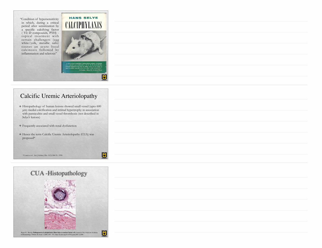
Calcific Uremic Arteriolopathy! Histopathology of human lesions showed small vessel (upto 600
µm) medial calcification and intimal hypertrophy in association with panniculitis and small vessel thrombosis (not described in Selye’s lesions)
! Frequently associated with renal dysfunction
! Hence the term Calcific Uremic Arteriolopathy (CUA) was proposed*
*Coates et al. Am J Kidney Dis. 32(3):384-91, 1998
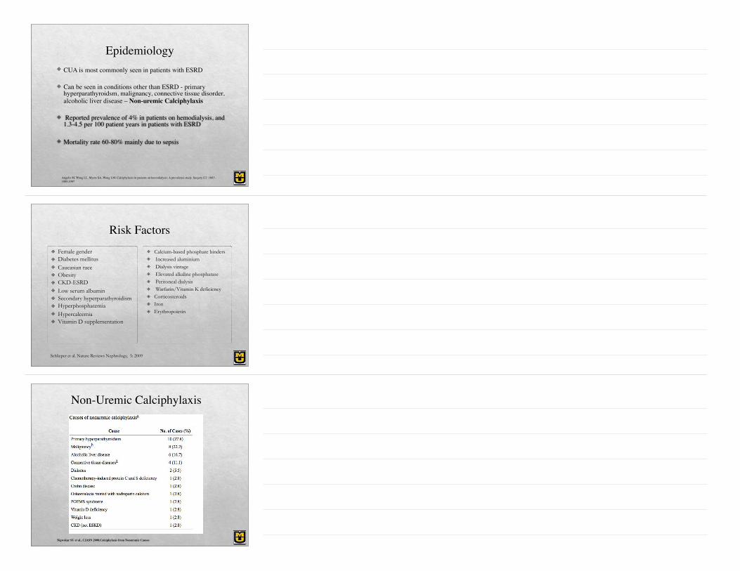
Roger H. Weenig. Pathogenesis of calciphylaxis: Hans Selye to nuclear factor κ-B. Journal of the American Academy of Dermatology, Volume 58, Issue 3, 2008, 458 – 471. http://dx.doi.org/10.1016/j.jaad.2007.12.006
CUA -Histopathology
Epidemiology! CUA is most commonly seen in patients with ESRD
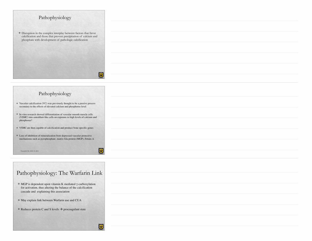
! Can be seen in conditions other than ESRD - primary hyperparathyroidsm, malignancy, connective tissue disorder, alcoholic liver disease – Non-uremic Calciphylaxis
! Reported prevalence of 4% in patients on hemodialysis, and 1.3-4.5 per 100 patient years in patients with ESRD
! Mortality rate 60-80% mainly due to sepsis
Angelis M, Wong LL, Myers SA, Wong LM: Calciphylaxis in patients on hemodialysis: A prevalence study. Surgery122 :1083– 1089,1997
Risk Factors! Female gender ! Diabetes mellitus ! Caucasian race ! Obesity ! CKD-ESRD ! Low serum albumin ! Secondary hyperparathyroidism ! Hyperphosphatemia ! Hypercalcemia ! Vitamin D supplementation
! Calcium-based phosphate binders! Increased aluminium! Dialysis vintage! Elevated alkaline phosphatase! Peritoneal dialysis! Warfarin/Vitamin K deficiency! Corticosteroids! Iron! Erythropoietin
Schlieper et al. Nature Reviews Nephrology, 5: 2009
Non-Uremic Calciphylaxis
Nigwekar SU et al., CJASN 2008.Calciphylaxis from Nonuremic Causes
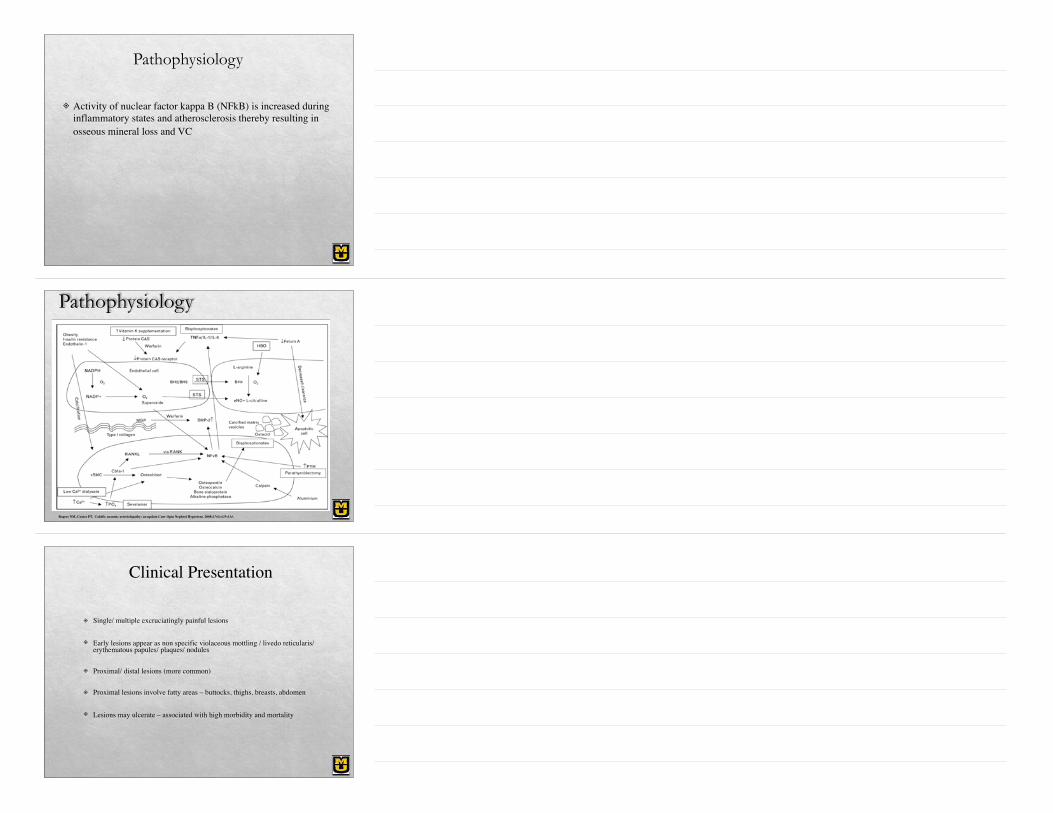
Pathophysiology
! Disruption in the complex interplay between factors that favor calcification and those that prevent precipitation of calcium and phosphate with development of pathologic calcification
Pathophysiology
! Vascular calcification (VC) was previously thought to be a passive process secondary to the effects of elevated calcium and phosphorus level
! In vitro research showed differentiation of vascular smooth muscle cells (VSMC) into osteoblast-like cells on exposure to high levels of calcium and phosphorus*
! VSMC are then capable of calcification and produce bone specific genes
! Loss of inhibition of mineralization from depressed vascular protective mechanisms such as pyrophosphate, matrix Gla protein (MGP), Fetuin-A
*Giachelli CM. JASN 15: 2004
Pathophysiology: The Warfarin Link
! MGP is dependent upon vitamin K mediated γ-carboxylation for activation, thus altering the balance of the calcification cascade and explaining this association
! May explain link between Warfarin use and CUA
! Reduces protein C and S levels ! procoagulant state
Pathophysiology
! Activity of nuclear factor kappa B (NFkB) is increased during inflammatory states and atherosclerosis thereby resulting in osseous mineral loss and VC
Rogers NM, Coates PT. Calcific uraemic arteriolopathy: an update.Curr Opin Nephrol Hypertens. 2008;17(6):629-634.
Pathophysiology
Clinical Presentation
! Single/ multiple excruciatingly painful lesions
! Early lesions appear as non specific violaceous mottling / livedo reticularis/ erythematous papules/ plaques/ nodules
! Proximal/ distal lesions (more common)
! Proximal lesions involve fatty areas – buttocks, thighs, breasts, abdomen
! Lesions may ulcerate – associated with high morbidity and mortality
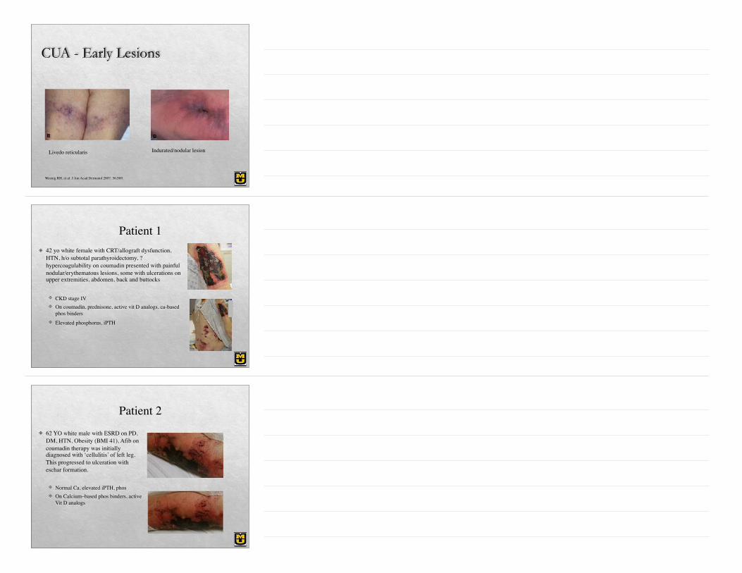
Livedo reticularis
CUA - Early Lesions
Indurated/nodular lesion
Weenig RH, et al. J Am Acad Dermatol 2007; 56:569.
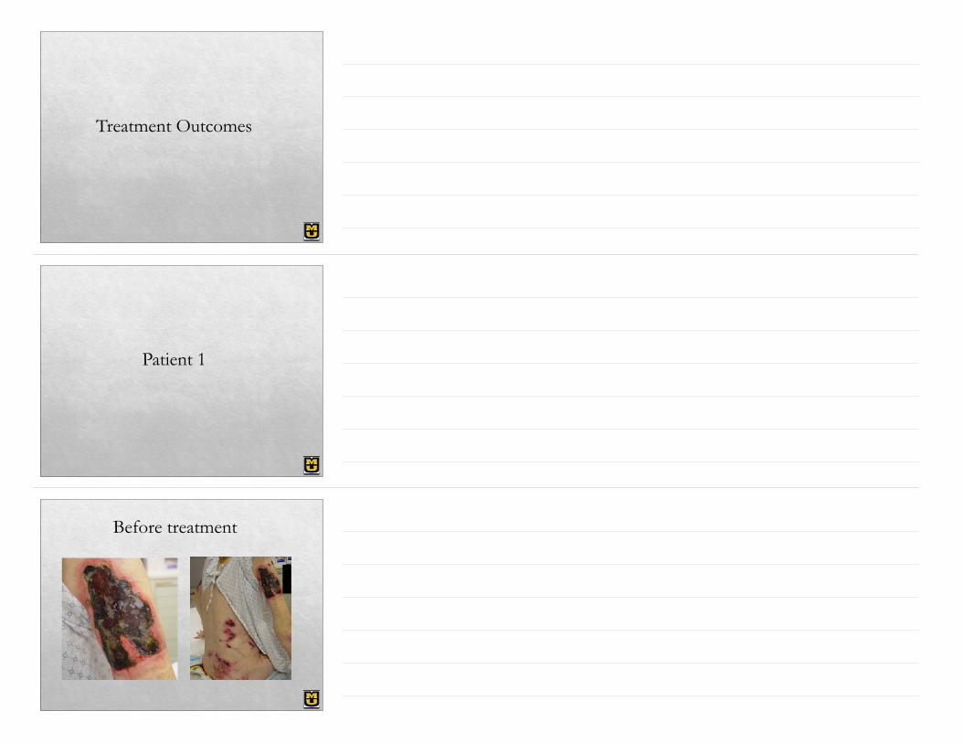
Patient 1! 42 yo white female with CRT/allograft dysfunction,
HTN, h/o subtotal parathyroidectomy, ?hypercoagulability on coumadin presented with painful nodular/erythematous lesions, some with ulcerations on upper extremities, abdomen, back and buttocks
" CKD stage IV" On coumadin, prednisone, active vit D analogs, ca-based
phos binders" Elevated phosphorus, iPTH
Patient 2! 62 YO white male with ESRD on PD,
DM, HTN, Obesity (BMI 41), Afib on coumadin therapy was initially diagnosed with ‘cellulitis’ of left leg. This progressed to ulceration with eschar formation.
" Normal Ca, elevated iPTH, phos" On Calcium–based phos binders, active
Vit D analogs
Differential Diagnosis
! Cholesterol embolization! Warfarin necrosis! Vasculitis! Cellulitis! Early lesions of Nephrogenic systemic fibrosis
Diagnosis! Typical clinical findings
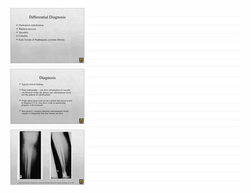
! Plain radiography – can show arborization of vascular calcification within the dermis and subcutaneous tissue – net-like pattern of calcifications
! Triple phase bone scan can be a useful non-invasive test to diagnose CUA; may have a role in monitoring progress with treatment
! Skin biopsy ( requires adequate subcutaneous tissue sample) is diagnostic but may initiate an ulcer
Fine A and Zacharias J. Calciphylaxis is usually non-ulcerating: Risk factors, outcome and therapy. KI, 61, 2002
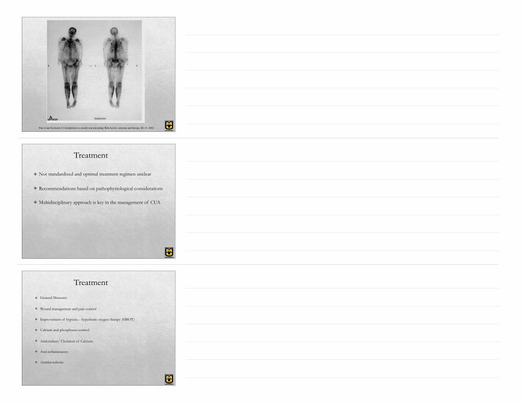
Fine A and Zacharias J. Calciphylaxis is usually non-ulcerating: Risk factors, outcome and therapy. KI, 61, 2002
Treatment
! Not standardized and optimal treatment regimen unclear
! Recommendations based on pathophysiological considerations
! Multidisciplinary approach is key in the management of CUA
Treatment
! General Measures
! Wound management and pain control
! Improvement of hypoxia – hyperbaric oxygen therapy (HBOT)
! Calcium and phosphorus control
! Antioxidant/ Chelation of Calcium
! Anti-inflammatory
! Antithrombotic
General Measures
! Eliminate trigger factors - ?corticosteroids, parenteral iron therapy, warfarin, calcium-based phosphorus binders, vitamin D supplements, erythropoietin
! Intensive nutritional support
! Antibiotics
! Avoid local tissue trauma including subcutaneous injections
Wound Management
! Proper wound care to prevent nodules from becoming necrotizing and spreading of existing necrosis
! Once CUA progresses to necrotic ulcer phase, mortality risk is significantly increased ranging from 30% to 80%
Surgical debridement
! Has been associated with improved survival*
! However this approach to wound management remains controversial
! Some reports suggest improved outcomes with atraumatic wound (regular wound cleansing and dressing) management compared to surgical debridement particularly in cases when the wound is dry and non-infected
*Weenig RH et al. J Am Acad Dermatol 56:2007
Sterile Maggot Debridement
! Sterile maggot debridement in CUA has been described in case reports*
! Option in patients who did not respond to, or those that are not candidates for surgical debridement
! Debridement of necrotic tissue by larval enzymes and potential antibacterial activity
! May be limited by pain, and effective pain management in essential
*Tittelbach J et al. J Dermatolog Treat 12: 2001 *Mason D and Best DS. Adv chr kid dis, 17 (5), 2010
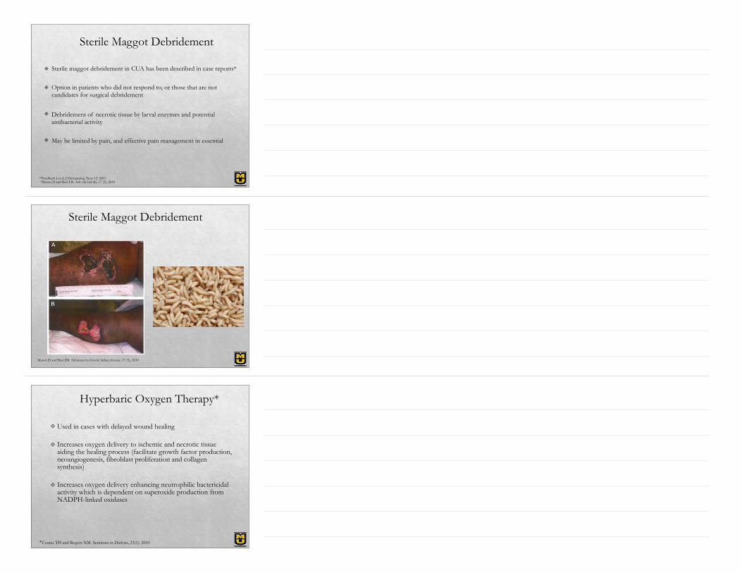
Sterile Maggot Debridement
Mason D and Best DS. Advances in chronic kidney disease. 17 (5), 2010
Hyperbaric Oxygen Therapy*
" Used in cases with delayed wound healing
" Increases oxygen delivery to ischemic and necrotic tissue aiding the healing process (facilitate growth factor production, neoangiogenesis, fibroblast proliferation and collagen synthesis)
" Increases oxygen delivery enhancing neutrophilic bactericidal activity which is dependent on superoxide production from NADPH-linked oxidases
*Coates TH and Rogers NM. Seminars in Dialysis, 23(1): 2010
Hyperbaric Oxygen Therapy*
" Optimal number of sessions required unknown, but case reports suggest 20-30 sessions
" Expensive, limited availability, increased pain after session
" Adverse events include development of seizures, worsening gangrene, death from ventricular arrhythmias
*Coates TH and Rogers NM. Seminars in Dialysis, 23(1): 2010
Calcium and phosphorus control
! Dialysis modification
! Avoid calcium-based phosphate binders, vitamin D
! Cinacalcet – Calcimimetic agent
! Parathyroidectomy
Dialysis modification ( Ca & P control )
" Lowering dialysate Ca concentration
" Increasing dialysis time
" Switching from PD to HD
Cinacalcet ( Ca & P control )
! Calcimimetic - decreases serum PTH, calcium, CaXP
! Increasing use – “medical parathyroidectomy”
! Reports of improvement in pain as early as 2 weeks and complete healing between 4-14 months
Parathyroidectomy ( Ca & P control )
! Role is unclear, especially since the advent of calcimimetics capable of ‘medical parathroidectomy’
! Should be considered in the presence of persistent hyperparathyroidism despite calcimimetic use
! Decreases calcium levels and possibly improve CUA lesions
! May cause severe postoperative hypocalcemia requiring aggressive replacement
Parathyroidectomy promotes wound healing and prolongs survival in patients with calciphylaxis from secondary hyperparathyroidism.Girotto JA et al., Surgery 2001
Parathyroidectomy
! *One study showed improved outcomes/resolution of CUA lesions with subtotal parathyoridectomy – before the era of calcimimetics/STS
! **Retrospective study – 7/16 pts with CUA had parathyroidectomy
! The median survival time for parathyroidectomy versus nonparathyroidectomy was 14.8 and 6.3 months (P =.22)
*Parathyroidectomy promotes wound healing and prolongs survival in patients with calciphylaxis from secondary hyperparathyroidism.Girotto JA et al., Surgery 2001
**Is calciphylaxis best treated surgically or medically? Kang AS et al., Surgery 2000
Parathyroidectomy
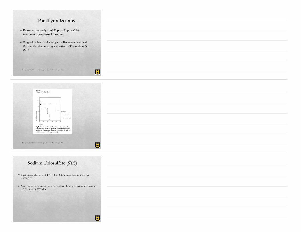
! Retrospective analysis of 35 pts – 23 pts (66%) underwent a parathyroid resection
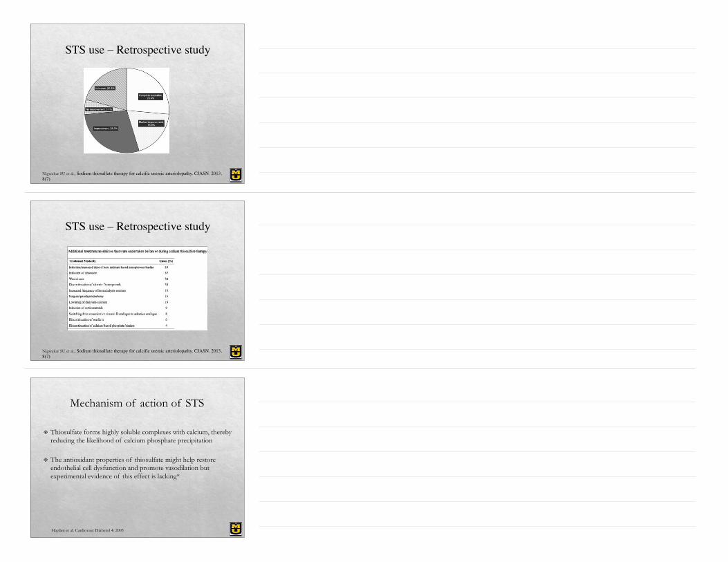
! Surgical patients had a longer median overall survival (80 months) than nonsurgical patients (35 months) (P<.001)
Therapy for calciphylaxis: an outcome analysis. Arch Ferrer JE et al., Surgery 2003
Therapy for calciphylaxis: an outcome analysis. Arch Ferrer JE et al., Surgery 2003
Sodium Thiosulfate (STS)
! First successful use of IV STS in CUA described in 2005 by Cicone et al.
! Multiple case reports/ case series describing successful treatment of CUA with STS since
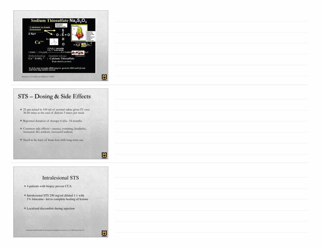
STS use – Retrospective study
Nigwekar SU et al., Sodium thiosulfate therapy for calcific uremic arteriolopathy. CJASN. 2013, 8(7)
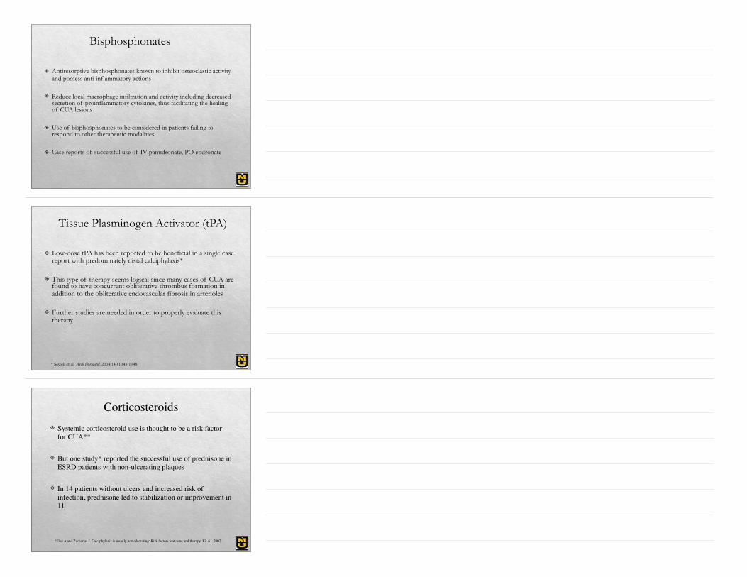
STS use – Retrospective study
Nigwekar SU et al., Sodium thiosulfate therapy for calcific uremic arteriolopathy. CJASN. 2013, 8(7)
Mechanism of action of STS
! Thiosulfate forms highly soluble complexes with calcium, thereby reducing the likelihood of calcium phosphate precipitation
! The antioxidant properties of thiosulfate might help restore endothelial cell dysfunction and promote vasodilation but experimental evidence of this effect is lacking*
Hayden et al. Cardiovasc Diabetol 4: 2005
Hayden et al. Cardiovasc Diabetol 4: 2005
! 25 gm mixed in 100 ml of normal saline given IV over 30-60 mins at the end of dialysis 3 times per week
! Reported duration of therapy 6 wks- 34 months
! Common side effects – nausea, vomiting, headache, increased AG acidosis, increased sodium
! Need to be wary of bone loss with long term use
STS – Dosing & Side Effects
Intralesional STS! 4 patients with biopsy proven CUA
! Intralesional STS 250 mg/ml diluted 1:1 with 1% lidocaine– led to complete healing of lesions
! Localized discomfort during injection
Intralesional sodium thiosulfate for the treatment of calciphylaxis. Strazzula L et al., JAMA Dermatology 2013
Bisphosphonates
! Antiresorptive bisphosphonates known to inhibit osteoclastic activity and possess anti-inflammatory actions
! Reduce local macrophage infiltration and activity including decreased secretion of proinflammatory cytokines, thus facilitating the healing of CUA lesions
! Use of bisphosphonates to be considered in patients failing to respond to other therapeutic modalities
! Case reports of successful use of IV pamidronate, PO etidronate
Tissue Plasminogen Activator (tPA)
! Low-dose tPA has been reported to be beneficial in a single case report with predominately distal calciphylaxis*
! This type of therapy seems logical since many cases of CUA are found to have concurrent obliterative thrombus formation in addition to the obliterative endovascular fibrosis in arterioles
! Further studies are needed in order to properly evaluate this therapy
* Sewell et al. Arch Dermatol. 2004;140:1045-1048
Corticosteroids! Systemic corticosteroid use is thought to be a risk factor
for CUA**
! But one study* reported the successful use of prednisone in ESRD patients with non-ulcerating plaques
! In 14 patients without ulcers and increased risk of infection, prednisone led to stabilization or improvement in 11
*Fine A and Zacharias J. Calciphylaxis is usually non-ulcerating: Risk factors, outcome and therapy. KI, 61, 2002
Treatment Outcomes
Patient 1
Before treatment
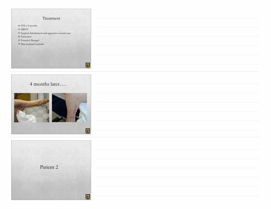
Treatment! STS x 5 months ! HBOT ! Surgical debridement and aggressive wound care ! Cinacalcet ! Fosrenol, Renagel ! Discontinued warfarin
4 months later….
Patient 2

Treatment! Switched from PD to HD ! STS x 2 months ! Wound care ! Cinacalcet ! Discontinued warfarin ! Pt’s lesions progressed with deterioration of his overall
condition. Expired 4 months after diagnosis
Summary
! CUA is a serious condition with high morbidity and mortality ! Multidisciplinary approach is key ! STS should be considered as first-line therapy in addition to
aggressive wound management and calcium-phosphorus control ! Other therapies such as bisphosphonates, corticosteroids, low dose
tPA may be tried if initial therapy fails ! Evolving role of Vitamin K supplementation ! High mortality despite treatment
Thank You!