Embed Size (px)
Citation preview
AbstrAct
Tuberculosis of the calvarium is an uncommon entity with increasing
incidence of late. Only anecdotal reports describing its imaging features have been previously published in the literature. We report the role of conventional radiogra-phy, CT and especially MRI findings in the evaluation of calvarial tuberculosis
in 3 cases. Presence of adjacent liquefied soft tissue with minimal hyperintensity on T1 weighted images may give a clue
to the diagnosis.
Key words: Calvarium, tuberculosis, radiography, CT scan, MRI
IntroductIon Skull tuberculosis is rare and is re-ported to occur in only 0.01% of patients with mycobacterial infections. Before the advent of effective chemotherapy, calvarial tuberculosis was estimated to represent 0.2-1.3% of all cases of skel-etal tuberculosis (1). Although rare, inci-dence of tuberculosis of calvaria is on rise in the developing countries, mainly attributed to rampant malnutrition, poor socioeconomic conditions, and immuno-deficiency syndromes. It most commonly involves the frontal and parietal bones (2). It is important to know imaging features of this condition, since early diagnosis can prevent unwanted neurological com-plications and effective therapy is avail-able, leading to complete cure.
cAse reports:
DR. JESHIL R. SHAH*, M.D.*Consultant Radiologist, G. T. Hospital, CT and MRI department,Mumbai, Maharashtra, India.E-MAIL – [email protected]
calvarial tuberculosis features in 3 cases
Case 1: A 28 year-old girl from middle class family presented with a midline parietal scalp swelling noticed inciden-tally, one month prior to admission, after a minor trauma. The swelling was 4 cm x 3cm in size, was non-tender and there were no signs of inflammation. The scalp over the swelling was normal. There was an impulse on coughing and a pal-pable calvarial defect. General physical and routine hematological examination revealed no abnormality. Erythrocyte sedimentation rate (ESR) was 32 mms in the first hour. HIV status was negative. Radiograph of the skull revealed a lytic area in the supero-posterior right parietal bone with sclerosis of edges. MRI (fig-ure 1) showed a bony defect involving both tables of the skull, just to the right of the sagittal suture, involving medial superior-posterior aspect of right pari-etal bone. A soft tissue collection was seen on both sides of calvarium, which appeared hyperintense on T2 weighted images and minimally hyperintense on T1 weighted images as well. Marrow hyperintensity was also noted in adjacent skull. There was a small epidural soft tis-sue component indenting the underlying
brain parenchyma. The superior sagittal sinus showed normal flow void. The lesion was exposed and ex-cised piecemeal. The contents of the lesion resembled the cheesy material. Defect in calvarial bone was repaired with iliac bone graft. Histo-pathological examination confirmed tuberculous osteomyelitis. The patient was treated with anti-tuberculous drugs. The patient recovered clinically and gained weight.
Case 2: A 10-year-old girl from lower socioeconomic strata presented with history of generalized tonic clonic con-vulsions and weakness in left side of the body, since 3 months. There was altered consciousness and drowsiness, since 2 days prior to admission. There was his-tory of development of swelling over the right parietal region, since 15 days, which measured 3 x 3 cms. History of progressive weight loss and low-grade intermittent fever for six months was noted. She had received several courses of oral antibiotics, anticonvulsants and local ointments from private practitio-ners, prior to reporting to our hospital. She was BCG vaccinated and there was no history of contact with tuberculo-sis in the family. Cervical and axillary lymphadenopathy was present. Power was reduced (3/5) in the left upper and lower limbs with positive Babinki’s sign.
WEMJ Volume 112 No 1 Article 2 March 2013
ADDRESS FOR CORRESPONDENCE:
Jeshil R. Shah, M.D.Consultant radiologist: Gokuldas Tejpal Spec-trum CT MRI Centre, Near Crawford Market, Mumbai 400001. Phone: 91-22-2262 6226/ 91-22-43538100Fax: 91-22-43538102Email: [email protected]: 39. Silver Gold, S.V. P. Road, Borivali (West), Mumbai 400092.Mobile: 91-9224455612, 91-9920087667
Fig. 1: Case 1: MR coronal T1 (A), STIR (B), FLAIR (C) and sagittal T2 (D) and T1 (E) weighted images, showing calvarial bony de-fect, marrow changes and adjacent external as well as extra-dural collections.
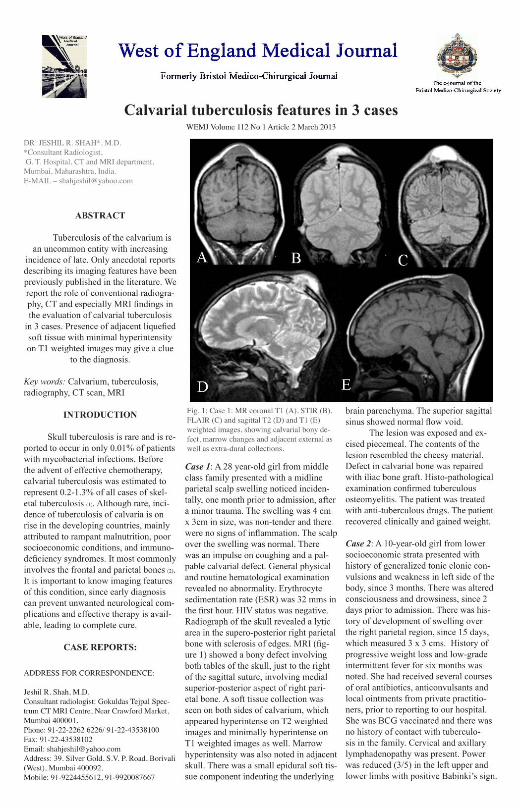
No cranial nerve involvement was seen. Laboratory data showed hemoglobin of 8 g/dl. ESR was elevated (92 / 1st hour) and HIV test was negative. Tuberculin skin test was positive with 20 _ 25 mm induration with vesiculation. Chest ra-diograph was normal. Skull radiograph showed osteoporosis and permeative type osteolytic lesions in the right pari-etal bone. MRI revealed a bunch of co-alescing lesions, appearing hypointense on T2 and iso to minimally hyperintense on T1 weighted images, showing intense contrast enhancement, suggestive of tuberculomas, in the right parietal lobe of the brain. There was marked perifocal edema, adjacent to these lesions, causing midline shift. There was irregularity of adjacent skull tables with sclerotic and hyperintense areas in the marrow Over-lying soft tissue swelling was seen over the right parietal bone region. FNAC of
this swelling was done and tubercular etiology was established. The patient was started on anti-tubercular treatment with mannitol, steroids and anti-convulsants. There was marked improvement in the patient’s condition over 20 days of hospital stay. She was dis-charged with advice to take anti Koch’s / tubercular treatment (AKT) and anticonvulsants. Frequency of convulsions has decreased, scalp swelling dis-appeared and she has gained weight, at 4 months follow up.
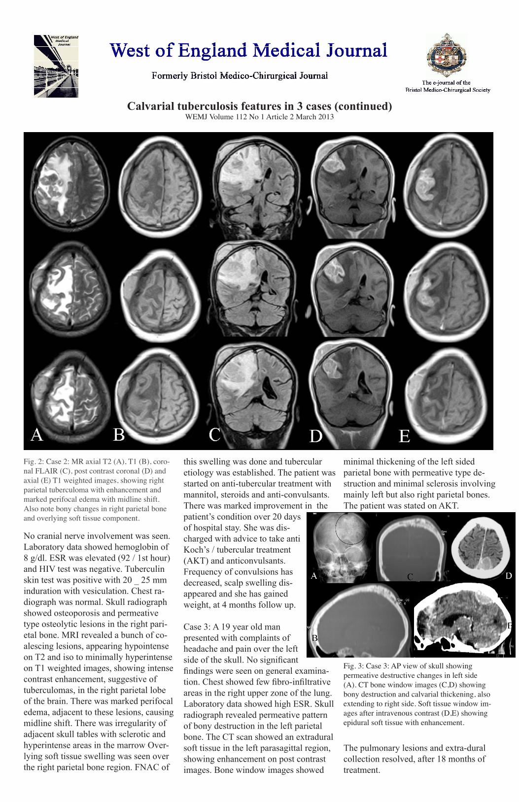
Case 3: A 19 year old man presented with complaints of headache and pain over the left side of the skull. No significant findings were seen on general examina-tion. Chest showed few fibro-infiltrative areas in the right upper zone of the lung. Laboratory data showed high ESR. Skull radiograph revealed permeative pattern of bony destruction in the left parietal bone. The CT scan showed an extradural soft tissue in the left parasagittal region, showing enhancement on post contrast images. Bone window images showed
minimal thickening of the left sided parietal bone with permeative type de-struction and minimal sclerosis involving mainly left but also right parietal bones. The patient was stated on AKT.
The pulmonary lesions and extra-dural collection resolved, after 18 months of treatment.
calvarial tuberculosis features in 3 cases (continued)WEMJ Volume 112 No 1 Article 2 March 2013
Fig. 2: Case 2: MR axial T2 (A), T1 (B), coro-nal FLAIR (C), post contrast coronal (D) and axial (E) T1 weighted images, showing right parietal tuberculoma with enhancement and marked perifocal edema with midline shift. Also note bony changes in right parietal bone and overlying soft tissue component.
Fig. 3: Case 3: AP view of skull showing permeative destructive changes in left side (A). CT bone window images (C,D) showing bony destruction and calvarial thickening, also extending to right side. Soft tissue window im-ages after intravenous contrast (D,E) showing epidural soft tissue with enhancement.
dIscussIon
Tuberculosis accounts for major health problems in developing countries with enormous social and economic impact. Also in developed countries, where tuber-culosis has been largely controlled, it poses fresh health care challenges due to migra-tion of people from the developing world and HIV infection. This has resulted in a worldwide resurgence of tuberculosis. 1.5% of the Indian population is infected with tuberculosis (2).
Tuberculosis of the skull, especially calva-rium is uncommon. Most cases occur in the first two decades; however infants are rarely affected, probably because of the paucity of cancellous bone in the skull (3). Clinical presentation depends on the immunity of the individual. Once the marrow of diploe is infected, the infection spreads towards inner and outer tables, causing bone destruc-tion and formation of granulation tissue. Extension of infection through the diploe is resisted by proliferation of encircling layer of fibrosis and if the process is not arrested, extension through either table occurs. If the process is rapid, sequestration with caseous material formation may occur. Involve-ment of the outer table is usually associ-ated with scalp swelling or a discharging sinus while involvement of the inner table results in extra-dural granulation tissue. The dura acts as a barrier to further spread; however, intradural involvement is occa-sionally seen. A good immunity will cause slow and restricted evolution of the lesion, while decreased resistance will rapidly lead to subgaleal (Pott’s puffy tumor) or extra-dural collections. Rare forms of presentation include seizures, motor deficits, meningitis, and non-specific headache (4,1). Different radiological forms of calvarial tuberculosis described are: circum-scribed lesions of the sclerotic and lytic type and diffuse tuberculosis of the cranium (3,5). “Perforating tuberculosis of the skull” is a term used to describe small punched-out le-sions with granulation tissue covering both the inner and outer tables of the calvaria. There is little tendency to spread and hence is not associated with a periosteal reaction. These lesions are commonly known as “cir-cumscribed lytic lesions.” Term “diffuse tuberculosis of the cranium” is used for le-sions causing widespread destruction of the inner table of the skull. When these lesions are associated with extradural granulation tissue, they have been redefined as “spread-ing-type” lesions. The least common lesion is the “circumscribed sclerotic type” with reactive sclerosis. Cold abscesses are com-
monly associated with this form. The differ-ential diagnosis of multiple osteolytic le-sions of the skull would include metastases, multiple myeloma, Paget’s disease, histio-cytosis, hyperparathyroidism and pyogenic, fungal, or tuberculous osteomyelitis (3,6). Investigations for these cases should include the work-up for primary malignancy, my-eloma, tuberculosis, or other systemic infec-tions and chronic inflammatory disease. CT can demonstrate soft tissue swelling with accompanying bony destruc-tion of one or both skull tables with bony sequestrum. It also shows disease spread to the extradural space, meninges and brain parenchyma. Epidural granulation tissue or abscess appears as crescentic or lentiform low-attenuation collection. The surround-ing meninges enhance intensely following contrast medium administration. CT also reveals evidence of meningitis and paren-chymal disease (3,6,7). MR imaging is much superior in demonstrating marrow involvement and soft tissue extent of the lesions. Proton density and T2-weighted images show a high-signal-intensity soft tissue mass within the defect in bone. This may project into the subgaleal and / or epidural spaces and show peripheral capsular enhancement on the contrast-enhanced image. MR imaging is sensitive in demonstrating changes in the meninges, ventricular walls and in detecting parenchymal foci of involvement (3,7,8). All our cases showed extensively liquefied soft tissue, appearing minimally hyperintense on T1 weighted images, which may give a clue to diagnosis of tuberculosis. Alternatively, rim of abscesses may be hyperintense on T1 weighted images. Bony fragments and calci-fied foci within it may produce hypointense areas within it.Because it is not always possible to reach a conclusive diagnosis on the basis of radio-logic and clinical findings, microbiologic or histologic confirmation is essential before starting chemotherapy. The demonstration of acid-fast bacilli in pus smear by using Kinyoun or Ziehl Nelsen stain or isola-tion of mycobacteria in Lowenstein-Jensen media culture is diagnostic. Microscopic examination reveals a preponderance of lymphocytes, Langhans giant cells, and multiple epitheloid and polymorphonuclear cells with proliferating blood vessels. The presence of caseous granulomas on histo-pathologic examination provides the most conclusive evidence of tuberculosis infec-tion. Chest radiographs are positive in only less than 50% of the cases. High ESR, Man-toux (PPD) skin test, serological tests (ELI-SA), amplification and detection of specific fragments of DNA by Polymerase Chain
Reaction (PCR), response to antitubercular treatment etc. can be used for making the final diagnosis (3,6,9).
Before the advent of antituberculosis chemotherapy, surgical excision was the mainstay of treatment of calvarial tubercu-losis. However, surgery is now performed in cases with large extradural collections caus-ing neurologic deficits or large scalp swell-ings with sinus formation and fulminant secondary infections (5). In such cases, com-plete excision of diseased bone and granula-tion tissue with extirpation of the sinus tract is recommended. Initial administration of five anti-tubercular drugs with continuation for at least 18-24 months is recommended (10). Because the role of anticonvulsants is controversial, its use in most cases is avoid-ed.
conclusIon
Alhough calvarial tuberculosis is rare, the incidence of this disease is on the rise, especially with increasing prevalence of immuno-deficiency syndromes. Imaging findings, in most cases, help in establishing the diagnosis. Scalp swelling, epidural com-ponent, bone destruction, marrow changes and associated meningeal and brain changes may be clearly seen on imaging.
references
1. Strauss DC. Tuberculosis of flat bones of the vault of the skull. Surg Gynecol Obstet 1933; 57: pp 384-398. 2. Raut AA, Nagar AM, Muzumdar D, Chawla AJ, Narlawar RS, Fattepurkar S, Bhatgadde VL. Imag-ing Features of Calvarial Tuberculosis: A Study of 42 Cases. Am J Neuroradiology 2004; 25: pp 409-414.3. Bhandari B, Mandowara SL, Harish J. Tubercu-lous osteomyelitis of skull. Indian J Pediatr 1981; 48: pp 113-115.4. Tata HR. Tuberculous osteomyelitis of the skull. Indian J Tuberculosis 1978; 25: pp 208-209. 5. Jadhav RN, Palande DA. Calvarial tuberculosis. Neurosurgery 1999; 45: pp 1345-1350.6. Samson SKG, Kulkarni V, Chacko AG. An un-usual presentation of calvarial tuberculosis. Postgr Med J 2002; 78: pp 188-189.7. Patankar T, Varma R, Krishnan A, Prasad S, Desai K, Castillo M. Radiographic findings in tuberculosis of the calvarium. Neuroradiology 2000; 42(7): pp 518-521.8. Wohaibi Al-M, Russel NA, Omojola M, Feriyan Al-A. Tuberculosis of the skull. J Neurosurg 2000; 92: pp 1065.9. Ravigilione MC, O’Brien RJ. Tuberculosis. In: Harrison’s Principles of Internal Medicine. Ed. Fauci SA, Longo DL, 14th edition, 1996, McGraw Hill publishers, Newyork, pp 1004-1014.10. Goel A, Pandya SK, Satoskar AR. Whether short course chemotherapy for tuberculous meningitis? Neurosurgery 1990; 27: pp 418 -421.
calvarial tuberculosis features in 3 cases (continued)WEMJ Volume 112 No 1 Article 2 March 2013