Embed Size (px)
Citation preview
This document consists of 9 printed pages and 3 blank pages.
DC (LK/FD) 113259/5© UCLES 2016 [Turn over
Cambridge International ExaminationsCambridge Ordinary Level
*3302704712*
BIOLOGY 5090/31
Paper 3 Practical Test October/November 2016
1 hour 15 minutes
Candidates answer on the Question Paper.
Additional Materials: As listed in the Confidential Instructions.
READ THESE INSTRUCTIONS FIRST
Write your Centre number, candidate number and name on all the work you hand in.Write in dark blue or black pen.You may use an HB pencil for any diagrams or graphs.Do not use staples, paper clips, glue or correction fluid.DO NOT WRITE IN ANY BARCODES.
Answer all questions.Write your answers in the spaces provided on the Question Paper.
Electronic calculators may be used.You may lose marks if you do not show your working or if you do not use appropriate units.
At the end of the examination, fasten all your work securely together.The number of marks is given in brackets [ ] at the end of each question or part question.
For Examiner’s Use
1
2
Total
2
5090/31/O/N/16© UCLES 2016
In order to plan the best use of your time, read through all the questions on this paper carefully before starting work.
1 In order to stay alive, cells depend on soluble molecules being able to enter and leave them.
You are going to use pieces of agar, a firm jelly, to represent three cells, A, B and C. The agar is coloured red with an indicator. You will place each piece in an acid solution. When the acid diffuses into the agar, the red indicator will change to a yellow colour.
You will measure the time it takes for each piece to completely change colour.
(a) (i) Complete the column headings in Table 1.1 ready to record your results.
Include:
• the time at which the agar pieces were placed in the acid solution
• the time at which the red colour had completely changed to yellow
• the time taken for the colour to change.
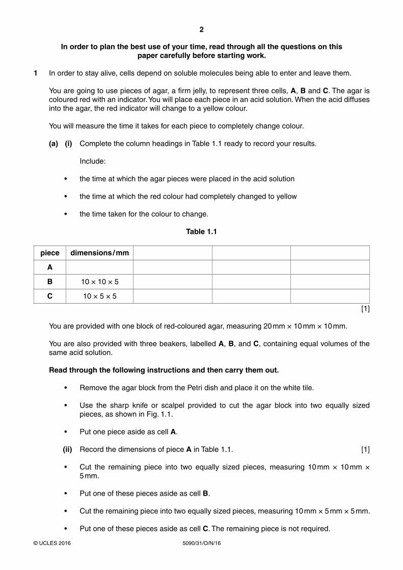
Table 1.1
piece dimensions / mm
A
B 10 × 10 × 5
C 10 × 5 × 5
[1]
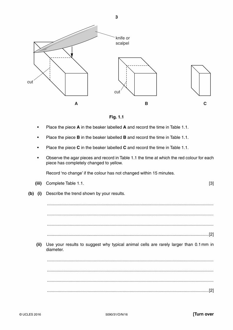
You are provided with one block of red-coloured agar, measuring 20 mm × 10 mm × 10 mm.
You are also provided with three beakers, labelled A, B, and C, containing equal volumes of the same acid solution.
Read through the following instructions and then carry them out.
• Remove the agar block from the Petri dish and place it on the white tile.
• Use the sharp knife or scalpel provided to cut the agar block into two equally sized pieces, as shown in Fig. 1.1.
• Put one piece aside as cell A.
(ii) Record the dimensions of piece A in Table 1.1. [1]
• Cut the remaining piece into two equally sized pieces, measuring 10 mm × 10 mm × 5 mm.
• Put one of these pieces aside as cell B.
• Cut the remaining piece into two equally sized pieces, measuring 10 mm × 5 mm × 5 mm.
• Put one of these pieces aside as cell C. The remaining piece is not required.
3
5090/31/O/N/16© UCLES 2016 [Turn over
Fig. 1.1
• Place the piece A in the beaker labelled A and record the time in Table 1.1.
• Place the piece B in the beaker labelled B and record the time in Table 1.1.
• Place the piece C in the beaker labelled C and record the time in Table 1.1.
• Observe the agar pieces and record in Table 1.1 the time at which the red colour for each piece has completely changed to yellow.
Record ‘no change’ if the colour has not changed within 15 minutes.
(iii) Complete Table 1.1. [3]
(b) (i) Describe the trend shown by your results.
...........................................................................................................................................
...........................................................................................................................................
...........................................................................................................................................
.......................................................................................................................................[2]
(ii) Use your results to suggest why typical animal cells are rarely larger than 0.1 mm in diameter.
...........................................................................................................................................
...........................................................................................................................................
...........................................................................................................................................
.......................................................................................................................................[2]
4
5090/31/O/N/16© UCLES 2016
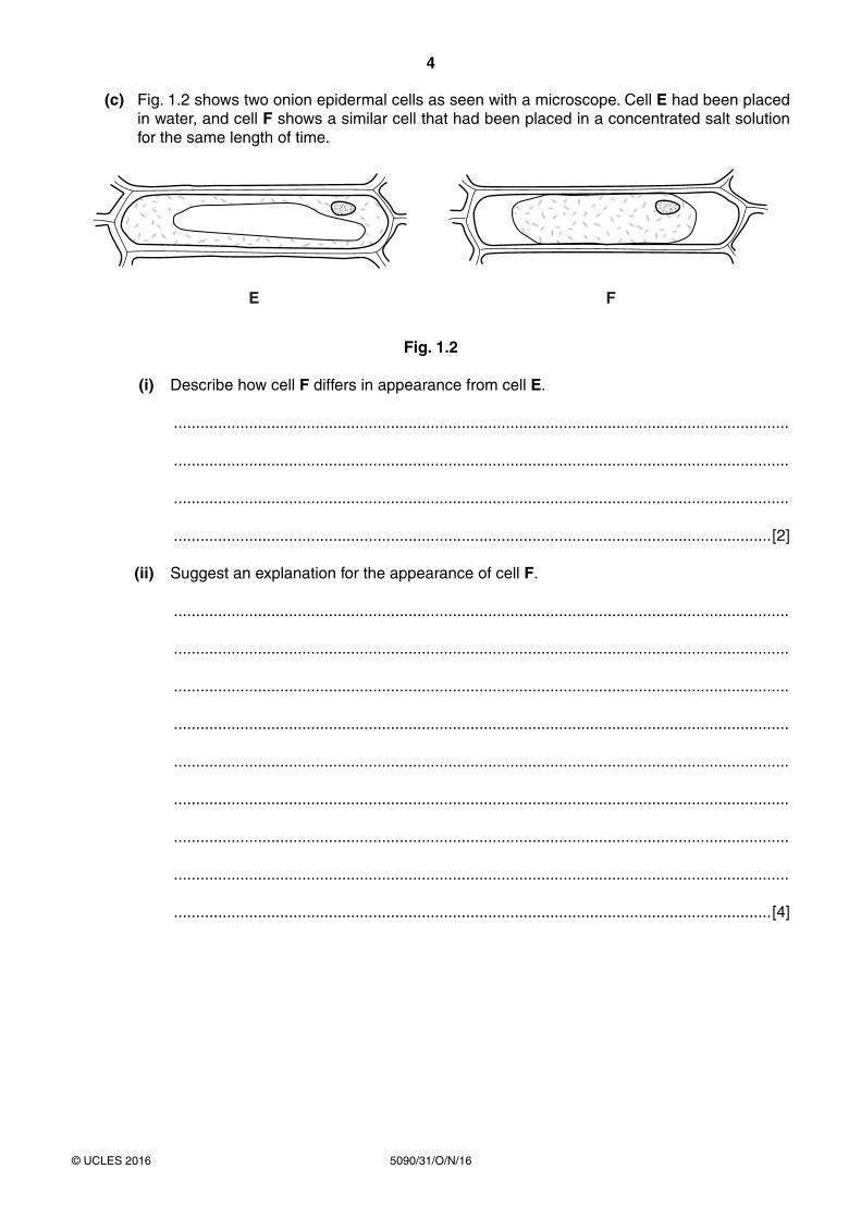
(c) Fig. 1.2 shows two onion epidermal cells as seen with a microscope. Cell E had been placed in water, and cell F shows a similar cell that had been placed in a concentrated salt solution for the same length of time.
Fig. 1.2
(i) Describe how cell F differs in appearance from cell E.
...........................................................................................................................................
...........................................................................................................................................
...........................................................................................................................................
.......................................................................................................................................[2]
(ii) Suggest an explanation for the appearance of cell F.
...........................................................................................................................................
...........................................................................................................................................
...........................................................................................................................................
...........................................................................................................................................
...........................................................................................................................................
...........................................................................................................................................
...........................................................................................................................................
...........................................................................................................................................
.......................................................................................................................................[4]

5
5090/31/O/N/16© UCLES 2016 [Turn over
(iii) Describe an investigation you could carry out to determine the concentration of salt solution that causes fresh onion epidermal cells to become like cell F.
...........................................................................................................................................
...........................................................................................................................................
...........................................................................................................................................
...........................................................................................................................................
...........................................................................................................................................
...........................................................................................................................................
...........................................................................................................................................
...........................................................................................................................................
...........................................................................................................................................
...........................................................................................................................................
...........................................................................................................................................
.......................................................................................................................................[4]
[Total: 19]
6
5090/31/O/N/16© UCLES 2016
2 Vegetarian sources of protein, for example beans and single cell proteins, are increasingly being used throughout the world.
You are provided with two bean seeds.
• Remove the testa (seed coat) carefully from one seed.
• Separate the two halves of the seed to observe the embryo.
(a) Make a large drawing of the exposed surface of the half of the bean seed that shows the embryo. Label the radicle and plumule.
[4]
(b) (i) Describe how you could test a seed for the presence of protein.
...........................................................................................................................................
...........................................................................................................................................
...........................................................................................................................................
...........................................................................................................................................
...........................................................................................................................................
...........................................................................................................................................
.......................................................................................................................................[3]
7
5090/31/O/N/16© UCLES 2016 [Turn over
(ii) Carry out the test that you have described using both of the bean seeds.
Record the result in Table 2.1.
Table 2.1
type of seed result
bean
maize
[1]
You are provided with two soaked maize grains.
• Remove the outer layers from both of the maize grains to expose the seed inside. Test the maize seeds for protein.
(iii) Record the result in Table 2.1. [1]
(iv) State your conclusion from your results.
...........................................................................................................................................
.......................................................................................................................................[1]
(v) Suggest how you could make this comparison of protein content more reliable.
...........................................................................................................................................
...........................................................................................................................................
...........................................................................................................................................
.......................................................................................................................................[2]

8
5090/31/O/N/16© UCLES 2016
Single cell protein can be produced from a fungus grown in large fermenters.
Fig. 2.1 shows some of a fungal culture as seen with a microscope, with a spore labelled Y.
Fig. 2.1
(c) (i) Measure and record the length of spore Y. ........................................................ mm [1]
(ii) Calculate the actual length of spore Y.
Show your working.
........................................................ mm [2]
Table 2.2 shows the protein content of some plant crops and single cell protein.
Table 2.2
source of protein protein content / g per 100 g
peanut 25.0
potato 2.2
rice 7.0
single cell protein 10.0
soya bean 33.7

9
5090/31/O/N/16© UCLES 2016
(d) (i) Construct a bar chart of the data in Table 2.2.
[4]
(ii) The average adult requires approximately 50 g of protein per day to maintain good health. Calculate the mass of single cell protein needed, per day, to provide 50 g of protein.
Show your working.
............................................................. g [2]
[Total: 21]

10
5090/31/O/N/16© UCLES 2016
BLANK PAGE

11
5090/31/O/N/16© UCLES 2016
BLANK PAGE

12
5090/31/O/N/16© UCLES 2016
BLANK PAGE
Permission to reproduce items where third-party owned material protected by copyright is included has been sought and cleared where possible. Every reasonable effort has been made by the publisher (UCLES) to trace copyright holders, but if any items requiring clearance have unwittingly been included, the publisher will be pleased to make amends at the earliest possible opportunity.
To avoid the issue of disclosure of answer-related information to candidates, all copyright acknowledgements are reproduced online in the Cambridge International Examinations Copyright Acknowledgements Booklet. This is produced for each series of examinations and is freely available to download at www.cie.org.uk after the live examination series.
Cambridge International Examinations is part of the Cambridge Assessment Group. Cambridge Assessment is the brand name of University of Cambridge Local Examinations Syndicate (UCLES), which is itself a department of the University of Cambridge.