Embed Size (px)
Citation preview
CASE REPORT Open Access
Candida pelliculosa endophthalmitis after cataractsurgery: a case reportHaluk Esgin*, Erkan Bulut and Çaglar Örüm
Abstract
Background: Here we report the first case of postoperative endophthalmitis due to Candida pelliculosa aftercataract surgery. We describe the clinical management of this type of candida infection in the eye.
Case presentation: A 57-year-old Turk man was seen at our clinic at the end of the first postoperative month aftercataract surgery. He presented with eye redness, pain and decreased visual acuity. His ophthalmologic examinationrevealed moderate tyndall and a mild flare in the anterior chamber. Hypopyon in the capsular bag posterior to theintraocular lens was seen in the second postoperative month. Despite topical and subconjunctival bacterialendophthalmitis treatment, there was no improvement in the clinical situation. Candida pelliculosa was isolatedfrom a sample culture obtained from the anterior chamber. Oral fluconazole could not be administered becauseof increased liver enzyme levels and intravenous amphotericin B could not be administered because of an allergicreaction. Intraocular lens explantation, pars plana vitrectomy and anterior chamber lavage by rupturing theposterior wall of the microabscesses were performed. Intravitreal and intracameral amphotericin B injections weregiven four times in addition to surgical interventions. The patient has been followed for 2 years and hisbest-corrected visual acuity was 0.4 at the last visit.
Conclusion: Nearly 1 month after cataract surgery, a patient presented with eye redness and blurred vision, withcorneal endothelial deposits, hypopyon in the capsular bag and microabscesses on the incision sites and cornealendothelium. Candida pelliculosa should be considered in patients showing these symptoms. Multiple intraocularamphotericin B (5 μg) administrations can be used safely even in cases with high sensitivity to systemic use.Rupturing the posterior wall of the abscesses on the corneal endothelium surgically with intraocular lensexplantation and pars plana vitrectomy are recommended.
BackgroundEndophthalmitis is a serious intraocular inflammatory dis-ease and the most common form is postoperative endoph-thalmitis. The incidence of post-surgical endophthalmitisis nearly 0.093% and the organisms responsible are gener-ally bacteria. Three percent of all cases with endophthal-mitis after cataract surgery are due to fungi [1,2]. Fungihave been isolated from 21.8% of all culture positive post-operative endophthalmitis cases, and mostly Aspergillusspp. and Candida spp. were determined to be the cause offungal endophthalmitis [3,4]. This is the first reported casewith endophthalmitis due to Candida pelliculosa.
Case presentationA 57-year-old Turk male was seen in our clinic with com-plaints of blurred vision in his left eye. His visual acuitywas 2/20 and he had a nucleocortical and posterior sub-capsular cataract. A history of systemic hypertension andpast coronary by-pass surgery was present. Uncomplicatedphacoemulsification surgery and foldable posterior cham-ber intraocular lens (IOL) implantation was performed.1 mg/0.1 ml intracameral cefuroxime sodium (Cefurol®1.5 g/15 ml flacon, I.E. Ulagay) was given for prophylaxis.The eye was closed with tobramycin ophthalmic ointment3 mg/g and dexamethasone ophthalmic ointment 1 mg/g.Postoperative best-corrected visual acuity (BCVA) was 16/20 with minimal corneal edema after the first week.
The patient complained of pain and eye redness for 4 dayson the 27th day after surgery. His BCVA was also decreasedto 10/20. Ophthalmic examination revealed ++++ Tyndall,
* Correspondence: [email protected] of Ophthalmology, Faculty of Medicine, Trakya University,Edirne, Turkey
© 2014 Esgin et al.; licensee BioMed Central Ltd. This is an Open Access article distributed under the terms of the CreativeCommons Attribution License (http://creativecommons.org/licenses/by/2.0), which permits unrestricted use, distribution, andreproduction in any medium, provided the original work is properly credited.
Esgin et al. BMC Research Notes 2014, 7:169http://www.biomedcentral.com/1756-0500/7/169
no hypopyon and ++ flare in the anterior chamber. In-traocular pressure was 14 mmHg OD; 17 mmHg OS.Fundus examination results were normal and the patienthad no pain. Late onset toxic anterior segment syndrome(TASS) was suspected. Dexamethasone sodium phosphateeye drops 0.1% (Onadron® collyrium 1 ml, I.E. Ulagay)were given hourly, netilmicin sulfate eye drops 0.38%(Netira® collyrium, SIFI) were given 4 times a day andcyclopentolate HCl eye drops 1% (Sikloplejin® col-lyrium, Abdi Ibrahim) were given 3 times a day as atopical treatment.
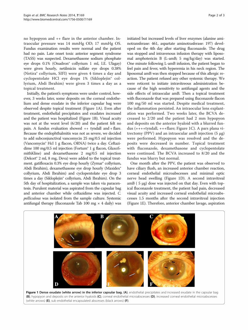
Initially, the patient’s symptoms were under control, how-ever, 3 weeks later, some deposits on the corneal endothe-lium and dense exudate in the inferior capsular bag wereobserved despite topical treatment (Figure 1A). Even aftertreatment, endothelial precipitates and exudates increasedand the patient was hospitalized (Figure 1B). Visual acuitywas not at the worst level (6/20) and the patient felt nopain. A fundus evaluation showed ++ tyndall and + flare.Because the endophthalmitis was not as severe, we decidedto add subconjunctival vancomycin 25 mg/0.5 ml injection(Vancomycin® Hcl 1 g flacon, ORNA) twice a day. Ceftazi-dime 100 mg/0.5 ml injection (Fortum® 1 g flacon, GlaxoS-mithKline) and dexamethasone 2 mg/0.5 ml injection(Dekort® 2 ml, 8 mg, Deva) were added to the topical treat-ment. gatifloxacin 0.3% eye drop hourly (Zymar® collyrium,Abdi Ibrahim), dexamethasone eye drop hourly (Maxidex®collyrium, Abdi Ibrahim) and cyclopentolate eye drop 3times a day (Sikloplejin® collyrium, Abdi Ibrahim). On the5th day of hospitalization, a sample was taken via paracen-tesis. Purulent material was aspirated from the capsular bagand anterior chamber while ceftazidime was injected. C.pelliculosa was isolated from the sample culture. Systemicantifungal therapy (fluconazole Tab 100 mg × 4 daily) was
initiated but increased levels of liver enzymes (alanine ami-notransferase: 461, aspartate aminotrasferase: 197) devel-oped on the 4th day after starting fluconazole. The drugwas stopped and intravenous infusion therapy with liposo-mal amphotericin B (L-amB; 5 mg/kg/day) was started.One minute following L-amB infusion, the patient began tofeel pain and fever, with hyperemia in his neck region. Theliposomal amB was then stopped because of this allergic re-action. The patient refused any other systemic therapy. Wewere reticent to initiate intravitreous administration be-cause of the high sensitivity to antifungal agents and theside effects of intraocular amB. Then a topical treatmentwith fluconazole that was prepared using fluconazole flacon100 mg/50 ml was started. Despite medical treatment,the inflammation persisted. An intraocular lens explant-ation was performed. Two weeks later, the BCVA de-creased to 2/20 and the patient had 2 mm hypopyonand deposits on the anterior hyaloid with a blurred fun-dus (++++tyndall, +++flare; Figure 1C). A pars plana vi-trectomy (PPV) and an intraocular amB injection (5 μg)were performed. Hypopyon was resolved and the de-posits were decreased in number. Topical treatmentwith fluconazole, dexamethasone and cyclopentolatewere continued. The BCVA increased to 8/20 and thefundus was blurry but normal.
One month after the PPV, the patient was observed tohave ciliary flush, an increased anterior chamber reaction,corneal endothelial microabscesses and minimal opticnerve head swelling (Figure 1D). A second intravitrealamB ( 5 μg) dose was injected on that day. Even with top-ical fluconazole treatment, the patient had pain, decreasedvisual acuity and increased corneal endothelial microabs-cesses 1.5 months after the second intravitreal injection(Figure 1E). Therefore, anterior chamber lavage, aspiration
Figure 1 Dense exudate (white arrow) in the inferior capsular bag. (A), endothelial precipitates and increased exudate in the capsular bag(B), hypopyon and deposits on the anterior hyaloids (C), corneal endothelial microabscesses (D), increased corneal endothelial microabscesses(white arrows) (E), sub endothelial encapsulated abscesses (black arrows) (F).
Esgin et al. BMC Research Notes 2014, 7:169 Page 2 of 3http://www.biomedcentral.com/1756-0500/7/169
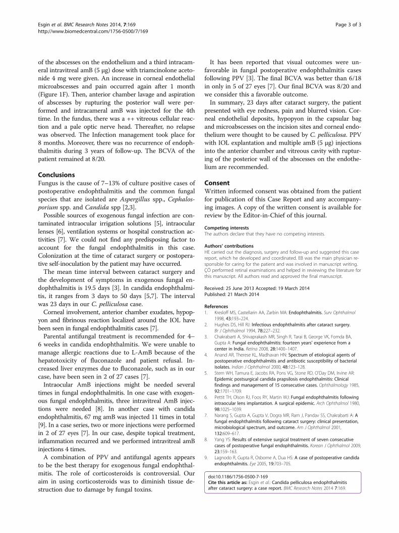
of the abscesses on the endothelium and a third intracam-eral intravitreal amB (5 μg) dose with triamcinolone aceto-nide 4 mg were given. An increase in corneal endothelialmicroabscesses and pain occurred again after 1 month(Figure 1F). Then, anterior chamber lavage and aspirationof abscesses by rupturing the posterior wall were per-formed and intracameral amB was injected for the 4thtime. In the fundus, there was a ++ vitreous cellular reac-tion and a pale optic nerve head. Thereafter, no relapsewas observed. The Infection management took place for8 months. Moreover, there was no recurrence of endoph-thalmitis during 3 years of follow-up. The BCVA of thepatient remained at 8/20.
ConclusionsFungus is the cause of 7–13% of culture positive cases ofpostoperative endophthalmitis and the common fungalspecies that are isolated are Aspergillus spp., Cephalos-porium spp. and Candida spp [2,3].
Possible sources of exogenous fungal infection are con-taminated intraocular irrigation solutions [5], intraocularlenses [6], ventilation systems or hospital construction ac-tivities [7]. We could not find any predisposing factor toaccount for the fungal endophthalmitis in this case.Colonization at the time of cataract surgery or postopera-tive self-inoculation by the patient may have occurred.
The mean time interval between cataract surgery andthe development of symptoms in exogenous fungal en-dophthalmitis is 19.5 days [3]. In candida endophthalmi-tis, it ranges from 3 days to 50 days [5,7]. The intervalwas 23 days in our C. pelliculosa case.
Corneal involvement, anterior chamber exudates, hypop-yon and fibrinous reaction localized around the IOL havebeen seen in fungal endophthalmitis cases [7].
Parental antifungal treatment is recommended for 4–6 weeks in candida endophthalmitis. We were unable tomanage allergic reactions due to L-AmB because of thehepatotoxicity of fluconazole and patient refusal. In-creased liver enzymes due to fluconazole, such as in ourcase, have been seen in 2 of 27 cases [7].
Intraocular AmB injections might be needed severaltimes in fungal endophthalmitis. In one case with exogen-ous fungal endophthalmitis, three intravitreal AmB injec-tions were needed [8]. In another case with candidaendophthalmitis, 67 mg amB was injected 11 times in total[9]. In a case series, two or more injections were performedin 2 of 27 eyes [7]. In our case, despite topical treatment,inflammation recurred and we performed intravitreal amBinjections 4 times.
A combination of PPV and antifungal agents appearsto be the best therapy for exogenous fungal endophthal-mitis. The role of corticosteroids is controversial. Ouraim in using corticosteroids was to diminish tissue de-struction due to damage by fungal toxins.
It has been reported that visual outcomes were un-favorable in fungal postoperative endophthalmitis casesfollowing PPV [3]. The final BCVA was better than 6/18in only in 5 of 27 eyes [7]. Our final BCVA was 8/20 andwe consider this a favorable outcome.
In summary, 23 days after cataract surgery, the patientpresented with eye redness, pain and blurred vision. Cor-neal endothelial deposits, hypopyon in the capsular bagand microabscesses on the incision sites and corneal endo-thelium were thought to be caused by C. pelliculosa. PPVwith IOL explantation and multiple amB (5 μg) injectionsinto the anterior chamber and vitreous cavity with ruptur-ing of the posterior wall of the abscesses on the endothe-lium are recommended.
ConsentWritten informed consent was obtained from the patientfor publication of this Case Report and any accompany-ing images. A copy of the written consent is available forreview by the Editor-in-Chief of this journal.
Competing interestsThe authors declare that they have no competing interests.
Authors’ contributionsHE carried out the diagnosis, surgery and follow-up and suggested this casereport, which he developed and coordinated. EB was the main physician re-sponsible for caring for the patient and was involved in manuscript writing.ÇÖ performed retinal examinations and helped in reviewing the literature forthis manuscript. All authors read and approved the final manuscript.
Received: 25 June 2013 Accepted: 19 March 2014Published: 21 March 2014
References1. Kresloff MS, Castellarin AA, Zarbin MA: Endophthalmitis. Surv Ophthalmol
1998, 43:193–224.2. Hughes DS, Hill RJ: Infectious endophthalmitis after cataract surgery.
Br J Ophthalmol 1994, 78:227–232.3. Chakrabarti A, Shivaprakash MR, Singh R, Tarai B, George VK, Fomda BA,
Gupta A: Fungal endophthalmitis: fourteen years' experience from acenter in India. Retina 2008, 28:1400–1407.
4. Anand AR, Therese KL, Madhavan HN: Spectrum of etiological agents ofpostoperative endophthalmitis and antibiotic susceptibility of bacterialisolates. Indian J Ophthalmol 2000, 48:123–128.
5. Stern WH, Tamura E, Jacobs RA, Pons VG, Stone RD, O’Day DM, Irvine AR:Epidemic postsurgical candida prapsilosis endophthalmitis: Clinicalfindings and management of 15 consecutive cases. Ophthalmology 1985,92:1701–1709.
6. Pettit TH, Olson RJ, Foos RY, Martin WJ: Fungal endophthalmitis followingintraocular lens implantation. A surgical epidemic. Arch Ophthalmol 1980,98:1025–1039.
7. Narang S, Gupta A, Gupta V, Dogra MR, Ram J, Pandav SS, Chakrabarti A: Afungal endophthalmitis following cataract surgery: clinical presentation,microbiological spectrum, and outcome. Am J Ophthalmol 2001,132:609–617.
8. Yang YS: Results of extensive surgical treatment of seven consecutivecases of postoperative fungal endophthalmitis. Korean J Ophthalmol 2009,23:159–163.
9. Lagnodo R, Gupta R, Osborne A, Dua HS: A case of postoperative candidaendophthalmitis. Eye 2005, 19:703–705.
doi:10.1186/1756-0500-7-169Cite this article as: Esgin et al.: Candida pelliculosa endophthalmitisafter cataract surgery: a case report. BMC Research Notes 2014 7:169.
Esgin et al. BMC Research Notes 2014, 7:169 Page 3 of 3http://www.biomedcentral.com/1756-0500/7/169