Embed Size (px)
Citation preview

The Plant Cell, Vol. 5, 953-961, August 1993 O 1993 American Society of Plant Physiologists
Carbon Fixation Gradients across Spinach Leaves Do Not Follow Interna1 Light Gradients
John N. Nishio,’ Jindong Sun, and Thomas C. Vogelmann
Department of Botany, University of Wyoming, Laramie, Wyoming 82071
In situ measurements of 14C-C02 incorporation into 40-pm paradermal leaf sections of sun- and shade-grown spinach leaves were determined. Chlorophyll, carotenoid, and ribulose-l,5-bisphosphate carboxylase/oxygenase (Rubisco) con- tent in similar 40-pm paradermal leaf sections was also measured. The carbon fixation gradient did not follow the leaf interna1 light gradient, which decreases exponentially across the leaf. Instead, the 14C-C02 fixation was higher in the middle of the leaf. Contrary to expectations, the distribution of carbon fixation across the leaf showed that the spongy mesophyll contributes significantly to the total carbon reduced. Approximately 60% of the carboxylation occurred in the palisade mesophyll and 40% occurred in the spongy mesophyll. Carbon reduction correlated well with Rubisco con- tent, and no correlation between chlorophyll and carotenoid content and Rubisco was observed in sun plants. The correlation among chlorophyll, carotenoids, Rubisco, and carbon fixation was higher in shade leaves than in sun leaves. The results are discussed in relation to leaf photosynthetic and biochemical measurements that generally consider the leaf as a single homogeneous unit.
INTRODUCTION
Leaves are composed of many different tissues (e.g., Fahn, 1974). The major photosynthetic tissue of C3 leaves is the mesophyll, which is separated into two types. The palisade mesophyll (PM), which may be one-to-severa1 cell layers thick, is composed of rod-shaped cells, with the long axis perpen- dicular to the leaf surface. Underlying the PM is the spongy mesophyll (SM), which is composed of loosely packed, irregu- larly shaped cells. The SM is typified by large air cavities.
Leaf anatomy and photosynthetic characteristics depend on the species and the light conditions during growth (Wylie, 1949, 1951; Loach, 1967; Alberte et al., 1976; Boardman, 1977; Anderson, 1986; Baker and McKiernan, 1988; Cui et al., 1991). Typically, leaves grown in the shade, compared to leaves grown in full sun, have fewer layers of PM and are thinner (Wylie, 1951; Cui et al., 1991). Additionally, shade leaves have less chlorophyll on an areal basis, a lower chlorophyll a/b ratio, lower rates of electron transport per unit of chlorophyll, lower COn fixation rates on both an areal and a chlorophyll basis, a lower light compensation point, and an increased photosystem II (PSl1)lphotosystem I (PSI) ratio (Boardman, 1977; Lichtenthaler et al., 1981; Barber, 1985; Anderson, 1986; Mustardy et al., 1990; McKiernan and Baker, 1991). It appears that the quality as well as the quantity of light influence leaf photosynthetic characteristics and anatomy, but the mechanisms by which the influence is exerted are not known. In general, the bio- chemical studies mentioned above treated the leaf as a unit and differences between the PM and SM were not emphasized.
To whom correspondence should be addressed.
Light impinging on the adaxial leaf surface is rapidly attenu- ated in the upper 20% of the leaf (Terashima and Saeki, 1983; Vogelmann et al., 1989; Cui et al., 1991). The rapid attenua- tion of light and change in light quality inside the leaf (Gates et al., 1965; Vogelmann et al., 1989) apparently affect the pho- tosynthetic characteristics of chloroplasts within the leaf, and there is evidence that shade-type chloroplasts exist in the abax- ia1 portion of sun leaves (Outlaw, 1987; Terashima, 1989). Thus, the measured photosynthetic characteristics of sun and shade leaves represent the sum of the dissimilar chloroplasts that developed in different light regimes within the leaf.
Light gradients across leaves are relatively steep, and models of carbon fixation across leaves generally predict that carbon fixation corresponds to the light gradient (Gutschik, 1984; Terashima, 1989; Fukshansky and von Remisowsky, 1992; Evans et al., 1993). In Contrast to the models that imply that the PM is the major site of carbon fixation, measurements of COn fixation in the SM and PM of intact leaves suggest that the SM can contribute up to 50% of the carbon fixation (Mokronosov et al., 1973; Outlaw and Fisher, 1975b). Other measurements have shown approximately three times more 14C-C02 incorporation into the PM than into the SM (Outlaw and Fisher, 1975a).
We are interested in how component mesophyll cell. layers adjust their metabolism to allow maximal performance under the saturating and nonsaturating light conditions in which plants exist. The regulation of the genes responsible for optimal per- formance is likely influenced by light and other signals. To properly address the problem, however, a more lucid view of
Dow
nloaded from https://academ
ic.oup.com/plcell/article/5/8/953/5984586 by guest on 16 February 2022
954 The Plant Cell
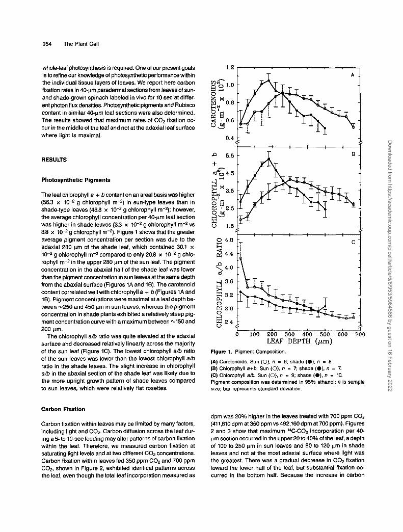
whole-leaf photosynthesis is required. One of our present goals is to refine our knowledge of photosynthetic performance within the individual tissue layers of leaves. We report here carbon fixation rates in 40-pm paradermal sections from leaves of sun- and shade-grown spinach labeled in vivo for 10 sec at differ- ent photon flux densities. Photosynthetic pigments and Rubisco content in similar 40ym leaf sections were also determined. The results showed that maximum rates of C02 fixation oc- cur in the middle of the leaf and not at the adaxial leaf surface where light is maximal.
RESULTS
Photosynthetic Pigments
The leaf chlorophyll a + b content on an areal basis was higher (56.3 x 10⁻² g chlorophyll m-2) in sun-type leaves than in shade-type leaves (48.8 x 10⁻² g chlorophyll m2); however, the average chlorophyll concentration per 40-pm leaf section was higher in shade leaves (3.3 x 10⁻² g chlorophyll m-2 vs 3.8 x 10⁻² g chlorophyll m2). Figure 1 shows that the greater average pigment concentration per section was due to the adaxial 280 pm of the shade leaf, which contained 30.1 x 10⁻² g chlorophyll m-2 compared to only 20.8 x 10⁻² g chlo- rophyll m2 in the upper 280 pm of the sun leaf. The pigment concentration in the abaxial half of the shade leaf was lower than the pigment concentration in sun leaves at the same depth from the abaxial surface (Figures 1A and 1B). The carotenoid content correlated well with chlorophyll a + b (Figures 1A and 16). Pigment concentrations were maximal ata leaf depth be- tween m250 and 450 pm in sun leaves, whereas the pigment concentration in shade plants exhibited a relatively steep pig- ment concentration curve with a maximum between 4 5 0 and 200 pm.
The chlorophyll a/b ratio was quite elevated at the adaxial surface and decreased relatively linearly across the majority of the sun leaf (Figure 1C). The lowest chlorophyll a/b ratio of the sun leaves was lower than the lowest chlorophyll a/b ratio in the shade leaves. The slight increase in chlorophyll a/b in the abaxial section of the shade leaf was likely due to the more upright growth pattern of shade leaves compared to sun leaves, which were relatively flat rosettes.
Carbon Fixation
Carbon fixation within leaves may be limited by many factors, including light and COn. Carbon diffusion across the leaf dur- ing a 5- to 10-sec feeding may alter patterns of carbon fixation within the leaf. Therefore, we measured carbon fixation at saturating light levels and at two different C02 concentrations. Carbon fixation within leaves fed 350 ppm C02 and 700 ppm C02, shown in Figure 2, exhibited identical patterns across the leaf, even though the total leaf incorporation measured as
1.2 T A
-F 0.4
5.5
n + cdNg 4.5
A d g N x 3.5 a' 2 E 2.5 O M c-7-
1.5
2 4.8 b 2 4 . 4
P \ 4.0 al L1
GJ 3.6 * 5: 3.2 D-4 O ffi 2.8 O J 5: 2.4 V
O 100 200 300 400 500 600 700 LEAF DEPTH (pm)
Figure 1. Pigment Composition.
(A) Carotenoids. Sun (O), n = 6; shade (O), n = 8. (E) Chlorophyll a+b. Sun (O), n = 7; shade (O), n = 7. (C) Chlorophyll a/b. Sun (O), n = 9; shade (O), n = 10. Pigment composition was determined in 95% ethanol; n is sample size; bar represents standard deviation.
dpm was 20% higher in the leaves treated with 700 ppm CO2 (411,810 dpm at 350 ppm vs 492,160 dpm at 700 ppm). Figures 2 and 3 show that maximum 14C-C02 incorporation per 40- pm section occurred in the upper 20 to 40% of the leaf, adepth of 100 to 250 pm in sun leaves and 80 to 120 pm in shade leaves and not at the most adaxial surface where light was the greatest. There was a gradual decrease in CO2 fixation toward the lower half of the leaf, but substantial fixation oc- curred in the bottom half. Because the increase in carbon
Dow
nloaded from https://academ
ic.oup.com/plcell/article/5/8/953/5984586 by guest on 16 February 2022

Photosynthetic Gradients across Spinach Leaves 955
n
3 j" ci 4 80 Q) p:
e, 1 0 0 r
W
60
.rl 4 ci 4 0 - X tz 0" 20
u
incorporation at 700 ppm was not accompanied by a shift in the pattern of fixation across the leaf, fixation under the steady state conditions used in our experiments does not appear to be limited by 14C-C02 diffusion.
To test the effect of light on the C02 fixation gradient, 14C- C02 incorporation at different light levels was assayed. Ac- tua1 incorporation rates showed that carbon reduction in sun plants measured at 800 and 2000 pmol photons of PAR m-2 secl (all further reference to photon flux is referred to as pmol PAR m-2 sec-l) were virtually identical, whereas there was a significant increase in fixation when shade plants were as- sayed at 2000 pmol PAR m-* secl compared to the growth conditions of 200 pmol PAR m-2 sec-l, as shown in Figure 3. Interestingly, the 10-fold increase in the photon flux density at which the C02 reduction in shade leaves was assayed in- creased C02 incorporation by only 6O%, as shown in Table 1, but did not alter the general fixation pattern across the leaf (Figure 3, closed symbols). The adaxial layers increased by -3Oo/o, whereas the abaxial layers (SM) exhibited a doubling of rates (Figure 3). In sun leaves, a 2.5-fold increase in assay light did not significantly increase the rate of fixation (Figure 3). The sun leaf is likely operating closer to saturation than a shade leaf.
-
-
-
Rubisco Content
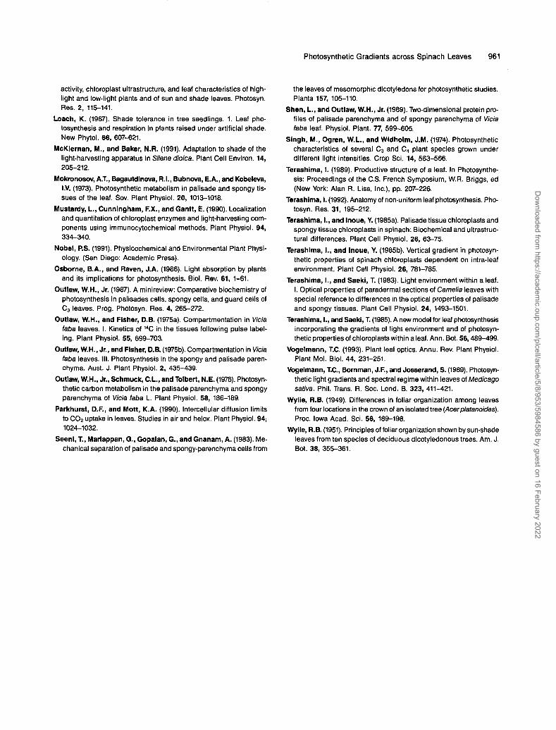
The large and small subunits (LSU and SSU) of Rubisco are easily recognizable in the polypeptide patterns from leaf sec- tions, as shown in Figure 4. The relative amount of Rubisco determined by densitometry of the Coomassie blue-stained gels, as shown in Figure 5, was similar to the data obtained
A
0; ' 20 ' ' 40 I ' 60 ' ' 80 I ' 100 I '
2-
Leaf Depth (Relative) Flgure 2. Effect of lncreased COn on Fixation Pattern across Sun Leaves of Spinach.
Sun plants were pretreated at growth conditions of 800 pmol PAR m-2 sec-l for 1 hr. A portion of the leaf was then labeled for 10 sec with either 350 ppm (O) or 700 ppm (A) 14C-C02 at 800 pmol PAR m-2 sec-l.
'0 l i 0 2d0 360 460 560 6dO 7AO LEAF DEPTH (pm)
Figure 3. COn Fixation across Sun and Shade Leaves of Spinach.
Sun plants were pretreated at growth conditions of 800 pmol PAR m2 ser1 for 1 hr. A portion of the leaf was then labeled for 10 sec with 14C-C02 at 800 pmol PAR m-p sec-1 (O), n = 10 and 2000 pmol PAR (A), n = 6. Shade plants were pretreated at growth conditions of 200 pmol PAR for 1 hr and then assayed at 200 pmol PAR m-2 sec-1 (O), n = 22 and 2000 pmol PAR (A), n = 5. Each point represents aver- age dpm f SD; sample sim given by n.
by counting SS-Met incorporated into the LSU of sun leaves, as illustrated in Figure 6. The distribution of Rubisco within the leaf was similar to the C02 fixation data, and the signifi- cant correlation between C02 fixation and Rubisco content is shown in Figure 7.
DISCUSSION
Where 1s Carbon Fixed within a Leaf?
Maximal C02 fixation occurred in the media1 section of the leaf. In contrast, the steep light gradient across leaves (Terashima and Saeki, 1983; Vogelmann et al., 1989; Cui et al., 1991) suggests that maximal C02 reduction would occur near the upper leaf surface. Carbon fixation was maximal at a depth of 100 to 250 pm in sun leaves and 80 to 120 pm in shade leaves (Figure 3). Forty percent of the carbon was fixed by the SM, and only -60% of the carbon fixation occurred in the PM, which comprises the upper 40% of the leaf. On a relative basis (percent of total carbon fixed), more fixation occurred in the PM of a shade leaf than in the PM of a sun leaf (Table 1). Studies with resolution at the SM and PM leve1 also showed a relatively high contribution of the SM to total C02 fixation when plants were labeled for 10 sec; no gradient of fixation across the leaves was presented (Mokronosov et al., 1973; Outlaw and Fisher, 1975b).
The off shifting of C02 fixation from the light gradient is sur- prising. An obvious explanation is that the C02 fixation data are flawed, the light gradient data are flawed, or both are flawed.
Dow
nloaded from https://academ
ic.oup.com/plcell/article/5/8/953/5984586 by guest on 16 February 2022

956 The Plant Cell
Table 1. Distribution of CO2 Fixation in Palisade Mesophyll andSpongy Mesophyll of Sun and Shade Leaves
GrowthConditionSunSunShadeShade
Treatment(jimolsPAR rrr2
seer1)for 1 hr
800800200200
AssayConditions
rrr2 seer1)800
2000200
2000
% TotalCO2 FixationPM
57526457
SM
43483643
It is possible that transport of products could account for thefixation in the bottom of the leaf. We attempted to minimizethe possibility of transport of fixed products by keeping fixa-tion times minimal (less than 10 sec). Conversely, we labeledlong enough to overcome diffusional limitations to CO2 andto allow significant measurable incorporation. A 5-sec pulse
followed by a 25-sec chase exhibited no statistically significantdifferences (data not shown) between a 5-sec pulse and 5-secchase. Labeling for 5 sec vs 10 sec did not alter the incorpora-tion pattern, which is consistent with calculations that indicatethat CO2 diffuses across leaves within fractions of a second(Nobel, 1991). In studies done on coltsfoot labeled for 10 secand then chased for 50 sec, the initial label in the PM decreasedonly ~20°/o (Mokronosov et al., 1973). Thus, it seems that diffu-sional limitations and transport artifacts are not influencingour labeling patterns.
Measurements from the upper leaf surface may be skewedby the paradermal sectioning technique and may account forthe lower fixation rate at the adaxial leaf surface. The data fromthe first paradermal cut represent ~80% of a full section. Theuppermost section also included the ~15-um thick epidermis,which is not photosynthetically active, so the first layer wouldbe expected to contain fewer counts. Even if we corrected forthe loss of 20% of the tissue or ignored the first two points,the CO2 fixation gradient would not be maximal at the upperleaf surface. Additionally, the pigment data collected by thesame methodology exhibit a smaller slope, and the first two
A 1 2 3 4 5 MW 6 7 8 9 10 11 12 13 14 MW15 16 17 18
bB 1 2 3 4 5 6 7 8 9 10 11 12 MW
SSl> - — _ _ -*"•
«•*
Figure 4. Rubisco Content across Sun and Shade Leaves.
(A) Sun leaf. Lanes 1 to 18 represent the most adaxial section to the most abaxial section, respectively. MW is molecular weight standards: 66,45, 36, 29, 24, 20, 14 kD.(B) Shade leaf. Lanes 1 to 12 represent the most adaxial section to the most abaxial section.Polypeptides from 40-nm paradermal leaf sections were separated by SDS-PAGE and stained with Coomassie blue (see Methods). The largeband represents LSU, and the distinct lower band represents SSU.
Dow
nloaded from https://academ
ic.oup.com/plcell/article/5/8/953/5984586 by guest on 16 February 2022

Photosynthetic Gradients across Spinach Leaves 957
n
$ .r(
100 90 80 70 60 50 O
U 40 VI E 30
Sun * Shode
'0' IÒ0 2ÔO 3dO 4ÒO 5ÒO 660 7d0 I Leaf Depth (pm)
Figure 5. Large Subunit of Rubisco across Sun and Shade Leaves.
Gels similar to the ones shown in Figure 4 were scanned, and the area under the curve representing the LSU was integrated. The maxi- mum amount of area was given a value of 100. The curves are trinomial fits (no significance to the curved tails) and represent ~ 9 3 % of the data for both sun and shade leaves.
data points fall nicely within the range of what would be predicted from the slope deeper within the leaf.
Blue light (480 nm), measured with afiber optic microprobe (Vogelmann et al., 1989; Cui et al., 1991) and by an image- analysis system (T.C. Vogelmann, J. Sun, D.A. Myers, and J.N. Nishio, unpublished data), is rapidly attenuated by the upper portion of the PM. Fiber optic measurements showed that 90% attenuation of blue and red light (680 nm) occurs within 140 pm of the adaxial leaf surface and that the SM is exposed to very low quantities of mostly green light (Cui et al., 1991). The mea- sured exponential decrease in light across leaves agrees closely with theoretical models of light gradients across leaves (Fukshansky and von Remisowsky, 1992). Thus, it seems un- likely that the deviation between COp fixation patterns and light gradients can be explained by artifacts in measuring the light gradient.
Another approach to examining the light gradient in leaves isto use the chlorophyll a/b as a bioassay of light quantity and quality. As expected, the chlorophyll a/b ratio decreased with increasing depth within the leaf. Earlier work showed that the chlorophyll a/b ratio is decreased in shade-grown plants (Lichtenthaler et al., 1981; Anderson, 1986; Baker and McKiernan, 1988) and that there is a decrease in the chlorophyll a/b ratio from the top of the leaf to the bottom, because the bottom is more shaded than the top (e.g., Outlaw, 1987; Terashima, 1989; Cui et al., 1991). If we used the chlorophyll a/b ratio as an indi- cator of the light gradient within the leaf, the light impinging on the shade leaf was similar to the light -150 pm deep in the sun leaf. Data collected by Cui et al. (1991), using a fiber optic microprobe, showed that red and blue light are attenu- ated by 90% within the first 150 pm of a sun leaf. Our sun leaves were grown at 800 pmol PAR m-2 sec-l. If the interna1 fluence rates (-2.3 times incident light at the leaf surface in spinach)
(Cui et al., 1991) for red, blue, and green light are combined, then at a depth of 150 pm there would be light approximately equivalent to the shade-grown plant (200 pmol PAR m-2 sec-l), with the red:far-red ratio being decreased. The rela- tively similar slopes in chlorophyll a/b vs leaf depth in sun and shade leaves suggest that after the initial90% attenuation of the light within the upper 150 pm of the leaf, further attenua- tion is minimal across the rest of the leaf. Only at the most abaxial section of the sun leaf did the chlorophyll a/b ratio be- come lower than in the shade leaf, suggesting that the bottom of the sun leaf received less light than the respective portion of the shade leaf. Indeed, the shade leaves grow with a slightly erect leaf orientation compared to sun leaves, which tend to grow in flat rosettes.
There appears to be little difference in the metabolic performance and capabilities of SM and PM of C3 plants (Mokronosov, et al., 1973; Outlaw and Fisher, 1975b; Outlaw et al., 1976; Seeni et al., 1983; Shen and Outlaw, 1989). Rela- tive fixation rates of 14C-C02 between the PM and SM suggest that the PM fixes more COp than the SM under light-limiting conditions (Outlaw and Fisher, 1975a), but the ratio approaches 1 at higher photon flux densities (Outlaw and Fisher, 1975b). Whereas Outlaw and Fisher (1975b) varied the light conditions under which the assays were made, another study varied the light conditions under which the plants were grown, and these investigators kept the measurement conditions constant (Seeni et al., 1983). Interestingly, the capacity for fixation (Seeni et al., 1983) followed the same pattern of fixation in the light satu- ration experiment of Outlaw and Fisher (1975b); that is, the PM:SM COn fixation ratio was higher in low light-grown plants compared to high light (full sun)-grown plants (Seeni et al.,
ld0 240 3ÒO 4ÒO 5;)O 6dO 7;O Leaf Depth (pm)
Figure 6. Large Subunit of Rubisco across Sun Leaves.
Leaf plugs were allowed to incorporate =S-Met for 1 hr, and polypep- tides from 40-pm paradermal leaf sections were separated by SDS-PAGE. incorporation into LSU was determined by cut- ting the LSU from gels and counting by liquid scintillation counting. The maxirnum amount of incorporation was given a value of 100. The synthesis of LSU corresponds well with the accumulated Rubisco shown in Figure 5.
Dow
nloaded from https://academ
ic.oup.com/plcell/article/5/8/953/5984586 by guest on 16 February 2022

958 The Plant Cell'
100 - 90- 80 70
A - -
.E 50 - af 40 - $ 30-
60 -
4 - 20 - v
o" 80 -
50 -
RUBISCO (Relative) Figure 7. Relation between 14C-COp lncorporation and Rubisco Con- tent across Sun and Shade Leaves of Spinach.
(A) Sun leaves. Regression is defined by the function, y = 1.27X - 25.6245, r2 = 0.923. (6) Shade leaves. y = l.lOX - 13.48, r2 = 0.866. Rubisco content determined by densitometry and 14C-C02 incorpo- ration (see Methods) were plotted on a relative basis with maximum amounts given a value of 100.
1983). Additionally, when 14C-C02 incorporation was deter- mined in isolated SM and PM under standard conditions, there was little difference in the fixation products and rates (Outlaw et al., 1976; Seeni et al., 1983). Analysis of specific enzymes and polypeptides in the SM and PM led to the conclusion that there is no significant difference in overall protein complement and photosynthetic carbon metabolism in the different meso- phyll cells of Vicia faba (Outlaw et al., 1976; Shen and Outlaw, 1989).
In contrast to our findings, it remains a general perception that the PM is the main site of carbon fixation. The rapid at- tenuation of light by chlorophyll has led to statements that light limits carbon reduction in the SM (Outlaw and Fisher, 1975b; Terashima and Saeki, 1985), so changes in incident flux would have a large effect on the top of the leaf but little effect in the underlying tissue (Osborne and Raven, 1986). Additionally, models of carbon fixation across a leaf generally predict that fixation follows the light gradient (e.g., Gutschik, 1984; Evans et al., 1993). In light of the work of Mokronosov et al. (1973),
Outlaw and Fisher (1975a, 1975b), and thevaried approaches to measuring the light gradient, we concluded that the measured gradients of C02 fixation are due to interactions be- tween light and other factors. New models of photosynthesis in leaves should incorporate the notion that the SM contrib- utes significantly to COn incorporation and that somehow the light gradient is disconnected from C02 fixation.
Does C02 Diffusion across the Leaf Limit C02 Fixation?
One of our experiments was aimed at determining whether the COrlabeling procedure was limited by COn diffusion across the leaf. The light gradient across leaves (Vogelmann, 1993) is steeper than the apparent COn gradient (Terashima, 1992). Typically, the PM has a greater exposed interna1 space than the SM, even though there are larger air spaces in the SM (Fahn, 1974; Nobel, 1991). Thus, the COn gradient across the leaf under atmospheric conditions may be insignificant in terms of carbon fixation limitations. Experiments with helox suggested that a COn diffusion limitation exists only at less than normal ambient CO2 concentrations (Parkhurst and Mott, 1990). If there is a physical limitation to carbon fixation across a leaf in air, then it would likely be due to a light limitation rather than a COn limitation, although the C02 gradient could affect enzyme distribution.
In our experiments, doubling the COn concentration from 350 to 700 increased the rate of incorporation but did not change the pattern of fixation. Our data clearly showed that there is a C02 limitation to total leaf fixation, but the pattern of fixation across the leaf is dueto an enzyme limitation, be- cause doubling the light did not increase total carbon reduction or affect the pattern of fixation across sun leaves. We concluded that under ambient, steady state conditions, COn diffusion does not limit carbon fixation in spinach. However, C02 gra- dients across leaves, if they exist, may be a factor that contributes to the Rubisco distribution within the leaf.
1s C02 Fixation Related to Rubisco and Pigment Content?
The Rubisco content exhibited a strong correlation between Coa fixation and the carboxylating enzyme (r2 E 0.92 for sun and 0.87 for shade leaves). The slope of the correlations, 1.0 for shade and 1.3 for sun plants, suggested that enzyme con- tent more than enzyme activation controls fixation across the leaf. In contrast, the correlation coefficient for COn fixation and chlorophyll was low (r2 = 0.024 for sun and 0.476 for shade). The correlation among chlorophyll, carotenoids, Rubisco, and carbon fixation was higher in shade leaves than in sun leaves.
Studies on C3 plants indicate that Rubisco activity in total leaf homogenates, on both an areal and a chlorophyll basis, generally decreases with decreasing light (Bjorkman, 1968; Bowes et al., 1972; Singh et al., 1974). In spinach, the amount
Dow
nloaded from https://academ
ic.oup.com/plcell/article/5/8/953/5984586 by guest on 16 February 2022

Photosynthetic Gradients across Spinach Leaves 959
of Rubisco was greater in the PM than in the SM (Terashima and Inoue, 1985a) and greater in the upper portion of the leaf than in the lower, shaded region of the leaf (Terashima and Inoue, 1985b). In V faba, however, the Rubisco activity on a chlorophyll basis was greater in the SM than in the PM (Outlaw et al., 1976). In contrast to c! faba, in spinach there was less Rubiscolchlorophyll in the lower part of the leaf.
considerations) that leads to optimum photosynthetic perfor- mance under saturating conditions in the sun and nonsaturat- ing conditions in the shade or on cloudy days. Developmental studies including investigations into how enzyme distribution in cells is regulated and how interna1 photosynthetic perfor- mance is influenced by environmental factors such as increased COp or UV light will be enlightening.
What 1s the Relation between Light and COn Fixation within a Leaf?
The lack of carboxylation at the site of highest photon flux is surprising. Possible photoinhibitory damage or down regula- tion at the upper leaf surface was investigated by fluorescence measurements with a pulse amplitude modulated fluorome- ter (model2000; Heinz Walz GmbH, Effeltrich, Germany). The upper part of the leaves was not damaged (Fv/FM = OS), yet relatively little carboxylation occurred at the adaxial leaf sur- face. Photosynthetic carbon fixation rates were maximal at the point at which -90% of the blue and red light are attenuated. Thus, green light is a particularly important light energy source deep within the leaf (T.C. Vogelmann, J. Sun, and J.N. Nishio, unpublished data). We concluded that the upper leaf surface is light saturated and enzyme limited.
Outlaw and coworkers investigated the possibility that ma- late may play a direct role in C3 plants similar to that in C4 plants (Outlaw et al., 1976). Using I4C-CO2 incorporation, they found that in the PM, 20% of the labeled carbon was associated with malate, whereas in the SM only 5% of the labeled carbon was incorporated into malate, but no significance was spec- ulated. Because they used isolated cells and made measure- ments of COn fixation under similar light conditions, they inadvertently eliminated the light gradient. The results sug- gest that the capacity for malate production in the light may be higher in the PM than in the SM. Actual assayed phos- phoenolpyruvate carboxylase activity (on a chlorophyll basis) was lower in the PM than in the SM; possibly the SM was phos- phoenolpyruvate limited in the light.
Light utilization deep within a leaf may be enhanced by altered photosynthetic systems. The increase in PSlllPSl stoichiometry associated with shade plants allows for equiv- alent quantum efficiency under far-red light-enhanced conditions (Chow et al., 1990) by changes in the light-harvest- ing complex that more efficiently funnel PSll light to PSI (McKiernan and Baker, 1991). However, by every measure, light appears to be very depleted in the lower region of the leaf.
The results raise many interesting questions about the re- lation between light in leaves and photosynthetic carbon fixation and electron transport. The methods for measuring Coe fix- ation and protein turnover across leaves now enable us to investigate fundamental processes within leaves. We can now test further the relationship between leaf anatomy (possibly related to leaf optics and transport and diffusion of metabo- lites) and metabolism (for example, enzyme distribution, gene expression, and light harvesting and electron transport
METHODS
Plant Growth Conditions
Spinach (Spinacia oleracea) was cultured hydroponically in controlled environment growth chambers. Lighting was provided by metal arc lamps plus incandescent lamps; PAR of sun condition was 800 pmol m+ sec-I; PAR of shade condition was 200 pmol m-2 sec-I obtained with NRPOSMARL film (3M; Minneapolis, MN). The film reduces the red:far-red ratio to -0.25, which is equivalent to deep shade. Five- to 6-week-old plants were sampled.
Chlorophyll and Carotenoid Determination
Four leaf discs were paradermally sectioned into 40-pm thick layers using a cold-stage microtome (Spencer Lens Co., Buffalo, NY); corre- sponding layers were pooled in a small via1 containing 1 mL 95% ethanol plus a little sodium ascorbate powder, which acts as an antioxidant. Samples were kept in the dark until analysis. Chlorophyll a and b and carotenoids were determined by measuring absorbency at 470,648.6, and 664.2 nm minus absorbance at 730 nm with a spec- trophotometer (Lambda 48; Perkin Elmer, Norwalk, CT) using the extinction coefficients determined by Lichtenthaler (1987).
14C-C02 Fixation
After I-hr illumination, a leaf was clamped in a small leaf chamber that allowed illumination from the top and gas exchange through ports on the bottom. The leaf in the chamber was illuminated by a quartz halogen light (21 volt, 150 W, EKE) directed with a fiber optic cable. The direction of illumination was perpendicular to the leaf surface, which is similar to the direction of light under which the sun leaves devel- oped. Because shade leaves grow upward ata slight angle, the direction of assay illumination was not exactly the same as that which the leaves experienced in the growth chamber. Such a change in angle will af- fect the horizontal distribution of light within acell and can affect light penetration. On average, the change in illumination angle does not affect our interpretation of the data. 14C-C02(g) was delivered with a syringe to the abaxial surface of the leaf for -8 to 10 sec. The total volume injected was 2 5 mL containing 700 ppm COn and I4C-CO2 with a total activity of 0.8 pCi. lmmediately after labeling, a leaf disc (0.5 cmn) was acquired with a precooled (N2(l)) paper punch. Actual freezing time with the cold punch was insignificant, but manipulation of tissue and the punch resulted in approximately a 5-sec delay, so leaves were labeled for -12 to 15 sec. The frozen leaf disc was then transferred onto the stage of a freezing microtome and cut parader- mally into 40-pm thick sections. Each section was immediately put into 1 mL 95% ethanol in a liquid scintillation via1 and kept at -2OOC unless immediately counted. Two drops, delivered by a graduated
Dow
nloaded from https://academ
ic.oup.com/plcell/article/5/8/953/5984586 by guest on 16 February 2022

960 The Plant Cell
disposable pipette (5 mL capacity). of -25% acetic acid were added to each sample, and the via1 was then heated in a boiling water bath (95%) for 1 min to release unincorporated 14c-C02 and to bleach the photosynthetic pigments. The radioactivity was determined in a liquid scintillation counter (TriCarb 4430; United Technologies Packard, Downers Grove, IL).
(No. 91-37l!30-6672) and National Science Foundation (No. IBN-9118729) and the University of Wyoming Research Office, Arts and Science Col- lege, and Department of Botany. Special thanks to the University of Wyoming Arts and Science College electronic and machine shops for building the necessary equipment.
Received April 12, 1993; accepted June 21, 1993. Rublsco Determinatlon
Polyacrylamlde Gel Electrophoresis
A leaf disc was sampled and microsectioned as described in the 14C- C02 fixation section above. Each section was put into a 1.5 mL microcentrifuge tube in a 67-pL solution containing 25 mM Hepes, pH 7.6, 0.5 mM Na2-EDTA, 0.5 mM MgCI2, 0.5 mM MnCI2, and 5 mM p-mercaptoethanol. Samples were then macerated with a micro- homogenizer and centrifuged at 12,0009 for 15 min at 4%. A 3OyL aliquot of supernatant was mixed with 15 pL of sample buffer contain- ing 100 mM Tris-HCI, pH 6.8, 25% (v/v) glycerol, 6% (g/lOO mL) SDS, 5% (91100 mL) 2-mercaptoethanol (Laemmli, 1970). Samples were heated at 100% for 2 min and centrifuged at 12,0009 for 4 min at 4%. The total sample was loaded on a 13% SDS-polyacrylamide gel. Af- ter electrophoresis. gels were stained with 0.25% Coomassie blue in 50% methanol and 10% acetic acid and destained with a solution con- taining 50% methanol and 10% acetic acid. Gels were then scanned at 565 nm on a Beckman DUlOOO adapted by Gilford (Oberlin, OH). The scans were integrated on an integrator (model 3396, series II; Hewlett-Packard, Palo Alto, CA).
3SS-Methionine Labeling
After leaves were light treated for 1 hr at growth conditions (either 200 pmol PAR m+ sec-l or 800 pmol PAR m-2 sec-'), a 0.5-cm2 leaf disc was placed upside down in a glass via1 containing 100 pL 20 mM Hepes, pH 7.7, 0.4 mM NarEDTA, 0.01% (v/v) Triton X-100, 0.4 mM MgCI2, 0.4 mM MnCI2, 40 mM NaHC03, 0.25 mM each amino acid minus me- thionine and cysteine, 100 pCi %-methionine. The 35S-methionine (ICN, Irvine, CA) contained 15% labeled cysteine. A leaf plug-sized piece of white filter paper was then placed over the sample to keep the sample immersed. Samples were then vacuum infiltrated and bot- tom illuminated (the upper surface of the leaf faces the light) with light equivalent to the growth condition. Samples were shaken every 3 to 5 min. After 30 min, the samples were again vacuum infiltrated and placed in the light for another 30-min period. The leaf plugs were then sectioned and separated by PAGE as described above. Protein was blotted to PVDF membrane at 1 A for 2 hr at 4%. The filters were dried and exposed to Kodak XAR film. Film was developed with GBX (Eastman-Kodak, Rochester, NY). The piece of blot containing the large subunit of Rubisco was cut from the gel and incorporation was deter- mined by liquid scintillation counting VriCarb 4430; United Technologies Packard).
ACKNOWLEDGMENTS
Thanks to Dr. David W. Lee (Florida lnternational University) for providing shade film, Dr. William K. Smith for useful discussions and use of equip ment, and Greg Martin for excellent technical assistance. The research was supported by grants from CSCR U.S. Department of Agriculture
REFERENCES
Alberte, R.S., McClure, P.R., and Thornber, J.P. (1976). Photosynthesis in trees. Plant Physiol. 58, 341-344.
Andenon, J.M. (1986). Photoregulation of the composition, function and structure of thylakoid membranes. Annu. Rev. Plant Physiol.
Baker, N.R., and McKiernan, M. (1988). Modifications to the photosyn- thetic apparatus of higher plants in response to changes in the light environment. Biol. J. Linn. SOC. 34, 193-203.
Barber, J. (1985). Thylakoid membrane structure and organisation of electron transport components. In Photosynthetic Mechanisms and the Environment, J. Barber, and N.R. Baker, eds (Amsterdam: El- sevier), pp. 91-134.
Bjorkman, O. (1968). Carboxydismutase activity in shade-adapted and sun-adapted species of higher plants. Plant Physiol. 21, 1-10,
Boardman, N.K. (1977). Comparative photosynthesis of sun and shade plants. Annu. Rev. Plant Physiol. 28, 355-377.
Bowes, G., Ogren, W.L., and Hageman, R.H. (1972). Light satura- tion, photosynthesis rate, RuDP carboxylase activity, and specific leaf weight in soybeans grown under different light intensities. Crop Sci. 12, 77-79.
Chow, W.S., Melis, A., and Andenon, J.M. (1990). Adjustments of photosystem stoichiometry in chloroplasts improve the quantum ef- ficiency of photosynthesis. Proc. Natl. Acad. Sci. USA87,7502-7506.
Cui, M., Smith, W.K., and Vogelmann, T.C. (1991). Chlorophyll and light gradients in sun and shade leaves of Spinacia oleracea. Plant Cell Environ. 14, 493-500.
Evans, J.R., Jakobsen, I., and Ogren, E. (1993). Photosynthetic light- response curves. 2. Gradients of light absorption and photosynthetic capacity. Planta 189, 191-200.
Fahn, A. (1974). Plant Anatomy, 2nd Edition. (New York: Pergamon Press).
Fukshansky, L., and von Remisowsky, M. (1992). A theoretical study of the light microenvironment in a leaf in relation to photosynthesis. Plant Sci. 86, 167-182.
Gates, D.M., Keegan, H.J., Schleter, J.C., and Weidner, V.R. (1965). Spectral properties of plants. Appl. Optics 4, 11-20.
Gutschik, V.P. (1984). Photosynthesis model for C3 leaves incorporat- ing C02 transport, propagation of radiation, and biochemistry. 2. Ecological and agricultura1 utility. Photosynthetica 18, 569-595.
Laemmli, U.K. (1970). Cleavage of structural proteins during the as- sembly of the head of bacteriophage T4. Nature 227, 680-685.
Lichtenthaler, H.K. (1987). Chlorophylls and carotenoids: Pigments of photosynthetic biomembranes. Meth. Enzymol. 148, 350-382.
Lichtenthaler, H.K., Buschmann, C., Doll, M., Fietz, H.J., Bach, T., Kozel, U., Meier, D., and Rahmsdorf, U. (1981). Photosynthetic
37, 93-136.
Dow
nloaded from https://academ
ic.oup.com/plcell/article/5/8/953/5984586 by guest on 16 February 2022
Photosynthetic Gradients across Spinach Leaves 961
activity, chloroplast ultrastructure, and leaf characteristics of high- light and low-light plants and of sun and shade leaves. Photosyn. Res. 2, 115-141.
Loach, K. (1967). Shade tolerance in tree seedlings. 1. Leaf pho- tosynthesis and respiration in plants raised under artificial shade. New Phytol. 66, 607-621.
McKiernan, M., and Baker, N.R. (1991). Adaptation to shade of the light-harvesting apparatus in Silene dioica. Plant Cell Environ. 14,
Mokmnosov, A.T., Bagautdinova, R.I., Bubnova, E.A., and Kobelwa, I.V. (1973). Photosynthetic metabolism in palisade and spongy tis- sues of the leaf. Sov. Plant Physiol. 20, 1013-1018.
Mustardy, L., Cunningham, F.X., and Gantt, E. (1990). Localization and quantitation of chloroplast enzymes and light-harvesting com- ponents using immunocytochemical methods. Plant Physiol. 94,
Nobel, P.S. (1991). Physicochemical and Environmental Plant Physi- ology. (San Diego: Academic Press).
Osborne, B.A., and Raven, J.A. (1986). Light absorption by plants and its implications for photosynthesis. Biol. Rev. 61, 1-61.
Outlaw, W.H., Jr. (1987). A minireview: Comparative biochemistry of photosynthesis in palisades cells, spongy cells, and guard cells of C3 leaves. Prog. Photosyn. Res. 4, 265-272.
Outlaw, W.H., and Flsher, D.B. (1975a). Compartmentation in Vicia faba leaves. I. Kinetics of I4C in the tissues following pulse label- ing. Plant Physiol. 55, 699-703.
Outlaw, W.H., Jr., and Fisher, D.B. (1975b). Compartmentation in Vicia faba leaves. 111. Photosynthesis in the spongy and palisade paren- chyma. Aust. J. Plant Physiol. 2, 435-439.
Outlaw, W.H., Jr., Schmuck, C.L., and Tolbert, N.E. (1976). Photosyn- thetic carbon metabolism in the palisade parenchyma and spongy parenchyma of Vicia faba L. Plant Physiol. 58, 186-189.
Parkhurst, D.F., and Mott, K.A. (1990). lntercellular diffusion limits to C02 uptake in leaves. Studies in air and helox. Plant Physiol. 94,
Seeni, T., Mariappan, G., Gopalan, G., and Gnanam, A. (1983). Me- chanical separation of palisade and spongy-parenchyma cells from
205-212.
334-340.
1024-1032.
the leaves of mesomorphic dicotyledons for photosynthetic studies. Planta 157, 105-110.
Shen, L., and Outlaw, W.H., Jr. (1989). Two-dimensional protein pro- files of palisade parenchyma and of spongy parenchyma of Vicia faba leaf. Physiol. Plant. 77, 599-605.
Singh, M., Ogren, W.L., and Widholm, J.M. (1974). Photosynthetic characteristics of severa1 C3 and C4 plant species grown under different light intensities. Crop Sci. 14, 563-566.
Terashima, 1. (1989). Productive structure of a leaf. In Photosynthe- sis: Proceedings of the C.S. French Symposium, W.R. Briggs, ed (New York: Alan R. Liss, Inc.), pp. 207-226.
Terashima, 1. (1992). Anatomy of non-uniform leaf photosynthesis. Pho- tosyn. Res. 31, 195-212.
Terashima, I., and Inoue, Y. (1985a). Palisade tissue chloroplasts and spongy tissue chloroplasts in spinach: Biochemical and ultrastruc- tural differences. Plant Cell Physiol. 26, 63-75.
Terashima, I., and Inoue, Y. (1985b). Vertical gradient in photosyn- thetic properties of spinach chloroplasts dependent on intra-leaf environment. Plant Cell Physiol. 26, 781-785.
Terashima, I., and Saeki, T. (1983). Light environment within a leaf. I. Optical properties of paradermal sections of Camelia leaves with special reference to differences in the optical properties of palisade and spongy tissues. Plant Cell Physiol. 24, 1493-1501.
Terashima, I. , and Saeki, T. (1985). A new model for leaf photosynthesis incorporating the gradients of light environment and of photosyn- thetic properties of chloroplasts within a leaf. Ann. Bot. 56,489-499.
Vogelmann, T.C. (1993). Plant leaf optics. Annu. Rev. Plant Physiol. Plant MOI. Biol. 44, 231-251.
Vogelmann, T.C., Bornman, J.F., and Josserand, S. (1989). Photosyn- thetic light gradients and spectral regime within leaves of Medicago sativa. Phil. Trans. R. SOC. Lond. B. 323, 411-421.
Wylie, R.B. (1949). Differences in foliar organization among leaves from four locations in the crown of an isolated tree (Acerplatanoides). Proc. lowa Acad. Sci. 56, 189-198.
Wylle, R.B. (1951). Principles of foliar organization shown by sun-shade leaves from ten species of deciduous dicotyledonous trees. Am. J. Bot. 38, 355-361.
Dow
nloaded from https://academ
ic.oup.com/plcell/article/5/8/953/5984586 by guest on 16 February 2022