Embed Size (px)
Citation preview

CARCINOMA COLON AND IT’S
MANAGEMENTDr. SHIV KISHOR
Moderator: Dr. H.S KHETARPAL DR HARPAL, DR
SANJEEV
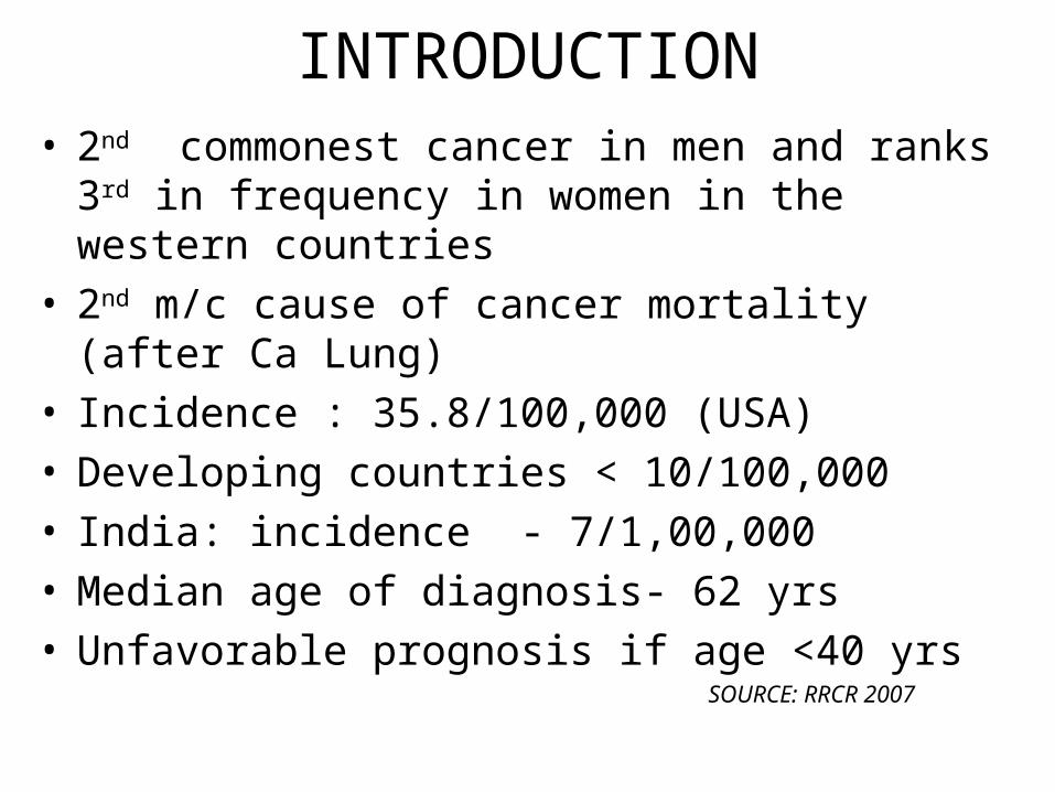
INTRODUCTION• 2nd commonest cancer in men and ranks 3rd in frequency
in women in the western countries
• 2nd m/c cause of cancer mortality (after Ca Lung)
• Incidence : 35.8/100,000 (USA)
• Developing countries < 10/100,000
• India: incidence - 7/1,00,000
• Median age of diagnosis- 62 yrs
• Unfavorable prognosis if age <40 yrs SOURCE: RRCR 2007
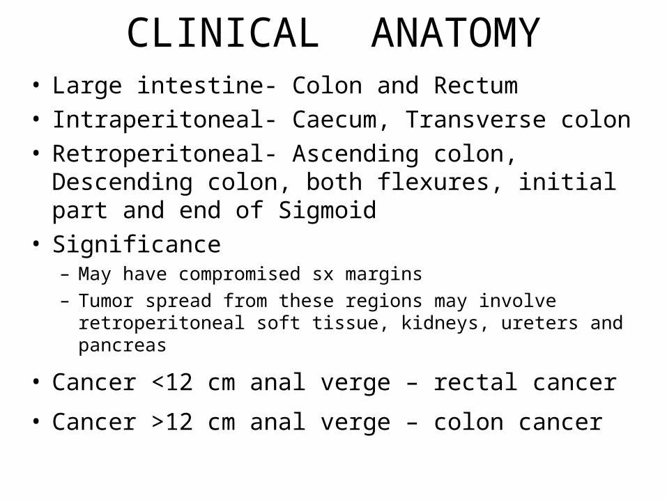
CLINICAL ANATOMY• Large intestine- Colon and Rectum
• Intraperitoneal- Caecum, Transverse colon
• Retroperitoneal- Ascending colon, Descending colon, both flexures, initial part and end of Sigmoid
• Significance– May have compromised sx margins
– Tumor spread from these regions may involve retroperitoneal soft tissue, kidneys, ureters and pancreas
• Cancer <12 cm anal verge – rectal cancer
• Cancer >12 cm anal verge – colon cancer
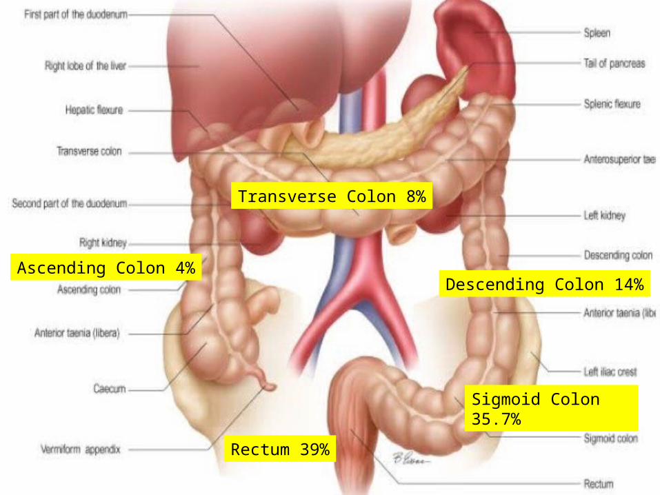
Ascending Colon 4%
Transverse Colon 8%
Descending Colon 14%
Sigmoid Colon 35.7%
Rectum 39%
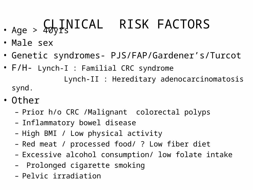
CLINICAL RISK FACTORS• Age > 40yrs
• Male sex
• Genetic syndromes- PJS/FAP/Gardener’s/Turcot • F/H- Lynch-I : Familial CRC syndrome
Lynch-II : Hereditary adenocarcinomatosis synd.
• Other– Prior h/o CRC /Malignant colorectal polyps
– Inflammatory bowel disease
– High BMI / Low physical activity
– Red meat / processed food/ ? Low fiber diet
– Excessive alcohol consumption/ low folate intake
– Prolonged cigarette smoking
– Pelvic irradiation
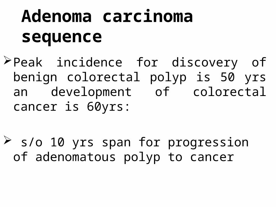
Adenoma carcinoma sequencePeak incidence for discovery of benign colorectal
polyp is 50 yrs an development of colorectal cancer is 60yrs:
s/o 10 yrs span for progression of adenomatous polyp to cancer
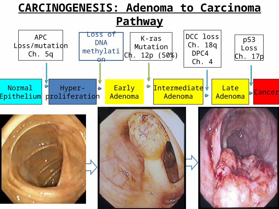
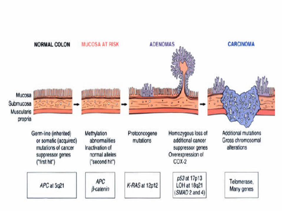
CARCINOGENESIS: Adenoma to Carcinoma Pathway
APCLoss/mutation
Ch. 5q
NormalEpithelium
EarlyAdenoma
CancerHyper-
proliferationIntermediate
AdenomaLate
Adenoma
K-rasMutation
Ch. 12p (50%)
DCC lossCh. 18qDPC4 Ch. 4
p53Loss
Ch. 17p
Loss of DNA methylation
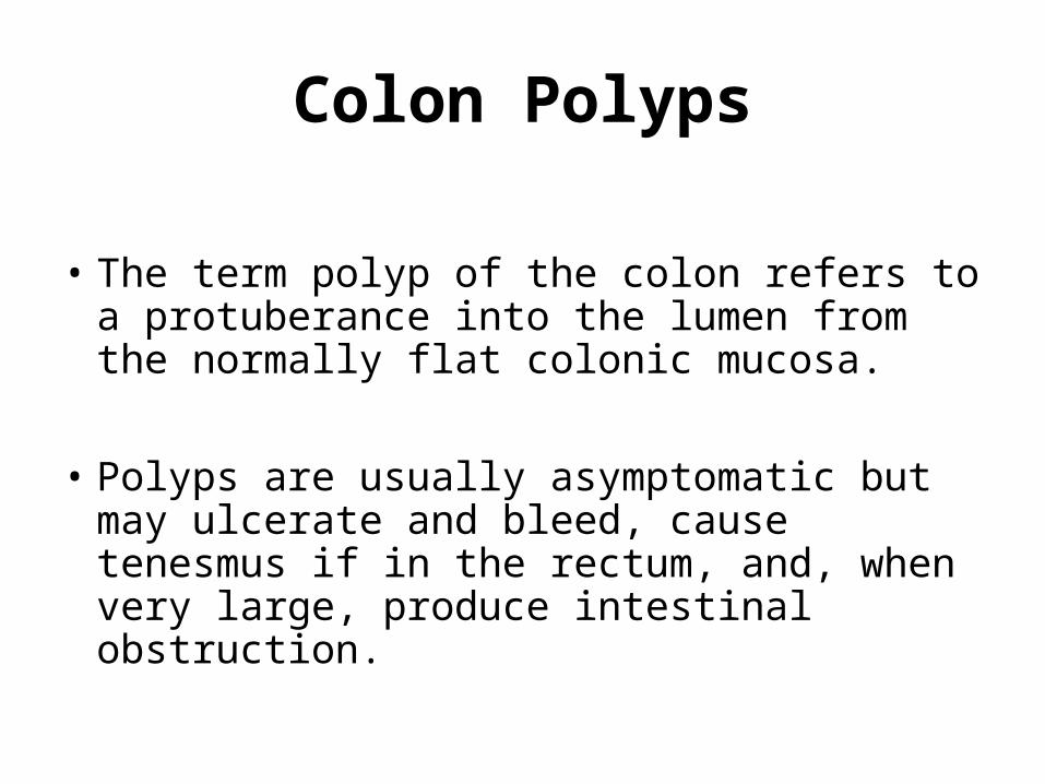
Colon Polyps
• The term polyp of the colon refers to a protuberance into the lumen from the normally flat colonic mucosa.
• Polyps are usually asymptomatic but may ulcerate and bleed, cause tenesmus if in the rectum, and, when very large, produce intestinal obstruction.
CLASSIFICATION
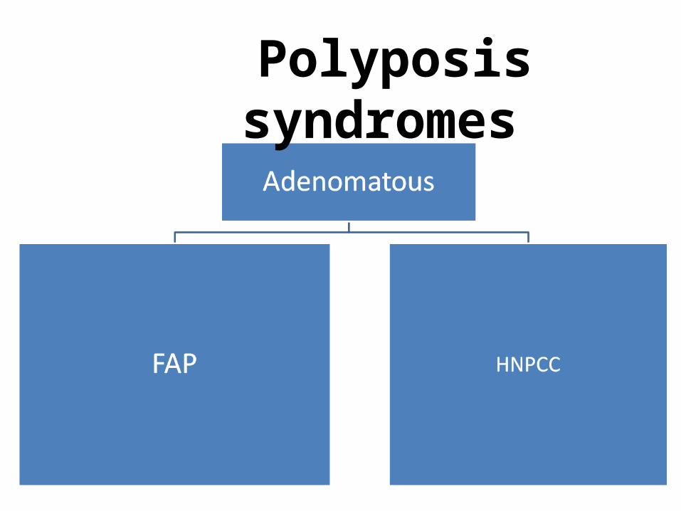
• Neoplastic (adenomas and carcinomas),• Hamartomatous, • Non-neoplastic, and • Submucosal (neoplastic / non-neoplastic).
Non-neoplastic polyps
• Hyperplastic • Mucosal • Inflammatory pseudopolyps • Submucosal
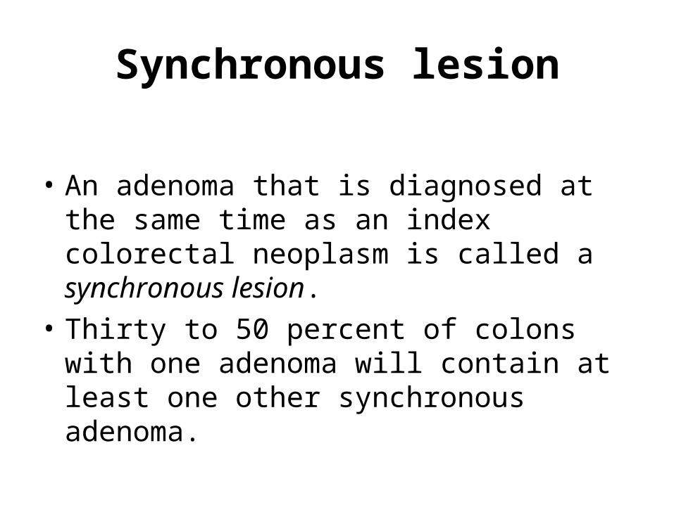
Synchronous lesion
• An adenoma that is diagnosed at the same time as an index colorectal neoplasm is called a synchronous lesion.
• Thirty to 50 percent of colons with one adenoma will contain at least one other synchronous adenoma.
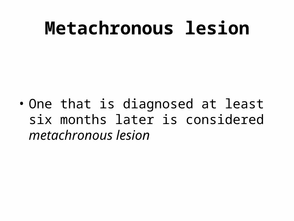
Metachronous lesion
• One that is diagnosed at least six months later is considered metachronous lesion
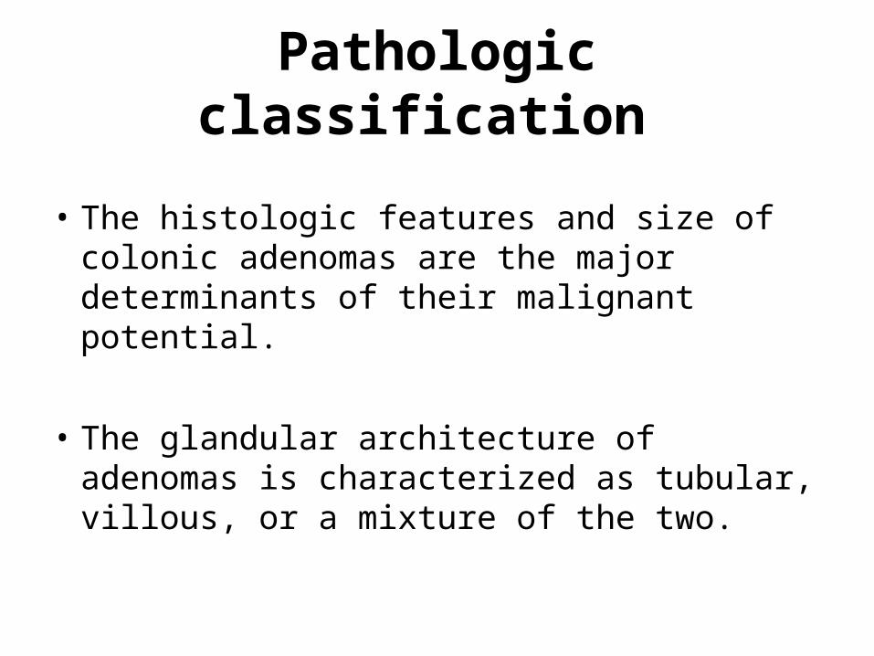
Pathologic classification
• The histologic features and size of colonic adenomas are the major determinants of their malignant potential.
• The glandular architecture of adenomas is characterized as tubular, villous, or a mixture of the two.
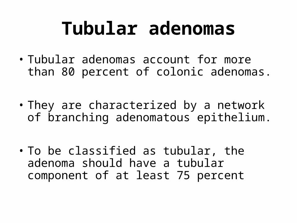
Tubular adenomas
• Tubular adenomas account for more than 80 percent of colonic adenomas.
• They are characterized by a network of branching adenomatous epithelium.
• To be classified as tubular, the adenoma should have a tubular component of at least 75 percent
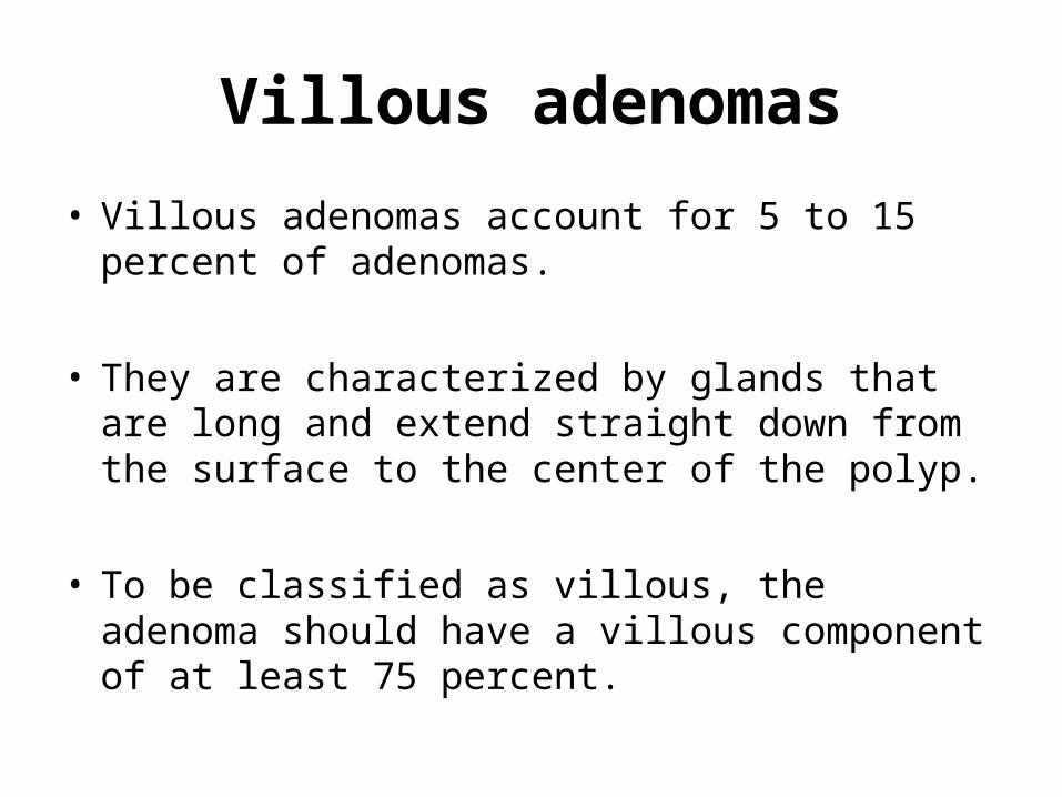
Villous adenomas
• Villous adenomas account for 5 to 15 percent of adenomas.
• They are characterized by glands that are long and extend straight down from the surface to the center of the polyp.
• To be classified as villous, the adenoma should have a villous component of at least 75 percent.
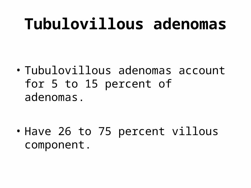
Tubulovillous adenomas
• Tubulovillous adenomas account for 5 to 15 percent of adenomas.
• Have 26 to 75 percent villous component.
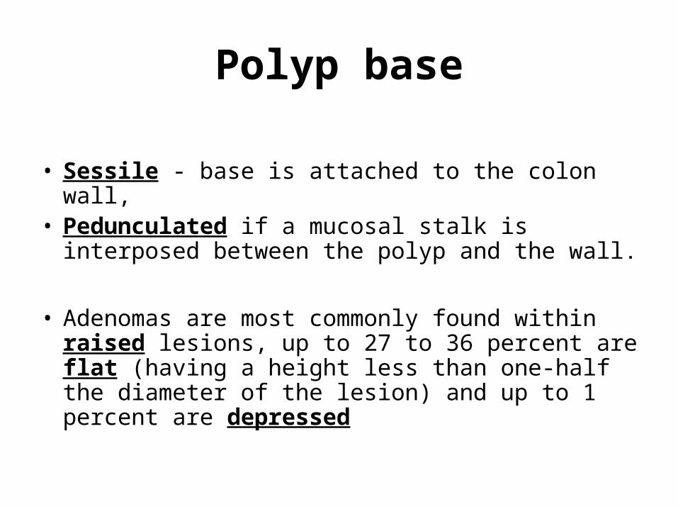
Polyp base
• Sessile - base is attached to the colon wall, • Pedunculated if a mucosal stalk is interposed
between the polyp and the wall.
• Adenomas are most commonly found within raised lesions, up to 27 to 36 percent are flat (having a height less than one-half the diameter of the lesion) and up to 1 percent are depressed
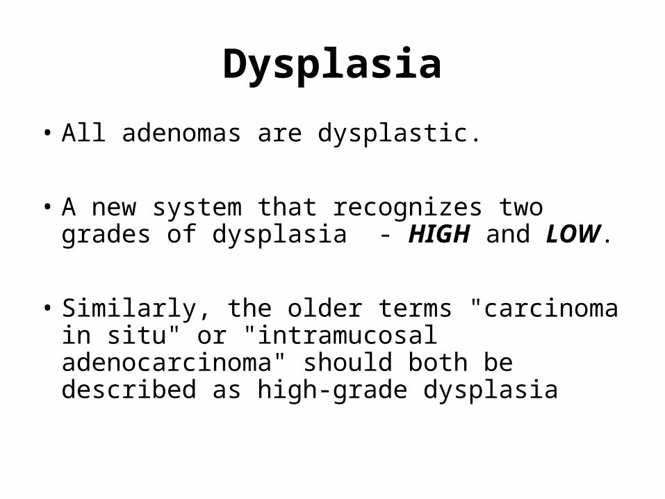
Dysplasia
• All adenomas are dysplastic.
• A new system that recognizes two grades of dysplasia - HIGH and LOW.
• Similarly, the older terms "carcinoma in situ" or "intramucosal adenocarcinoma" should both be described as high-grade dysplasia
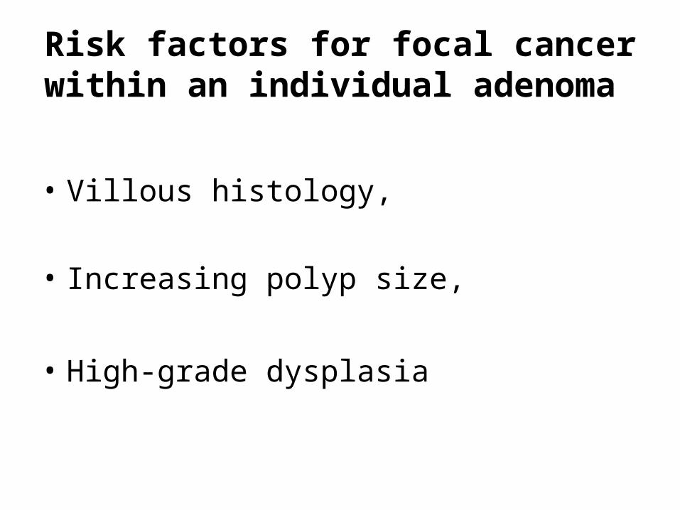
Risk factors for focal cancer within an individual adenoma
• Villous histology, • Increasing polyp size,
• High-grade dysplasia
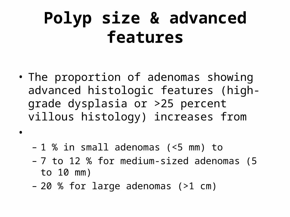
Polyp size & advanced features
• The proportion of adenomas showing advanced histologic features (high-grade dysplasia or >25 percent villous histology) increases from
• – 1 % in small adenomas (<5 mm) to – 7 to 12 % for medium-sized adenomas (5 to 10 mm) – 20 % for large adenomas (>1 cm)
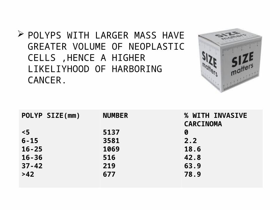
POLYPS WITH LARGER MASS HAVE GREATER VOLUME OF NEOPLASTIC CELLS ,HENCE A HIGHER LIKELIYHOOD OF HARBORING CANCER.
POLYP SIZE(mm)
<56-1516-2516-3637-42>42
NUMBER
513735811069516219677
% WITH INVASIVE CARCINOMA02.218.642.863.978.9
Age & advanced features
• Older age is also associated with high-grade dysplasia within an adenoma, independent of size and histology
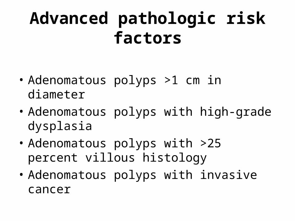
Advanced pathologic risk factors
• Adenomatous polyps >1 cm in diameter • Adenomatous polyps with high-grade
dysplasia • Adenomatous polyps with >25 percent villous
histology • Adenomatous polyps with invasive cancer
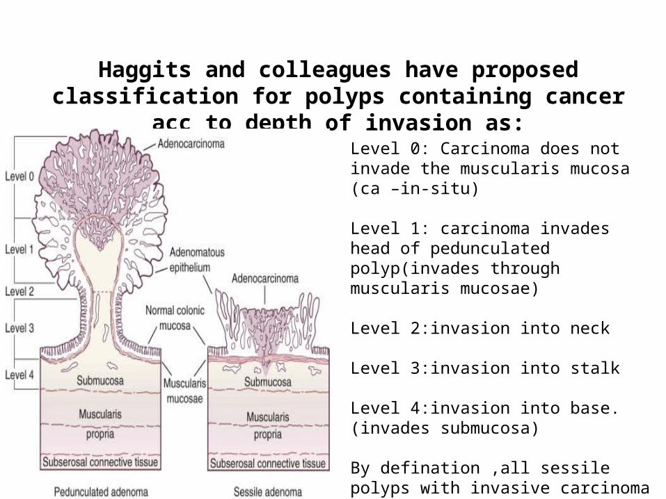
Haggits and colleagues have proposed classification for polyps containing cancer acc to depth of invasion as:
Level 0: Carcinoma does not invade the muscularis mucosa (ca –in-situ)
Level 1: carcinoma invades head of pedunculated polyp(invades through muscularis mucosae)
Level 2:invasion into neck
Level 3:invasion into stalk
Level 4:invasion into base.(invades submucosa)
By defination ,all sessile polyps with invasive carcinoma are level 4.
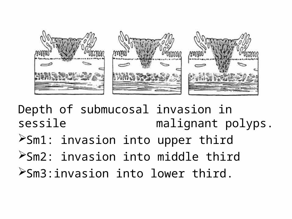
Depth of submucosal invasion in sessile malignant polyps. Sm1: invasion into upper third Sm2: invasion into middle thirdSm3:invasion into lower third.
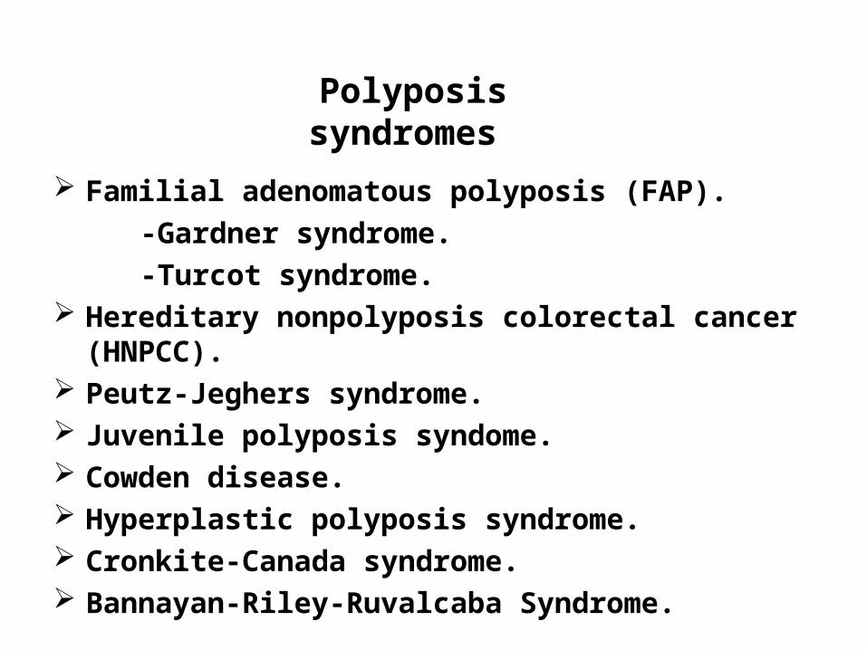
Polyposis syndromes
Familial adenomatous polyposis (FAP).
-Gardner syndrome.
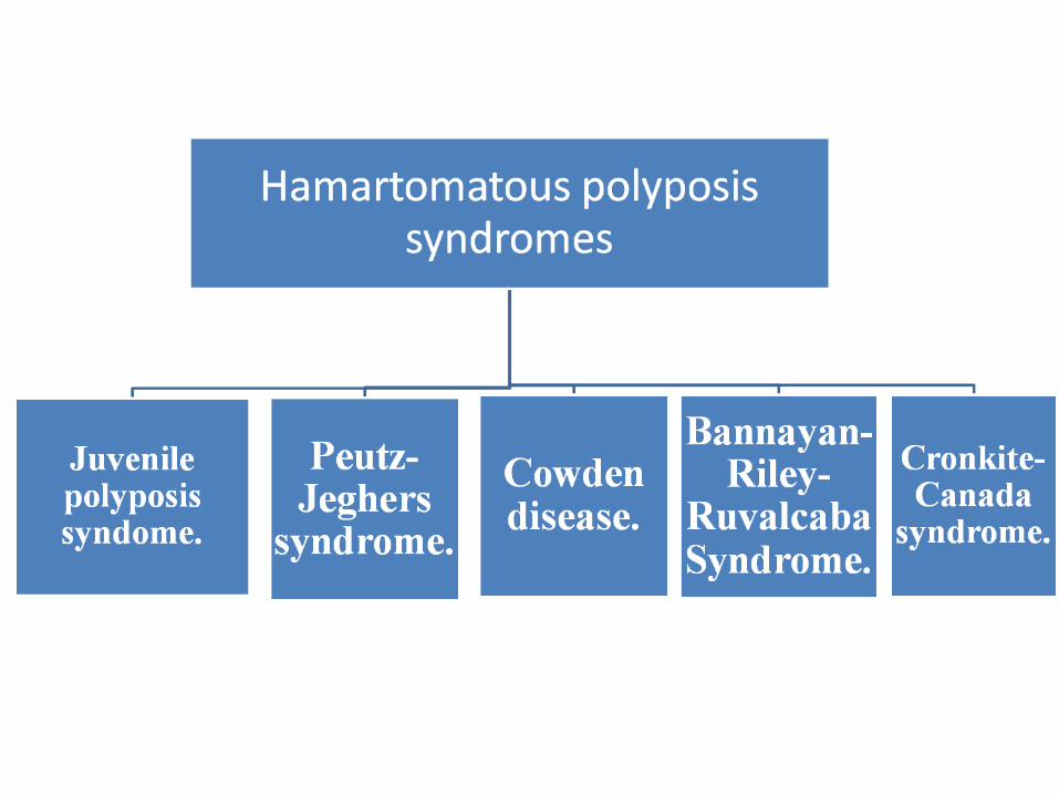
-Turcot syndrome. Hereditary nonpolyposis colorectal cancer (HNPCC). Peutz-Jeghers syndrome. Juvenile polyposis syndome. Cowden disease. Hyperplastic polyposis syndrome. Cronkite-Canada syndrome. Bannayan-Riley-Ruvalcaba Syndrome.
Polyposis syndromes
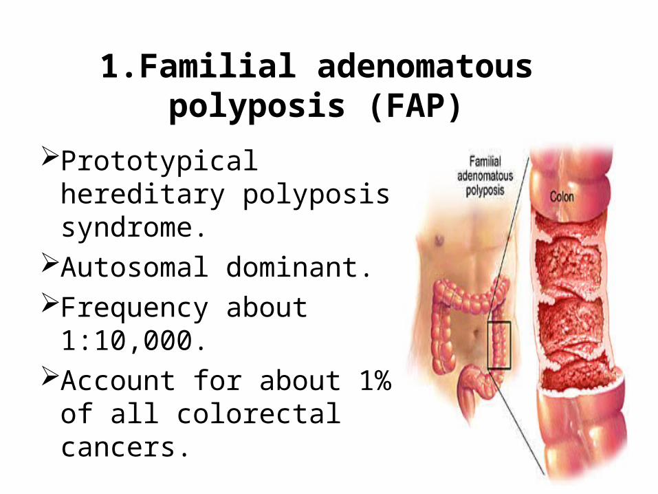
1.Familial adenomatous polyposis (FAP)
Prototypical hereditary polyposis syndrome.
Autosomal dominant.Frequency about
1:10,000.Account for about 1% of
all colorectal cancers.
The APC gene:The adenomatous polyposis coli (APC) gene is a tumour suppressor gene located on chromosome 5q21. Mutation in APC gene is genetic basis attributed to a truncating mutation in the germ-line APC gene.The gene expression is 100% in patients with the mutation.
GENETICS
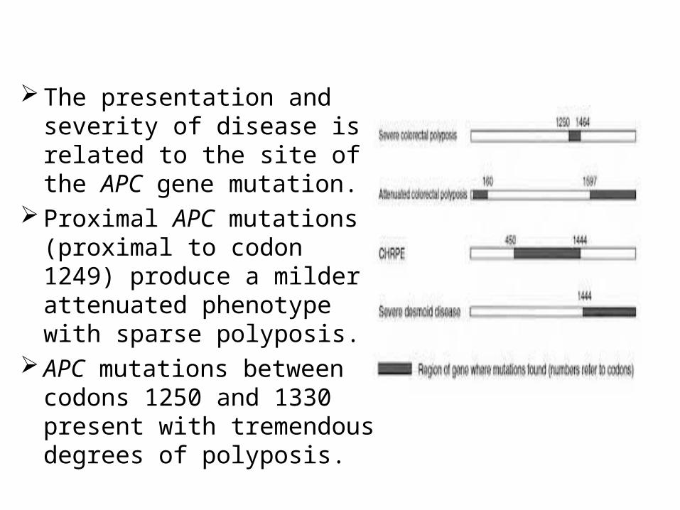
The presentation and severity of disease is related to the site of the APC gene mutation.
Proximal APC mutations (proximal to codon 1249) produce a milder attenuated phenotype with sparse polyposis.
APC mutations between codons 1250 and 1330 present with tremendous degrees of polyposis.
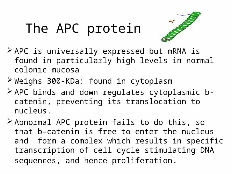
APC is universally expressed but mRNA is found in particularly high levels in normal colonic mucosa
Weighs 300-KDa: found in cytoplasm APC binds and down regulates cytoplasmic b-catenin,
preventing its translocation to nucleus. Abnormal APC protein fails to do this, so that b-catenin is
free to enter the nucleus and form a complex which results in specific transcription of cell cycle stimulating DNA sequences, and hence proliferation.
The APC protein
Common expression of syndrome is: Multiple colonic polyps(>100) Polyps start after age 10–20, cancer in 100% at age 40.All patients will develop cancer of colon if left untreated.
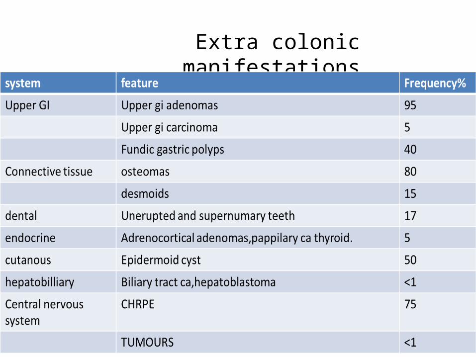
Extra colonic manifestations
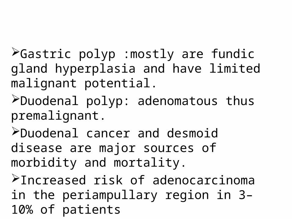
Gastric polyp :mostly are fundic gland hyperplasia and have limited malignant potential.Duodenal polyp: adenomatous thus premalignant.Duodenal cancer and desmoid disease are major sources of morbidity and mortality.Increased risk of adenocarcinoma in the periampullary region in 3–10% of patients
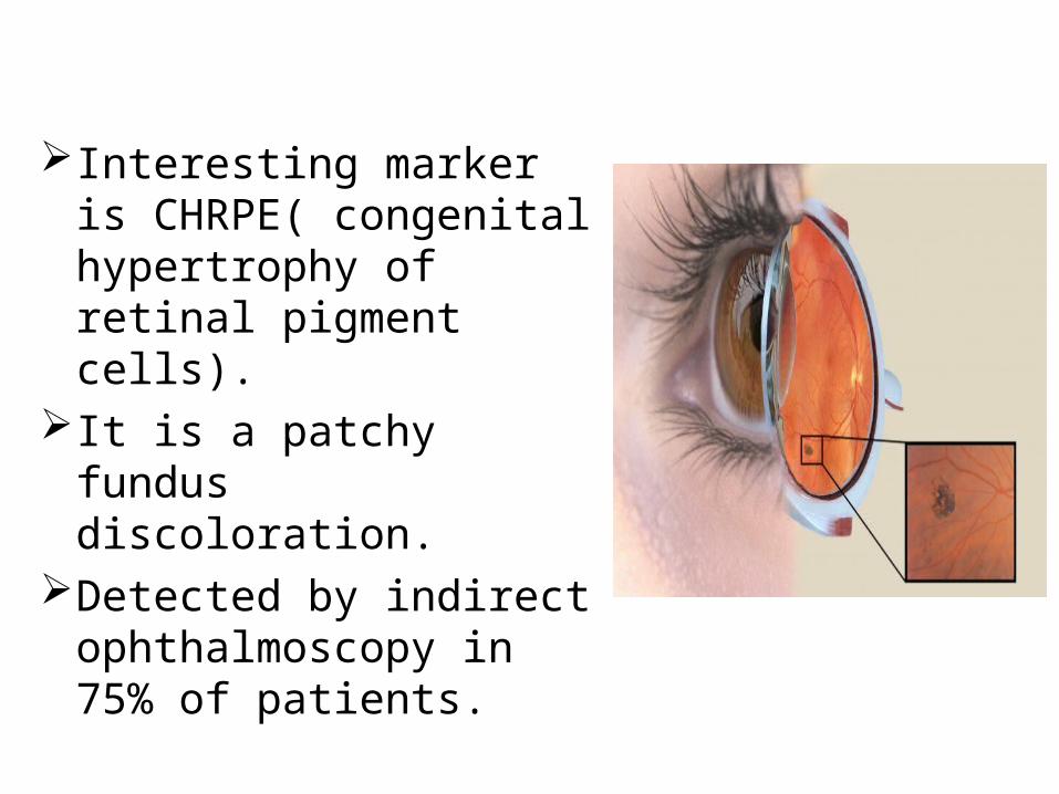
Interesting marker is CHRPE( congenital hypertrophy of retinal pigment cells).
It is a patchy fundus discoloration.
Detected by indirect ophthalmoscopy in 75% of patients.
Polyposis syndromes recognized to belong to general disorder of FAP include Gardners syndrome. Turcots syndrome.
Gardner syndrome (GS)Characterized byColonic adenomatous polyposis Osteomas: usually present in skull, mandible, and tibia
– They are virtually always benign.Soft tissue tumours like epidermoid cysts, fibromas, desmoid tumors.
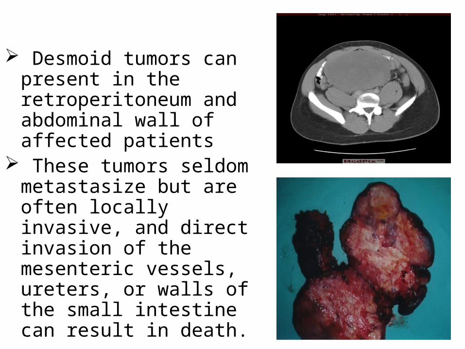
Desmoid tumors can present in the retroperitoneum and abdominal wall of affected patients
These tumors seldom metastasize but are often locally invasive, and direct invasion of the mesenteric vessels, ureters, or walls of the small intestine can result in death.
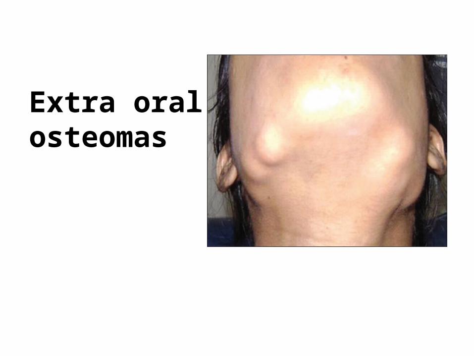
Extra oral osteomas
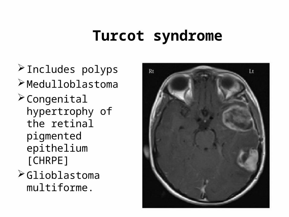
Turcot syndrome
Includes polypsMedulloblastomaCongenital
hypertrophy of the retinal pigmented epithelium [CHRPE]
Glioblastoma multiforme.
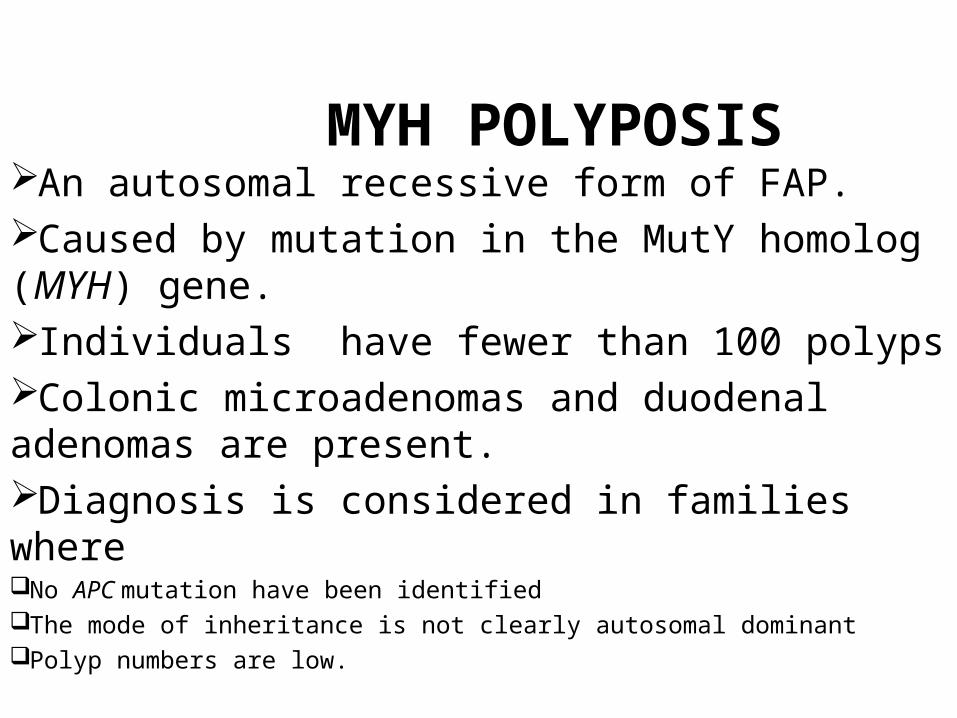
MYH POLYPOSISAn autosomal recessive form of FAP.Caused by mutation in the MutY homolog (MYH) gene.Individuals have fewer than 100 polypsColonic microadenomas and duodenal adenomas are present.Diagnosis is considered in families where No APC mutation have been identifiedThe mode of inheritance is not clearly autosomal dominantPolyp numbers are low.
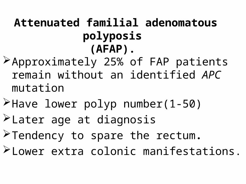
Attenuated familial adenomatous polyposis(AFAP).
Approximately 25% of FAP patients remain without an identified APC mutation
Have lower polyp number(1-50)Later age at diagnosisTendency to spare the rectum.Lower extra colonic manifestations.
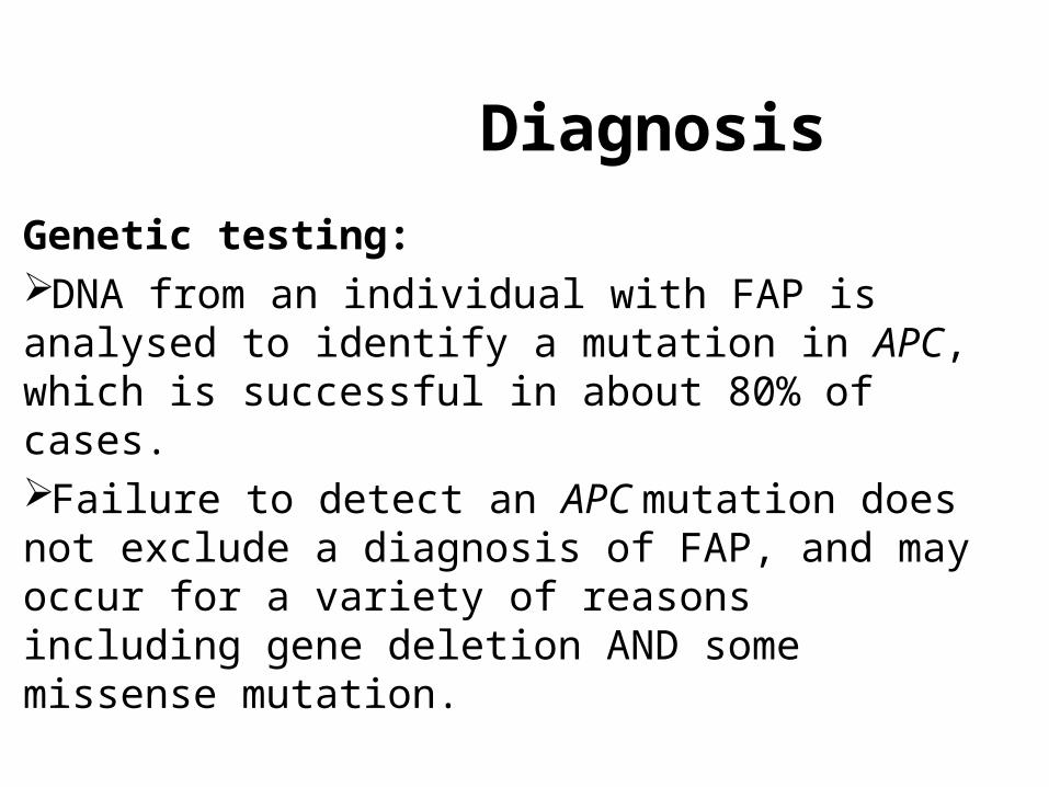
Diagnosis
Genetic testing:DNA from an individual with FAP is analysed to identify a mutation in APC, which is successful in about 80% of cases. Failure to detect an APC mutation does not exclude a diagnosis of FAP, and may occur for a variety of reasons including gene deletion AND some missense mutation.
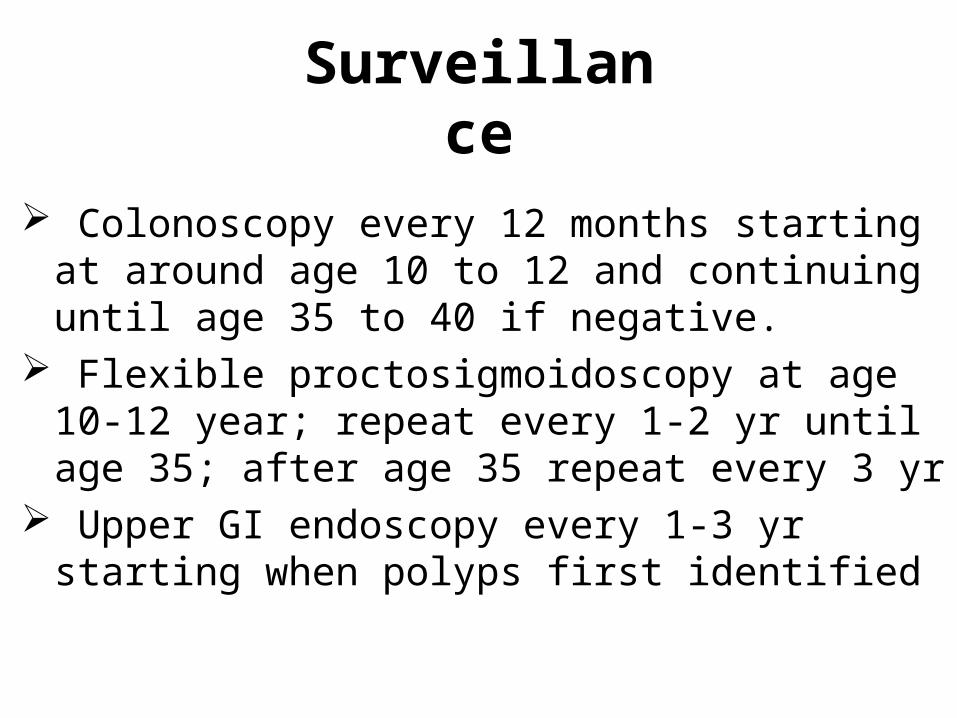
Surveillance
Colonoscopy every 12 months starting at around age 10 to 12 and continuing until age 35 to 40 if negative.
Flexible proctosigmoidoscopy at age 10-12 year; repeat every 1-2 yr until age 35; after age 35 repeat every 3 yr
Upper GI endoscopy every 1-3 yr starting when polyps first identified
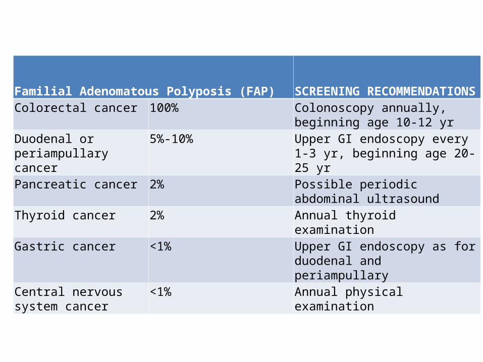
Familial Adenomatous Polyposis (FAP) SCREENING RECOMMENDATIONSColorectal cancer 100% Colonoscopy annually, beginning age
10-12 yrDuodenal or periampullary cancer
5%-10% Upper GI endoscopy every 1-3 yr, beginning age 20-25 yr
Pancreatic cancer 2% Possible periodic abdominal ultrasound
Thyroid cancer 2% Annual thyroid examination
Gastric cancer <1% Upper GI endoscopy as for duodenal and periampullary
Central nervous system cancer
<1% Annual physical examination
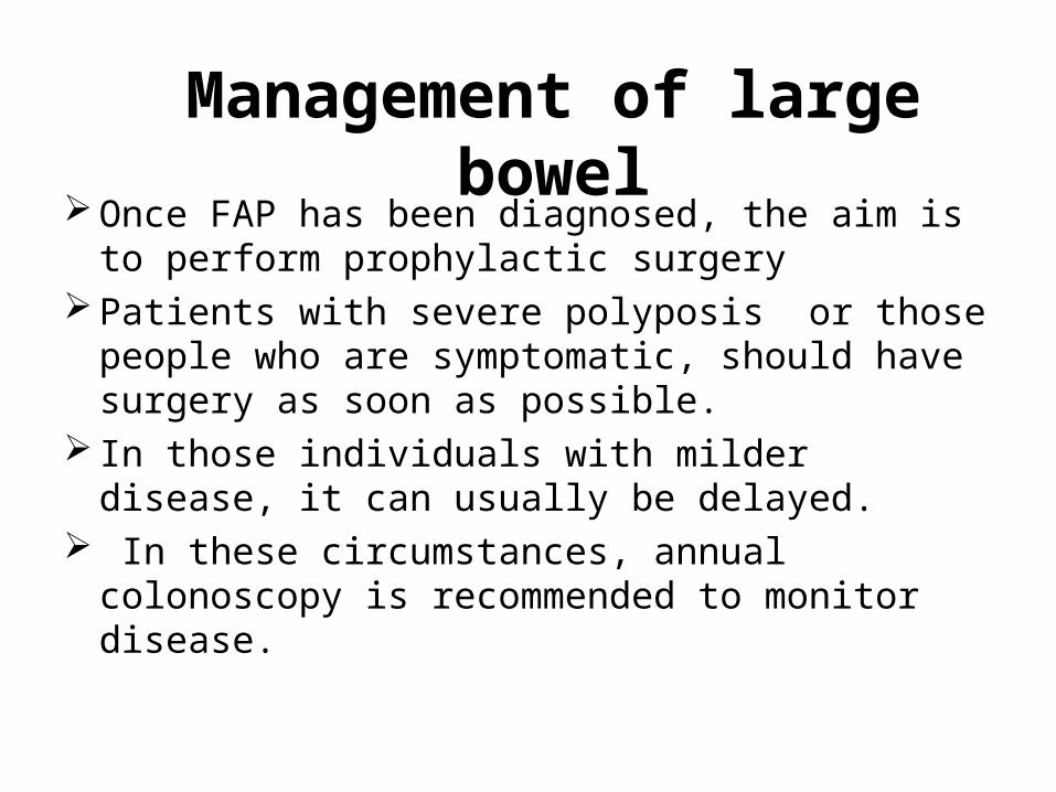
Management of large bowel Once FAP has been diagnosed, the aim is to perform
prophylactic surgery Patients with severe polyposis or those people who
are symptomatic, should have surgery as soon as possible.
In those individuals with milder disease, it can usually be delayed.
In these circumstances, annual colonoscopy is recommended to monitor disease.
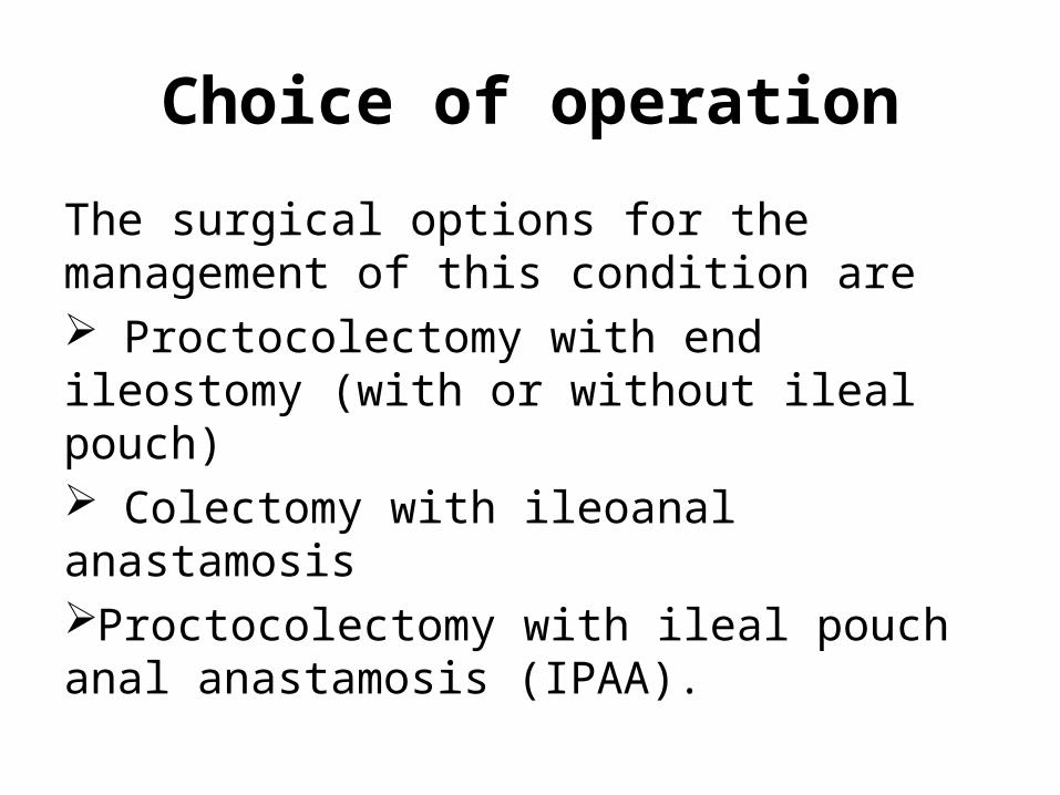
Choice of operation
The surgical options for the management of this condition are Proctocolectomy with end ileostomy (with or without ileal pouch) Colectomy with ileoanal anastamosis Proctocolectomy with ileal pouch anal anastamosis (IPAA).
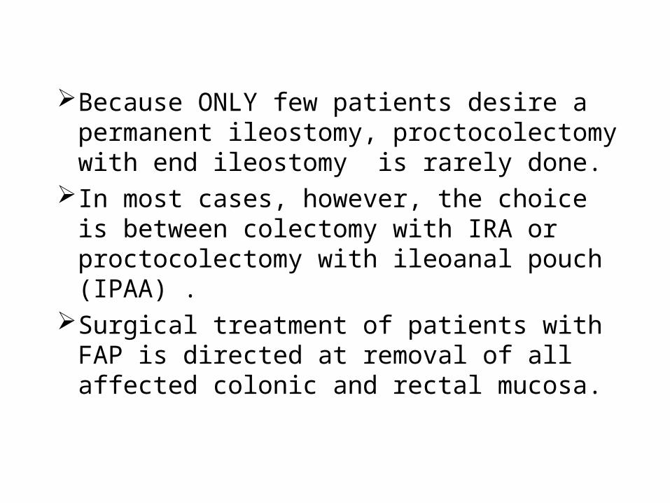
Because ONLY few patients desire a permanent ileostomy, proctocolectomy with end ileostomy is rarely done.
In most cases, however, the choice is between colectomy with IRA or proctocolectomy with ileoanal pouch (IPAA) .
Surgical treatment of patients with FAP is directed at removal of all affected colonic and rectal mucosa.
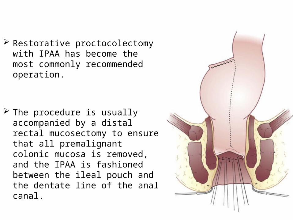
Restorative proctocolectomy with IPAA has become the most commonly recommended operation.
The procedure is usually accompanied by a distal rectal mucosectomy to ensure that all premalignant colonic mucosa is removed, and the IPAA is fashioned between the ileal pouch and the dentate line of the anal canal.
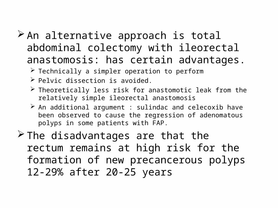
An alternative approach is total abdominal colectomy with ileorectal anastomosis: has certain advantages. Technically a simpler operation to perform Pelvic dissection is avoided. Theoretically less risk for anastomotic leak from the relatively simple
ileorectal anastomosis An additional argument : sulindac and celecoxib have been observed to
cause the regression of adenomatous polyps in some patients with FAP.
The disadvantages are that the rectum remains at high risk for the formation of new precancerous polyps 12-29% after 20-25 years
Patients with Gardner syndrome require surgical treatment of
Cutaneous cysts
Symptomatic dental anomalies and osteomas Biopsy and resection for malignancies, including
hepatoblastoma, thyroid carcinoma, osteocarcinoma, gastric carcinoma, periampullary carcinoma, and biliary tract carcinoma
Liver transplantation may be required in patients with hepatoblastoma
Patients with Turcot syndrome require
surgical intervention for diagnosis and management of CNS lesions, gastric lesions and hepatic lesions
Postop surveillanceAfter IRA, the retained rectum should be
examined using a flexible sigmoidoscope, every 6–12 months.
Polyps larger than 5 mm should be removed If severe dysplasia or uncontrolled polyposis
develops, completion proctectomy with or without ileoanal pouch formation is indicated.
In patients who have had IPAA, the pouch should be examined by flexible endoscopy annually, and a careful digital examination of the anorectal transition zone should be performed.
Chemoprevention
Have reduced the number and size of colorectal adenomas
THESE AREi. (NSAID) –sulindacii. The COX-2 inhibitor celecoxib
2.Hereditary non-polyposis colon cancer (HNPCC)
HNPCC is the most frequently occurring hereditary colorectal cancer syndrome
Autosomal dominant. It also known as Lynch I and II syndromes.The Lynch I variants describe patients with
predominantly colorectal cancer at a young ageLynch II: those with both colorectal and
extracolonic cancers.
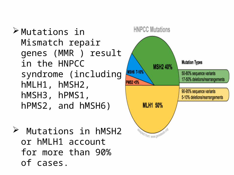
Mutations in Mismatch repair genes (MMR ) result in the HNPCC syndrome (including hMLH1, hMSH2, hMSH3, hPMS1, hPMS2, and hMSH6)
Mutations in hMSH2 or hMLH1 account for more than 90% of cases.
These mutations produce microsatellite instability which result in errors in S phase when DNA is newly synthesized and copied.
Patients with hMSH2 mutation tend to develop extracolonic cancers, in particular endometrial cancer, as compared with hMLH1 mutation carriers
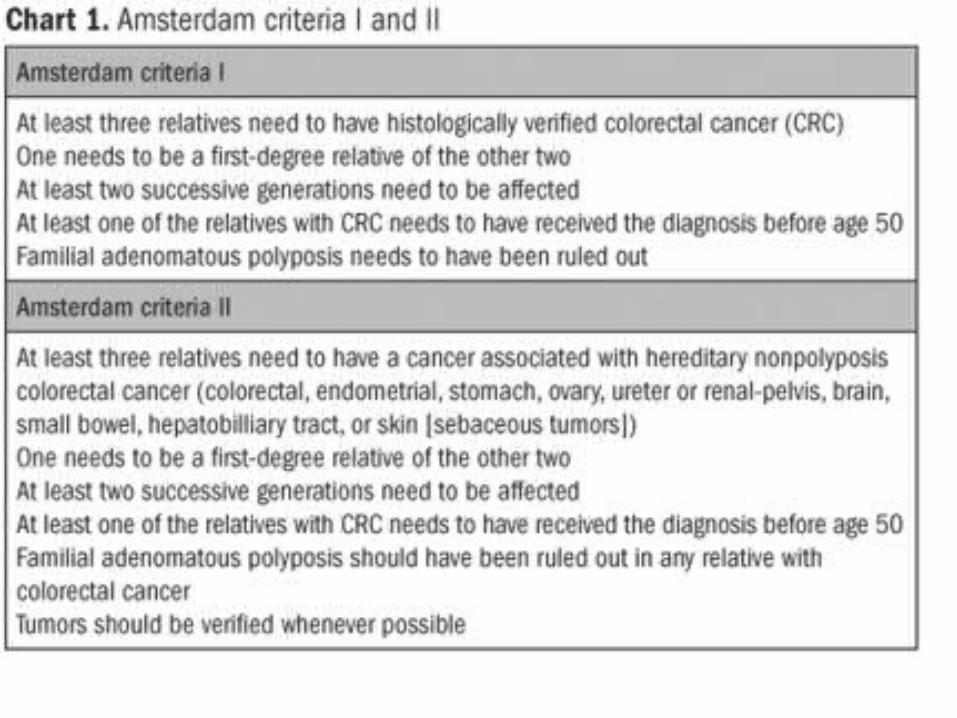
To facilitate the clinical diagnosis of HNPCC, the International Collaborative Group on HNPCC (ICG-HNPCC) proposed the Amsterdam Criteria in 1990.
Further liberalization for identifying patients with HNPCC occurred with the introduction of the Bethesda criteria
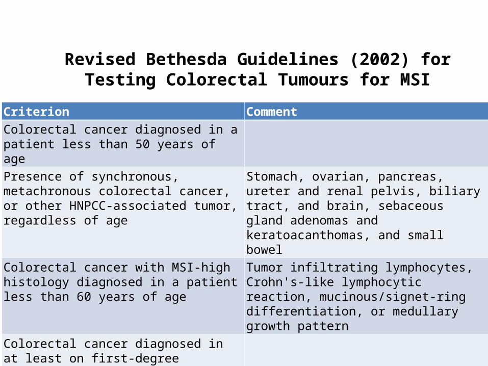
Revised Bethesda Guidelines (2002) for Testing Colorectal Tumours for MSI
Criterion CommentColorectal cancer diagnosed in a patient less than 50 years of agePresence of synchronous, metachronous colorectal cancer, or other HNPCC-associated tumor, regardless of age
Stomach, ovarian, pancreas, ureter and renal pelvis, biliary tract, and brain, sebaceous gland adenomas and keratoacanthomas, and small bowel
Colorectal cancer with MSI-high histology diagnosed in a patient less than 60 years of age
Tumor infiltrating lymphocytes, Crohn's-like lymphocytic reaction, mucinous/signet-ring differentiation, or medullary growth pattern
Colorectal cancer diagnosed in at least on first-degree relative with an HNPCC-related tumor diagnosed under age 50Colorectal cancer diagnosed in two or more first or second-degree relatives with HNPCC-related tumors, regardless of age.
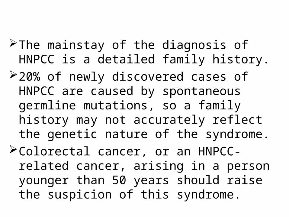
The mainstay of the diagnosis of HNPCC is a detailed family history.
20% of newly discovered cases of HNPCC are caused by spontaneous germline mutations, so a family history may not accurately reflect the genetic nature of the syndrome.
Colorectal cancer, or an HNPCC-related cancer, arising in a person younger than 50 years should raise the suspicion of this syndrome.
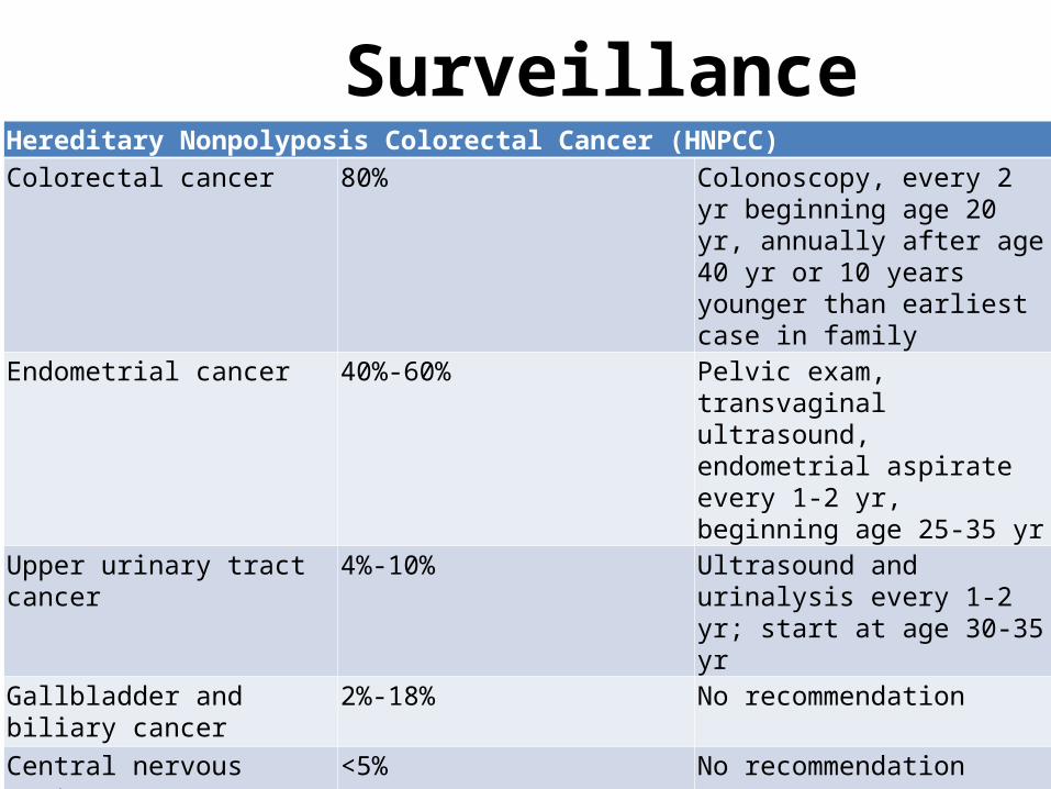
SurveillanceHereditary Nonpolyposis Colorectal Cancer (HNPCC)Colorectal cancer 80% Colonoscopy, every 2 yr
beginning age 20 yr, annually after age 40 yr or 10 years younger than earliest case in family
Endometrial cancer 40%-60% Pelvic exam, transvaginal ultrasound, endometrial aspirate every 1-2 yr, beginning age 25-35 yr
Upper urinary tract cancer 4%-10% Ultrasound and urinalysis every 1-2 yr; start at age 30-35 yr
Gallbladder and biliary cancer 2%-18% No recommendation
Central nervous system cancer <5% No recommendation
Small bowel cancer <5% No recommendation
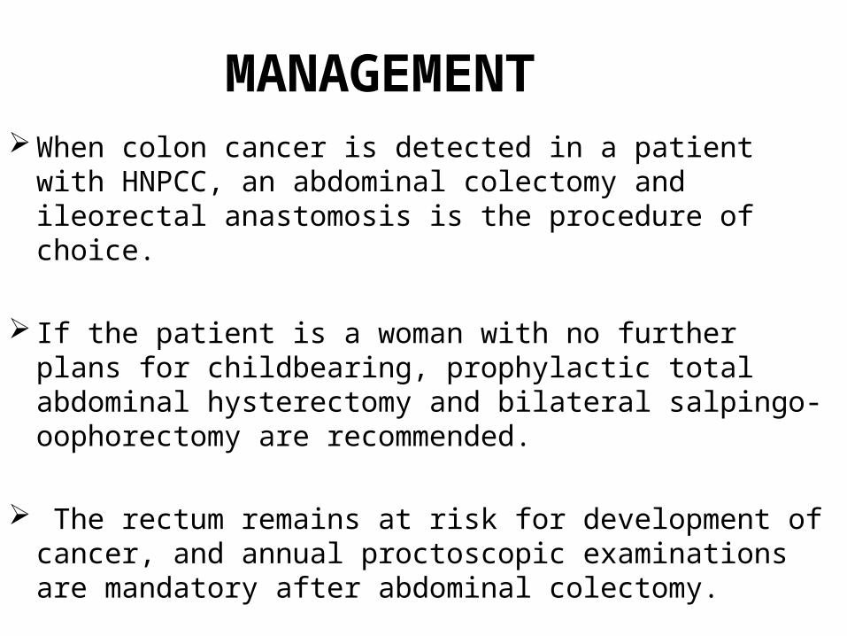
MANAGEMENT When colon cancer is detected in a patient with HNPCC, an
abdominal colectomy and ileorectal anastomosis is the procedure of choice.
If the patient is a woman with no further plans for childbearing, prophylactic total abdominal hysterectomy and bilateral salpingo-oophorectomy are recommended.
The rectum remains at risk for development of cancer, and annual proctoscopic examinations are mandatory after abdominal colectomy.
Other forms of cancer associated with HNPCC are treated according to the same criteria as in nonhereditary cases.
The role of prophylactic colectomy for patients with HNPCC has been considered in some instances, but this concept has not received universal acceptance.
It is an interesting but well-documented fact that the prognosis is better for cancer patients with HNPCC than for non-HNPCC patients with cancer of the same stage.
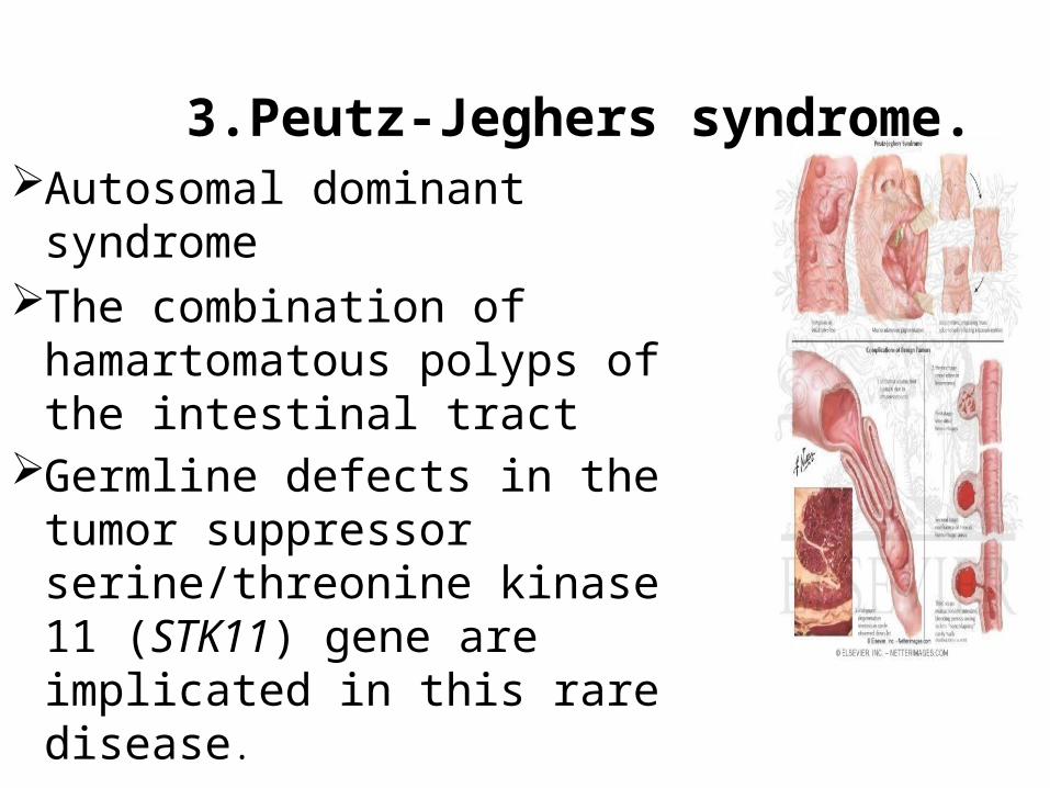
3.Peutz-Jeghers syndrome.Autosomal dominant syndrome The combination of
hamartomatous polyps of the intestinal tract
Germline defects in the tumor suppressor serine/threonine kinase 11 (STK11) gene are implicated in this rare disease.
Symptoms include:• GI bleeding• Intussusception• Rectal prolapse• Nasal polyposis (chronic sinusitis) Pigmented macules on the lips
and digits• Gynecomastia
The most common location of Peutz-Jeghers polyps is in the upper gastrointestinal tract, specifically the upper jejunum.
There is also an increased risk for extraintestinal malignancies includingcancer of the breast, ovary ,Cervix, fallopian tubesThyroidLungGallbladderbile ductspancreas testicles.
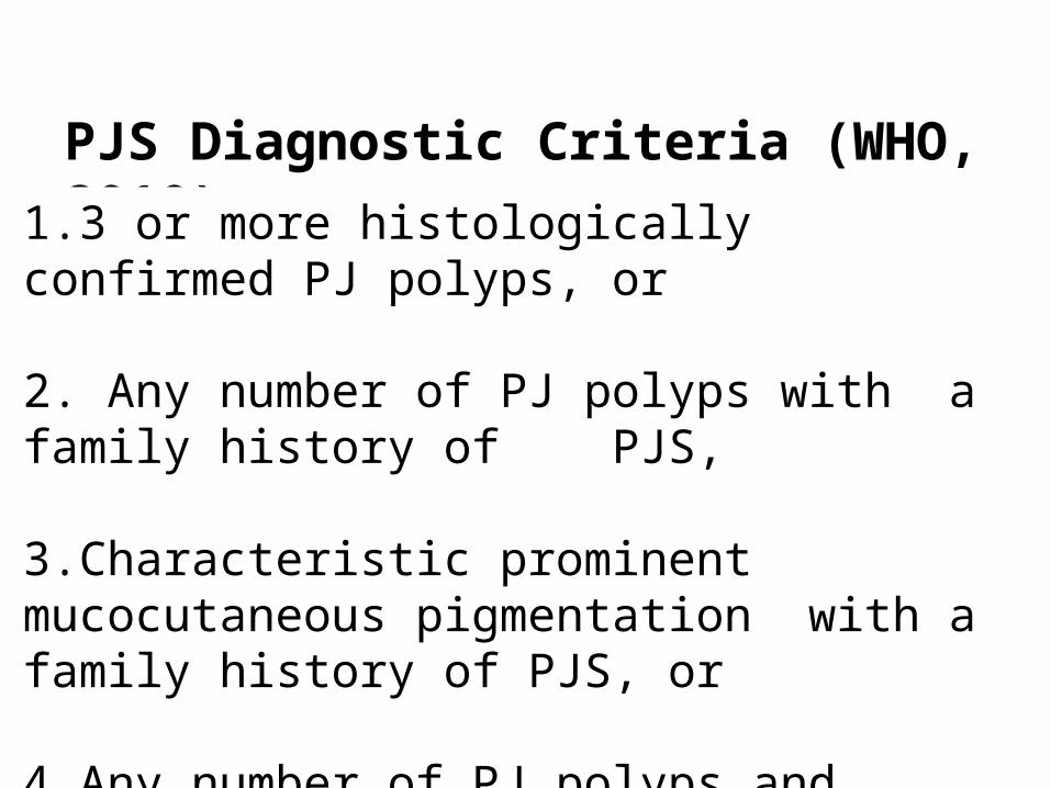
PJS Diagnostic Criteria (WHO, 2010)
1.3 or more histologically confirmed PJ polyps, or
2. Any number of PJ polyps with a family history of PJS,
3.Characteristic prominent mucocutaneous pigmentation with a family history of PJS, or
4.Any number of PJ polyps and characteristic prominent mucocutaneous pigmentation.

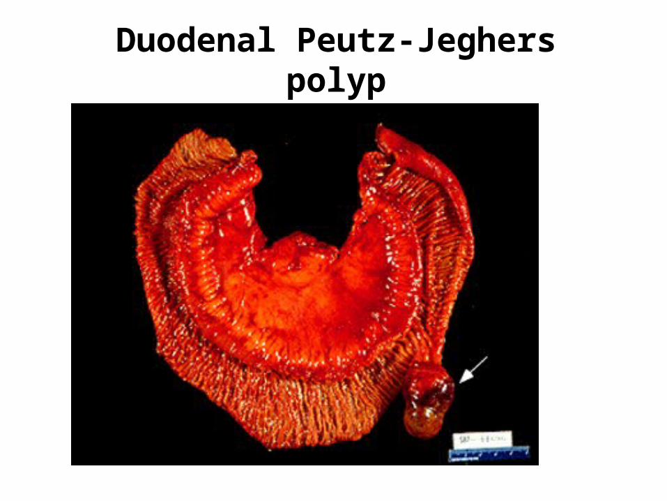
Duodenal Peutz-Jeghers polyp
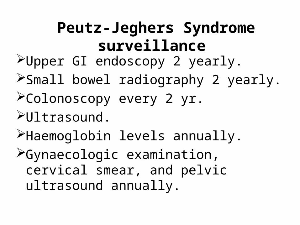
Peutz-Jeghers Syndrome surveillance
Upper GI endoscopy 2 yearly.Small bowel radiography 2 yearly.Colonoscopy every 2 yr.Ultrasound.Haemoglobin levels annually.Gynaecologic examination, cervical
smear, and pelvic ultrasound annually.
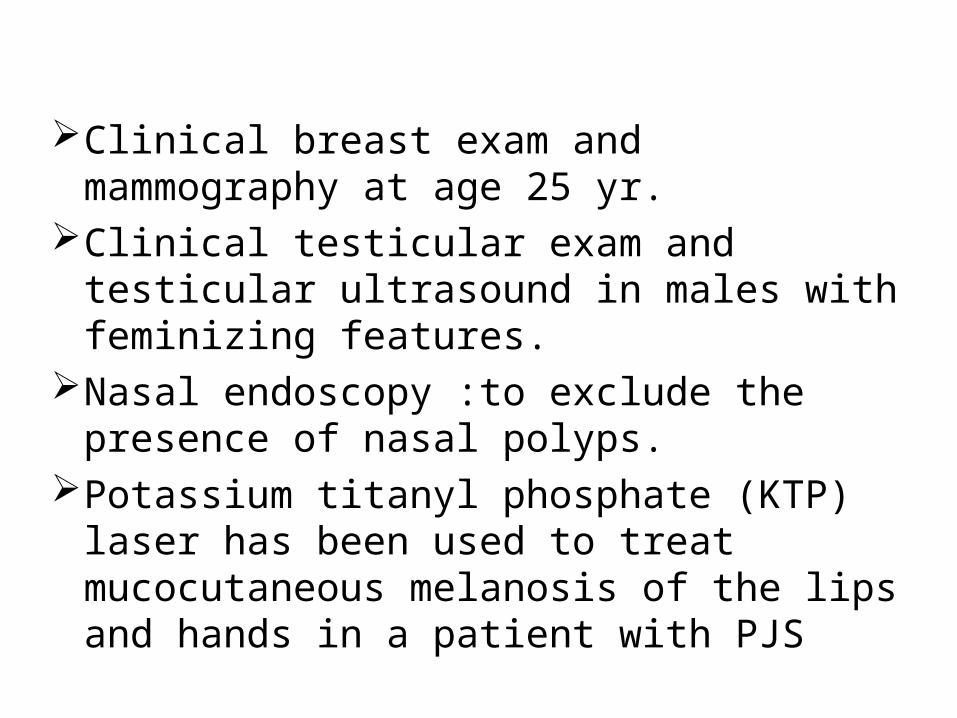
Clinical breast exam and mammography at age 25 yr.
Clinical testicular exam and testicular ultrasound in males with feminizing features.
Nasal endoscopy :to exclude the presence of nasal polyps.
Potassium titanyl phosphate (KTP) laser has been used to treat mucocutaneous melanosis of the lips and hands in a patient with PJS
4.Juvenile polyposis syndrome (JPS)
Most common hamartomatous syndrome Inherited as an autosomal dominant trait.A germ-line mutation in the SMAD-4 gene
(18q21) accounts for approximately 50% of
the reported cases of the syndrome.The term "juvenile" refers to the type of
polyp, not the age of onset of polyps.
Characterized by predisposition for hamartomatous polyps in the (GI) tract, specifically in the stomach, small intestine, colon, and rectum.
The average age of onset is approximately 18 years.
Associated with congenital birth defects (15%-20%) of patients including malrotation, hydrocephalus, cardiac lesions, Meckel's diverticulum, and mesenteric lymphangioma
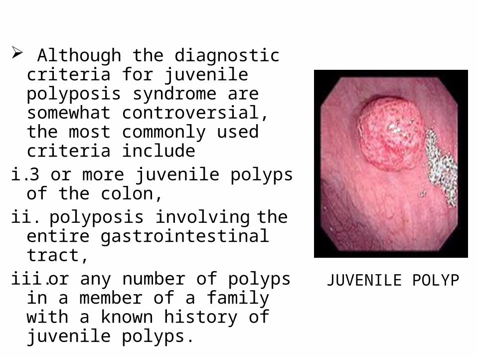
Although the diagnostic criteria for juvenile polyposis syndrome are somewhat controversial, the most commonly used criteria include
i. 3 or more juvenile polyps of the colon,
ii. polyposis involving the entire gastrointestinal tract,
iii.or any number of polyps in a member of a family with a known history of juvenile polyps.
JUVENILE POLYP
In infancy, patients may present with acute or chronic gastrointestinal bleeding,
intussusception, rectal prolapse, or a protein-losing enteropathy.
In adulthood, patients commonly present with either acute or chronic gastrointestinal blood loss.
Polyps are located most frequently in the recto sigmoid region.
Some individuals may only have four or five polyps over their lifetimes, whereas others in the same family may have over a hundred.
Most juvenile polyps are benign; however, malignant transformation can occur.
Estimates of developing GI cancers in families with JPS range from 9-50%.
Juvenile Polyposis screening
Screening by age 12 yr if symptoms have not yet arisen
Colonoscopy with multiple random biopsies every several years
5.Cowden syndrome Also known as multiple hamartoma-
neoplasia syndrome. It is an autosomal dominant condition
Complete penetrance by the age 20.Germ-line mutations in the PTEN tumor
suppressor gene located at 10q22. Polyps arise more commonly from
ectodermal rather than endodermal elements.
80% of patients present with benign tumor of the hair shaft.
CNS is the second most involved system, with approx 40% having macrocephaly.
The majority of patients with Cowden's disease suffer from benign thyroid or breast disease- projected lifetime risk of 10% for thyroid cancer and of 30–50% for breast cancer.
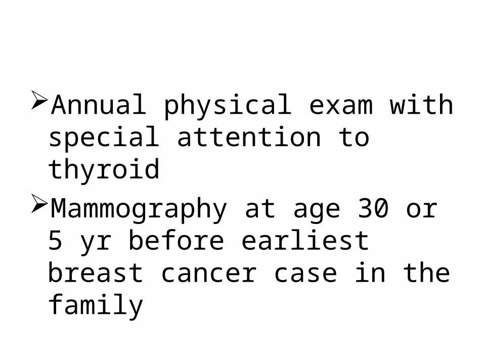
Annual physical exam with special attention to thyroid
Mammography at age 30 or 5 yr before earliest breast cancer case in the family
6.Hyperplastic polyposis syndrome
Hyperplastic polyps are found commonly in the large bowel, predominantly in the rectum and sigmoid.
Because of their small size, hyperplastic polyps rarely cause symptoms.
However, large or multiple hyperplastic polyps occasionally can be responsible for gastrointestinal symptoms.
HPS is a rare conditionCharacterized by numerous hyperplastic polyps throughout the large bowel that give the mucosa a "studded" look. The endoscopic and radiologic appearance of the mucosal abnormalities closely resembles FAP, but hyperplastic polyposis is not heritable and does not have any extraintestinal manifestations.
7.Cronkite-Canada syndrome Characterized by diffuse hamartomatous
polyposis The polyps are Ectodermal abnormalities such
as alopecia, onychodystrophy, and skin hyperpigmentation.
The syndrome can be distinguished by the diffuse distribution of polyps throughout the entire gastrointestinal tract with exception of the esophagus, which is spared.
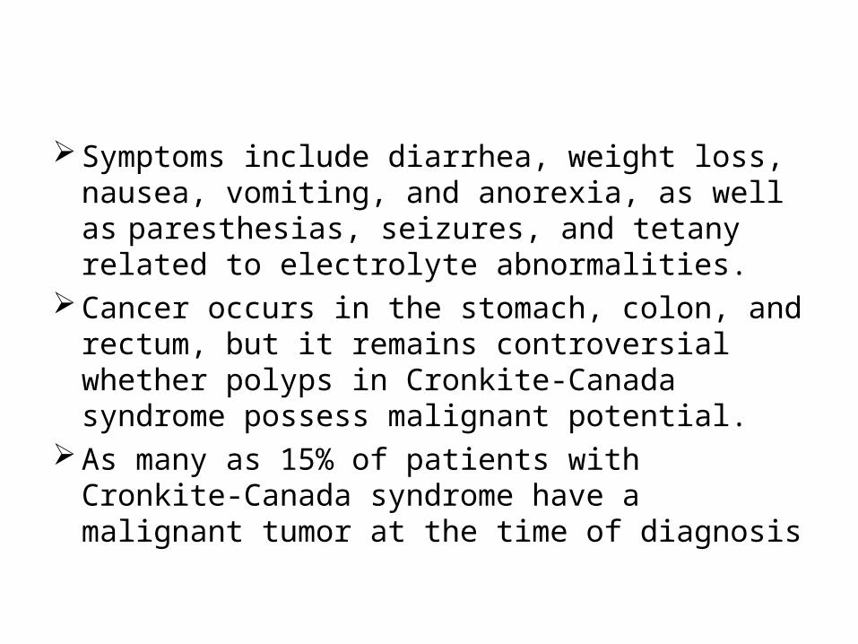
Symptoms include diarrhea, weight loss, nausea, vomiting, and anorexia, as well as paresthesias, seizures, and tetany related to electrolyte abnormalities.
Cancer occurs in the stomach, colon, and rectum, but it remains controversial whether polyps in Cronkite-Canada syndrome possess malignant potential.
As many as 15% of patients with Cronkite-Canada syndrome have a malignant tumor at the time of diagnosis
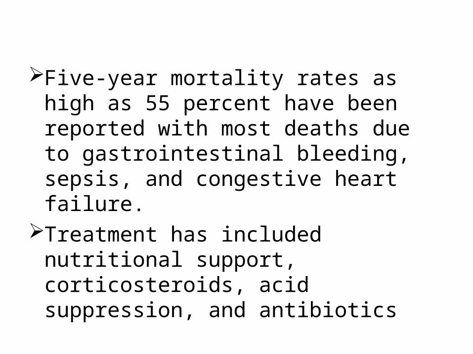
Five-year mortality rates as high as 55 percent have been reported with most deaths due to gastrointestinal bleeding, sepsis, and congestive heart failure.
Treatment has included nutritional support, corticosteroids, acid suppression, and antibiotics
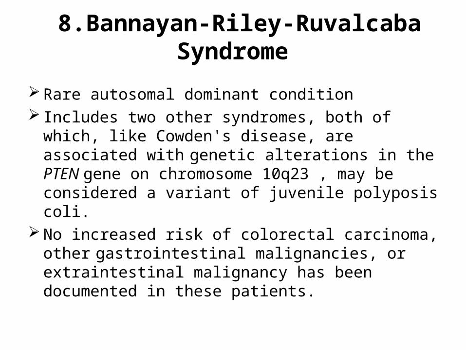
8.Bannayan-Riley-Ruvalcaba Syndrome
Rare autosomal dominant condition Includes two other syndromes, both of which, like
Cowden's disease, are associated with genetic alterations in the PTEN gene on chromosome 10q23 , may be considered a variant of juvenile polyposis coli.
No increased risk of colorectal carcinoma, other
gastrointestinal malignancies, or extraintestinal malignancy has been documented in these patients.
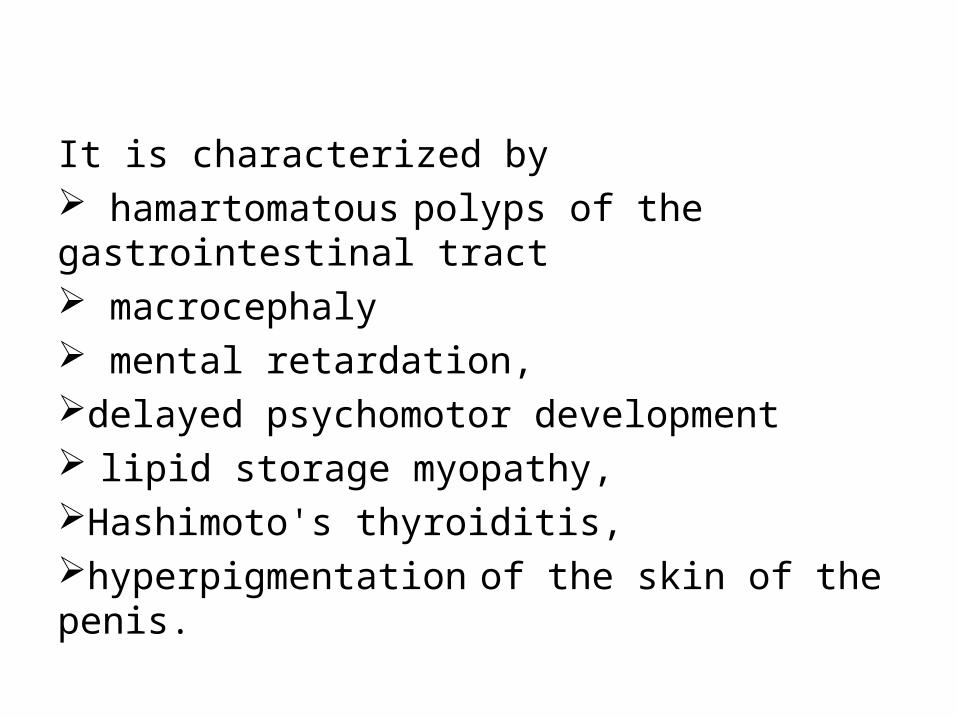
It is characterized by hamartomatous polyps of the gastrointestinal tract macrocephaly mental retardation,delayed psychomotor development lipid storage myopathy,Hashimoto's thyroiditis,hyperpigmentation of the skin of the penis.
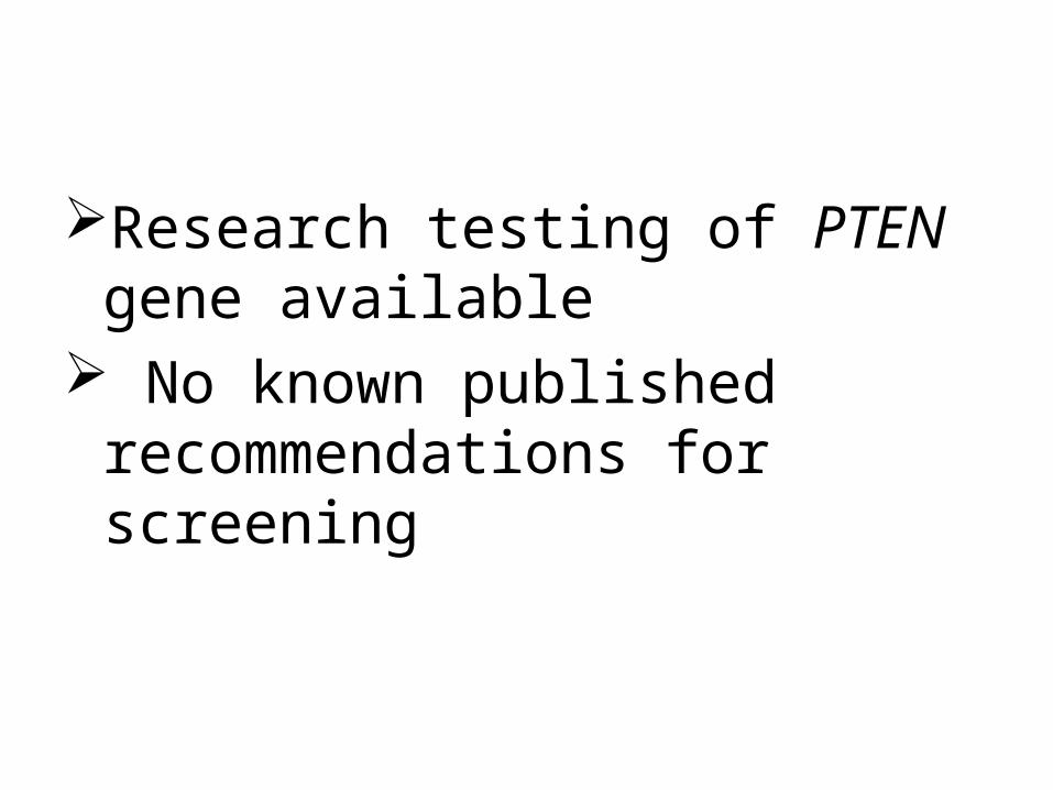
Research testing of PTEN gene available
No known published recommendations for screening
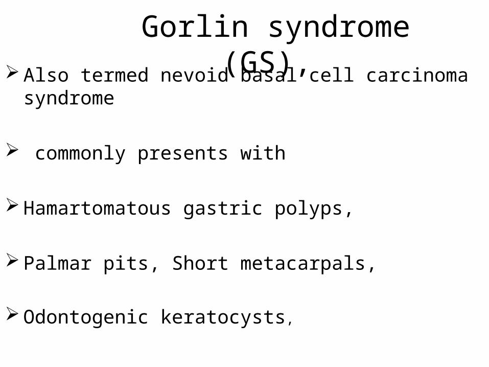
Gorlin syndrome (GS), Also termed nevoid basal cell carcinoma syndrome
commonly presents with
Hamartomatous gastric polyps,
Palmar pits, Short metacarpals,
Odontogenic keratocysts,
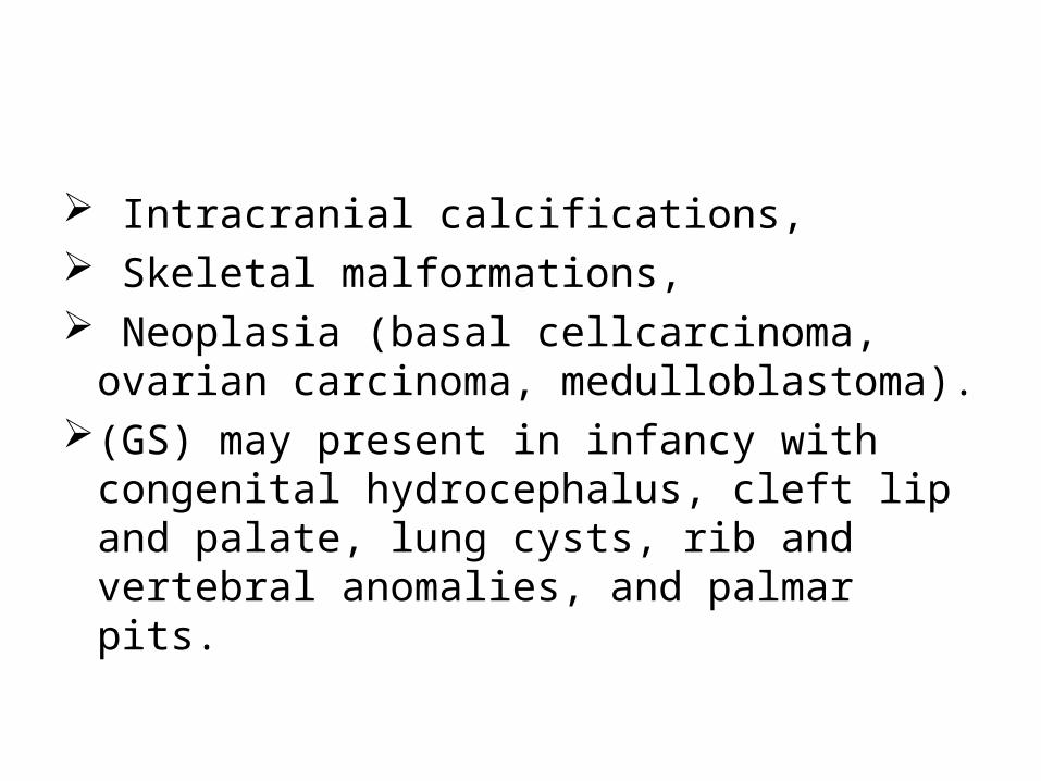
Intracranial calcifications, Skeletal malformations, Neoplasia (basal cellcarcinoma, ovarian
carcinoma, medulloblastoma). (GS) may present in infancy with congenital
hydrocephalus, cleft lip and palate, lung cysts, rib and vertebral anomalies, and palmar pits.
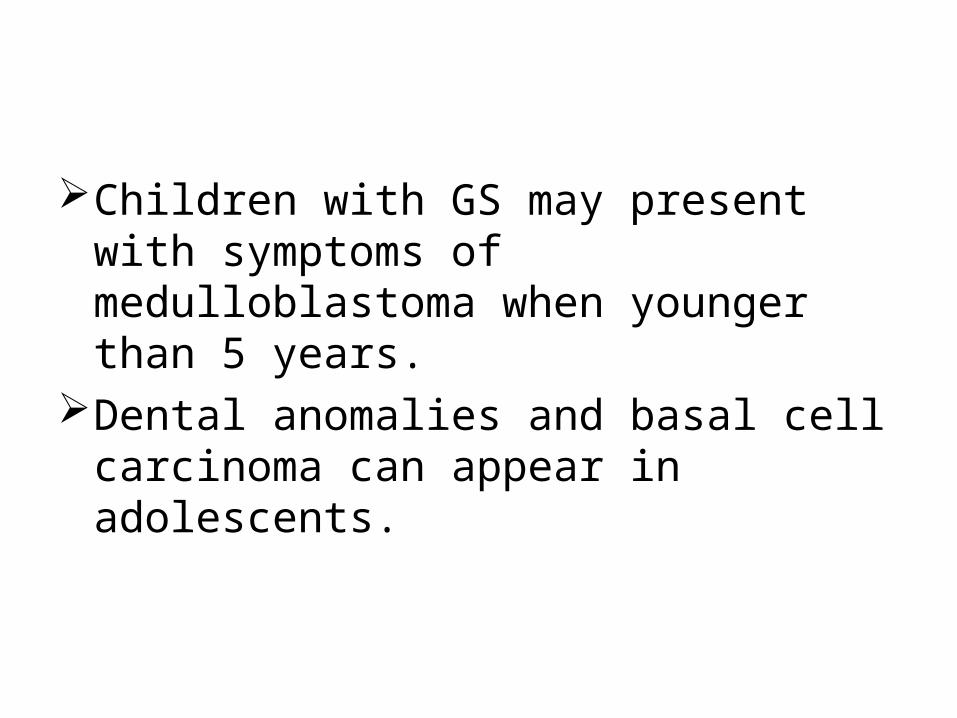
Children with GS may present with symptoms of medulloblastoma when younger than 5 years.
Dental anomalies and basal cell carcinoma can appear in adolescents.
• Patients with GS may require surgical management for the following:
• Craniofacial lesions (cleft lip and palate, jaw cysts, other mandibular lesions)
• Abdominal masses (mesenteric cysts, lymphatic cysts, ovarian fibromas)
• Diagnostic and therapeutic interventions for potential neoplasia within the CNS (medulloblastoma), skin (basal cell carcinoma), jaw (fibrosarcoma), ovaries (fibrosarcoma), and endometrium (adenocarcinoma)
•
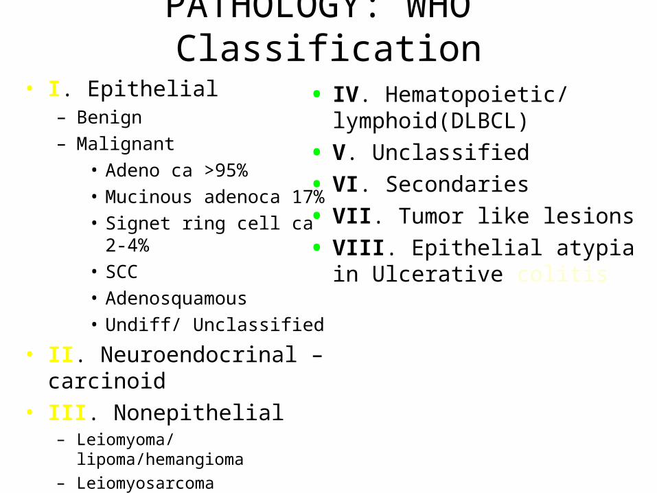
PATHOLOGY: WHO Classification
• I. Epithelial– Benign
– Malignant
• Adeno ca >95%
• Mucinous adenoca 17%
• Signet ring cell ca 2-4%
• SCC
• Adenosquamous
• Undiff/ Unclassified
• II. Neuroendocrinal – carcinoid
• III. Nonepithelial– Leiomyoma/ lipoma/hemangioma
– Leiomyosarcoma
• IV. Hematopoietic/ lymphoid(DLBCL)
• V. Unclassified
• VI. Secondaries
• VII. Tumor like lesions
• VIII. Epithelial atypia in Ulcerative colitis
Prevention
• Guidelines proposed by American College of Gastroenterology (ACG):
• A diet that is low in fat and high in fruits, vegetables, and fiber. There may be advantages with cruciferous vegetables and unprocessed forms of cereal fiber.
• Maintenance of normal body weight through regular exercise and caloric restriction.
• Avoidance of smoking and excessive alcohol use, especially beer.
• Dietary supplementation with 3 g of Calcium Carbonate.
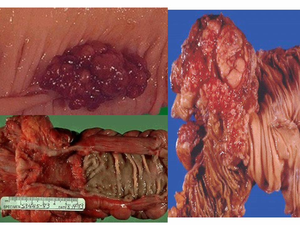
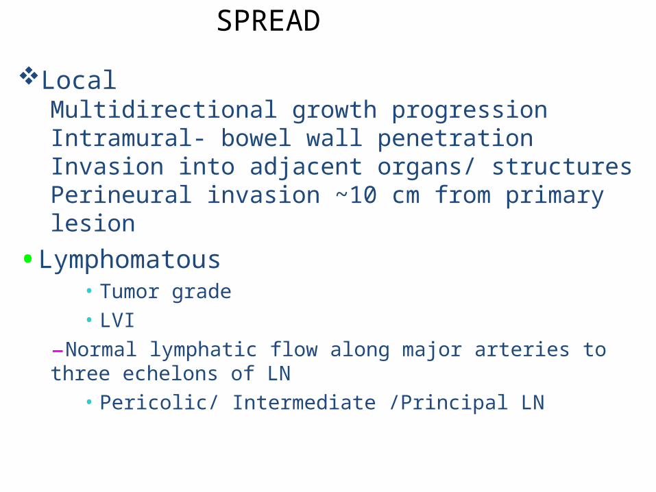
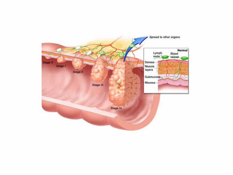
SPREAD
LocalMultidirectional growth progressionIntramural- bowel wall penetrationInvasion into adjacent organs/ structuresPerineural invasion ~10 cm from primary lesion
•Lymphomatous• Tumor grade
• LVI
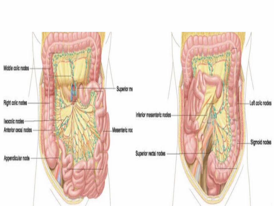
–Normal lymphatic flow along major arteries to three echelons of LN
• Pericolic/ Intermediate /Principal LN
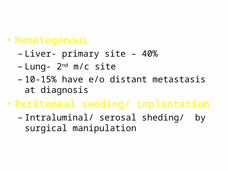
• Hematogenous– Liver- primary site – 40%
– Lung- 2nd m/c site
– 10-15% have e/o distant metastasis at diagnosis
• Peritoneal seeding/ implantation– Intraluminal/ serosal sheding/ by surgical
manipulation
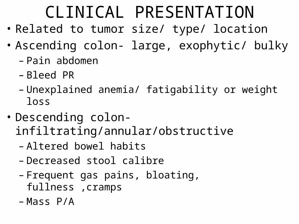
CLINICAL PRESENTATION• Related to tumor size/ type/ location
• Ascending colon- large, exophytic/ bulky– Pain abdomen
– Bleed PR
– Unexplained anemia/ fatigability or weight loss
• Descending colon- infiltrating/annular/obstructive– Altered bowel habits
– Decreased stool calibre
– Frequent gas pains, bloating, fullness ,cramps
– Mass P/A
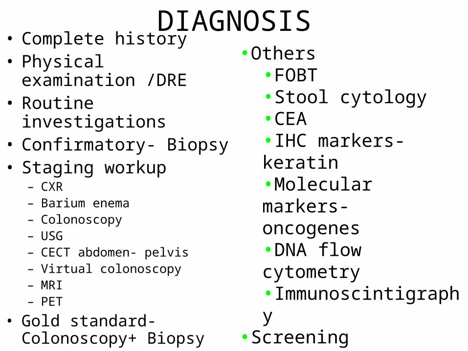
DIAGNOSIS• Complete history• Physical examination /DRE• Routine investigations• Confirmatory- Biopsy• Staging workup
– CXR– Barium enema– Colonoscopy– USG– CECT abdomen- pelvis– Virtual colonoscopy– MRI– PET
• Gold standard- Colonoscopy+ Biopsy
•Others•FOBT•Stool cytology •CEA•IHC markers- keratin•Molecular markers- oncogenes•DNA flow cytometry •Immunoscintigraphy
•Screening investigations
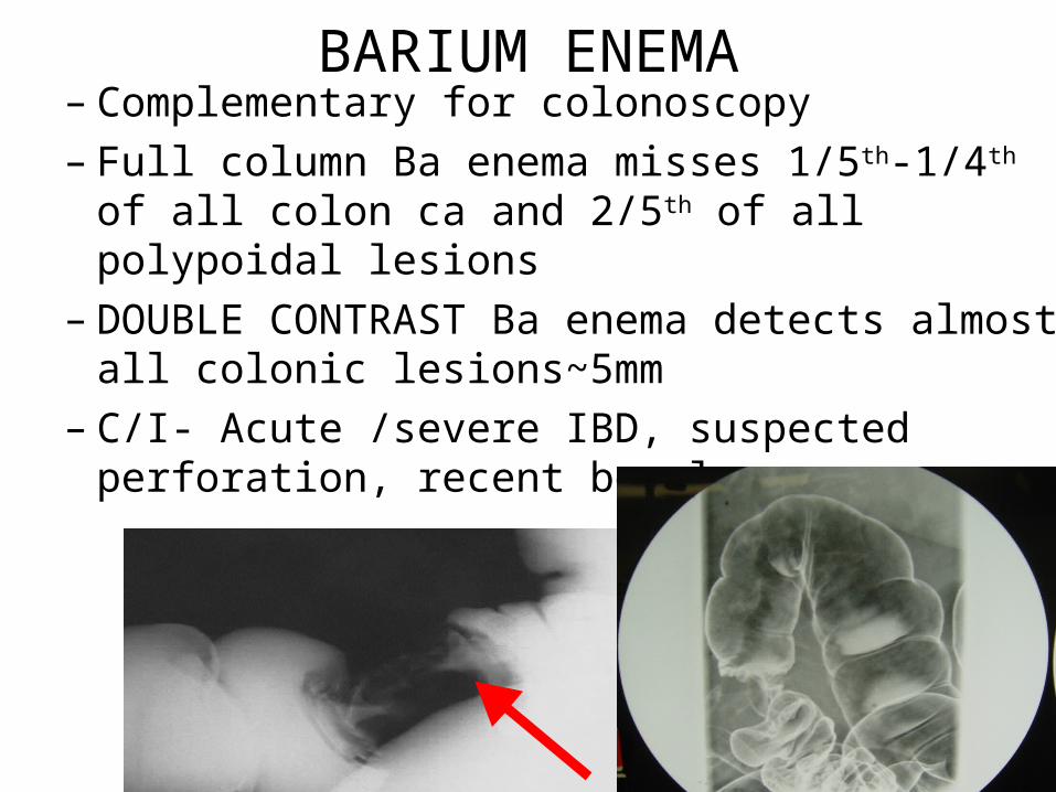
BARIUM ENEMA– Complementary for colonoscopy
– Full column Ba enema misses 1/5th-1/4th of all colon ca and 2/5th of all polypoidal lesions
– DOUBLE CONTRAST Ba enema detects almost all colonic lesions~5mm
– C/I- Acute /severe IBD, suspected perforation, recent bowel surgery
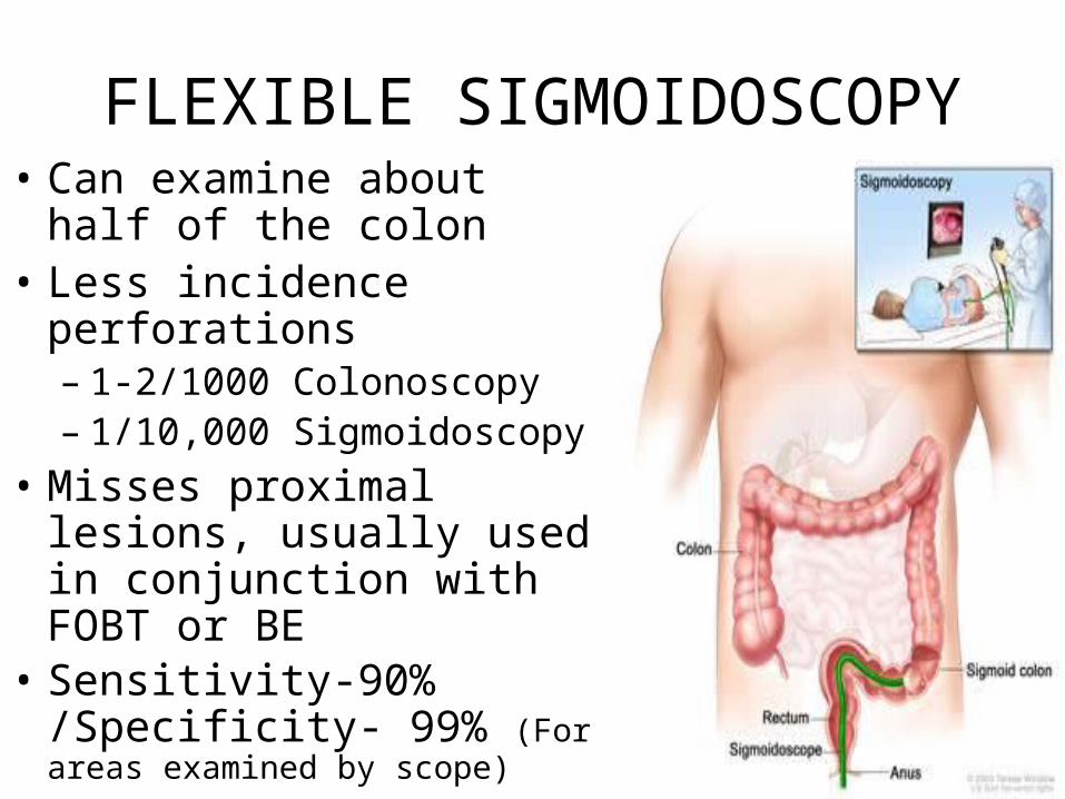
FLEXIBLE SIGMOIDOSCOPY• Can examine about half of
the colon• Less incidence perforations
– 1-2/1000 Colonoscopy– 1/10,000 Sigmoidoscopy
• Misses proximal lesions, usually used in conjunction with FOBT or BE
• Sensitivity-90% /Specificity- 99% (For areas examined by scope)
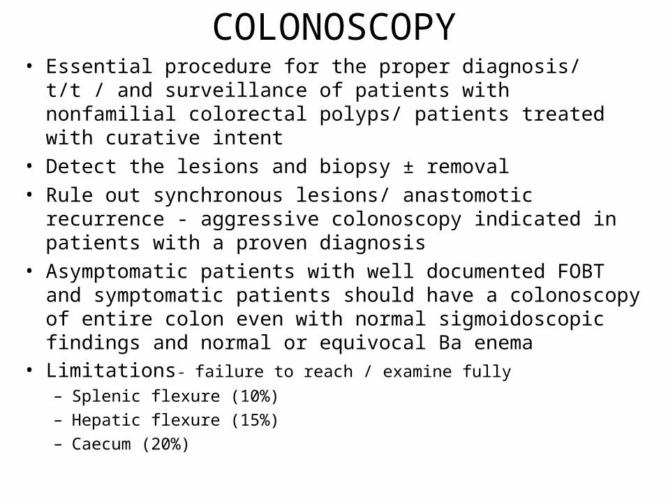
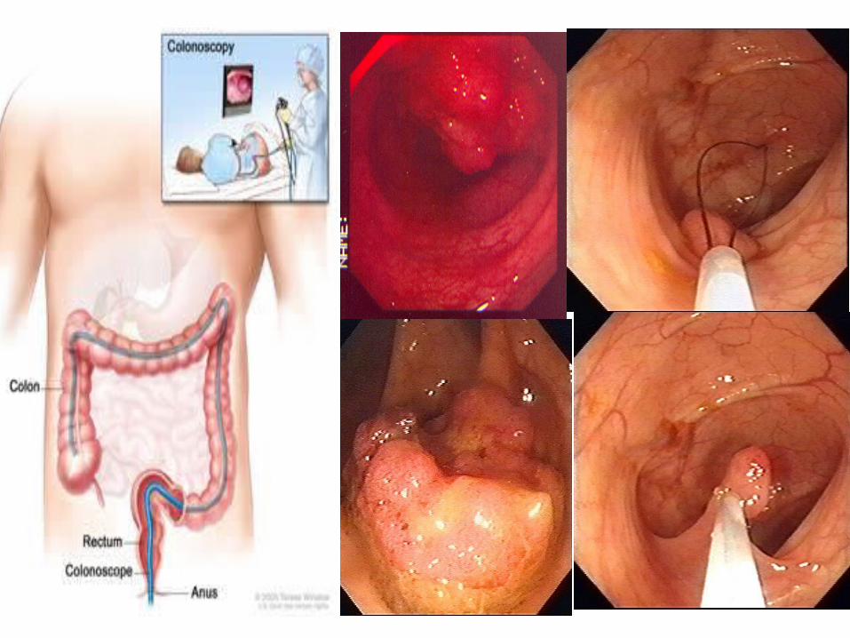
COLONOSCOPY• Essential procedure for the proper diagnosis/ t/t / and surveillance
of patients with nonfamilial colorectal polyps/ patients treated with curative intent
• Detect the lesions and biopsy ± removal
• Rule out synchronous lesions/ anastomotic recurrence - aggressive colonoscopy indicated in patients with a proven diagnosis
• Asymptomatic patients with well documented FOBT and symptomatic patients should have a colonoscopy of entire colon even with normal sigmoidoscopic findings and normal or equivocal Ba enema
• Limitations- failure to reach / examine fully
– Splenic flexure (10%)
– Hepatic flexure (15%)
– Caecum (20%)
• VIRTUAL COLONOSCOPY :Thin section helical CT using air contrast and glucagon for bowel sedation and has same efficacy to standard colonoscopy in detecting lesions≥ 6mm.
• USG: minimal role of TAUSG– Useful when augmented with retrograde instillation of
water into colon k/a HYDROCOLONIC SONOGRAPHY
which permits detailed evaluation of bowel wall permitting more precise preop staging
CECT ABDOMEN/ PELVIS • More accurate for T4 than T2/3 lesions
• Asses extraluminal extent of locally advanced dis.
• Limitation: can't detect– Tumor infiltration of pericolic fat
– Focal tumor spread external to muscularis propria
– Pericolonic LN <1 cm
– Liver mets <1cm
• Overall accuracy staging primary – 70%
• Sensitivity LN detection- 45%
• FDG-PET – To asses response to RT/CCT
– Detect occult areas of recurrent CRC patients considered for re exploration
• MRI- not superior to CT
• TUMOR MARKER : CEA– No role in diagnosis/ screening of ca colon
– Correlate with tumor burden and prognosis
– Monitoring tool for patients treated with curative intent
– Post op CEA level is more sensitive indicator of recurrence
– Normal :
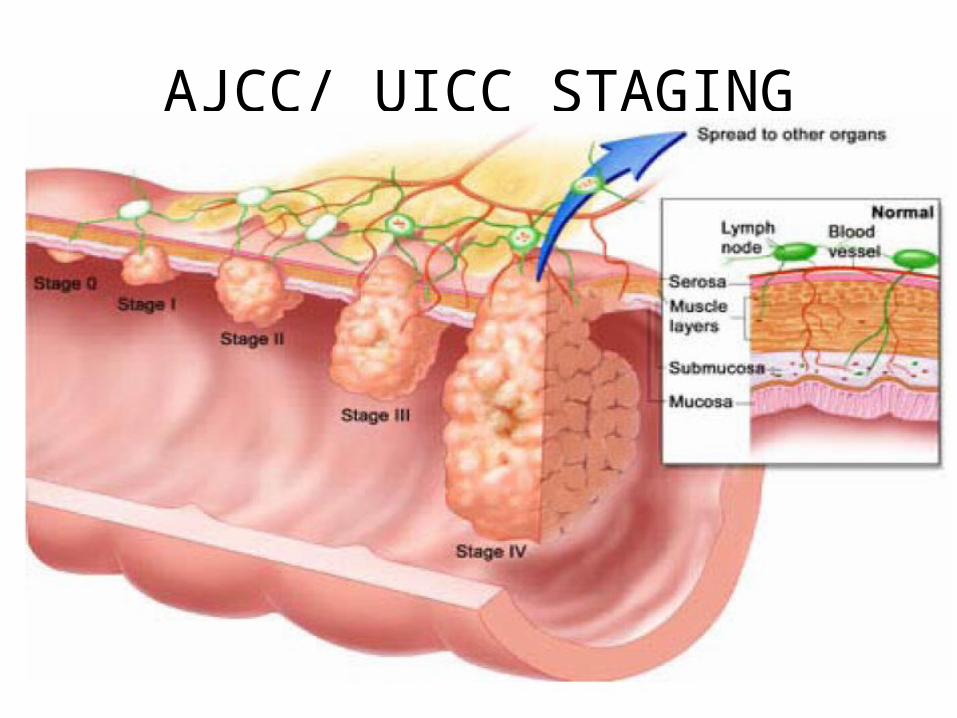
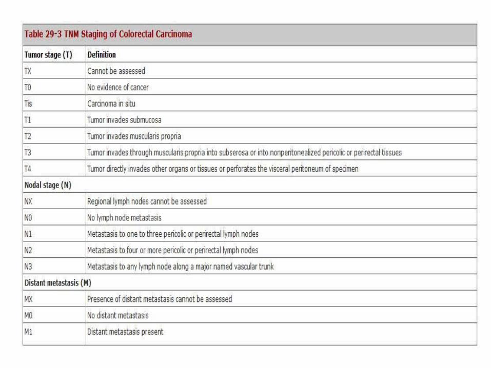
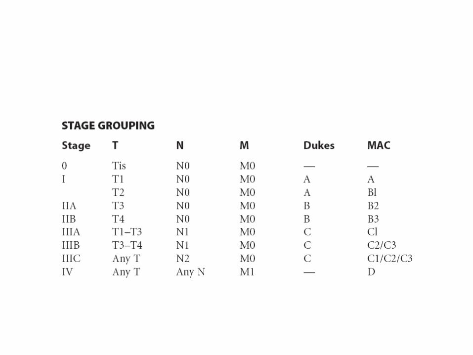
AJCC/ UICC STAGING
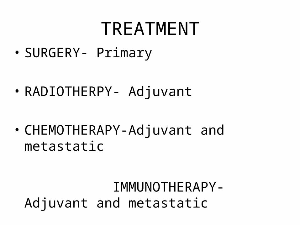
TREATMENT• SURGERY- Primary
• RADIOTHERPY- Adjuvant
• CHEMOTHERAPY-Adjuvant and metastatic
• TARGETED / IMMUNOTHERAPY- Adjuvant and metastatic
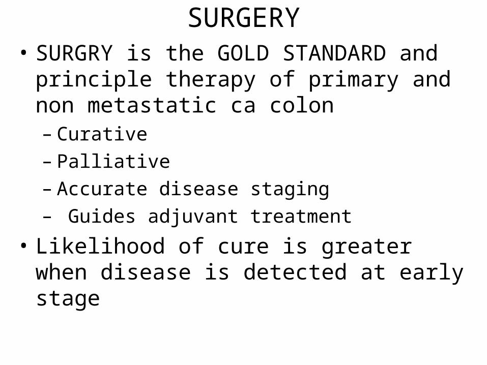
SURGERY• SURGRY is the GOLD STANDARD and
principle therapy of primary and non metastatic ca colon– Curative
– Palliative
– Accurate disease staging
– Guides adjuvant treatment
• Likelihood of cure is greater when disease is detected at early stage
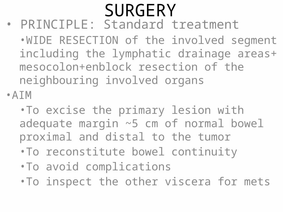
SURGERY• PRINCIPLE: Standard treatment
•WIDE RESECTION of the involved segment including the lymphatic drainage areas+ mesocolon+enblock resection of the neighbouring involved organs
•AIM•To excise the primary lesion with adequate margin ~5 cm of normal bowel proximal and distal to the tumor•To reconstitute bowel continuity •To avoid complications•To inspect the other viscera for mets
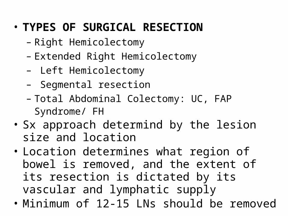
• TYPES OF SURGICAL RESECTION– Right Hemicolectomy
– Extended Right Hemicolectomy
– Left Hemicolectomy
– Segmental resection
– Total Abdominal Colectomy: UC, FAP Syndrome/ FH • Sx approach determind by the lesion size and
location• Location determines what region of bowel is
removed, and the extent of its resection is dictated by its vascular and lymphatic supply
• Minimum of 12-15 LNs should be removed
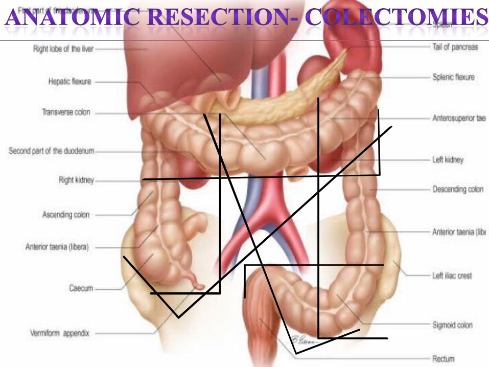
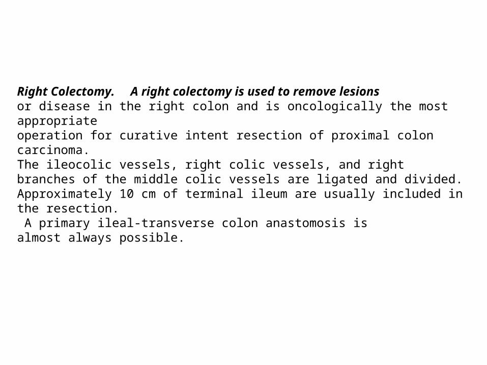
Right Colectomy. A right colectomy is used to remove lesionsor disease in the right colon and is oncologically the most appropriateoperation for curative intent resection of proximal coloncarcinoma. The ileocolic vessels, right colic vessels, and rightbranches of the middle colic vessels are ligated and divided.Approximately 10 cm of terminal ileum are usually included inthe resection. A primary ileal-transverse colon anastomosis isalmost always possible.
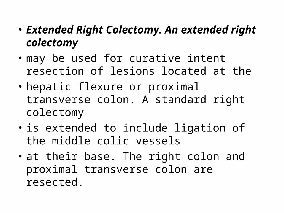
• Extended Right Colectomy. An extended right colectomy
• may be used for curative intent resection of lesions located at the
• hepatic flexure or proximal transverse colon. A standard right colectomy
• is extended to include ligation of the middle colic vessels
• at their base. The right colon and proximal transverse colon are resected.
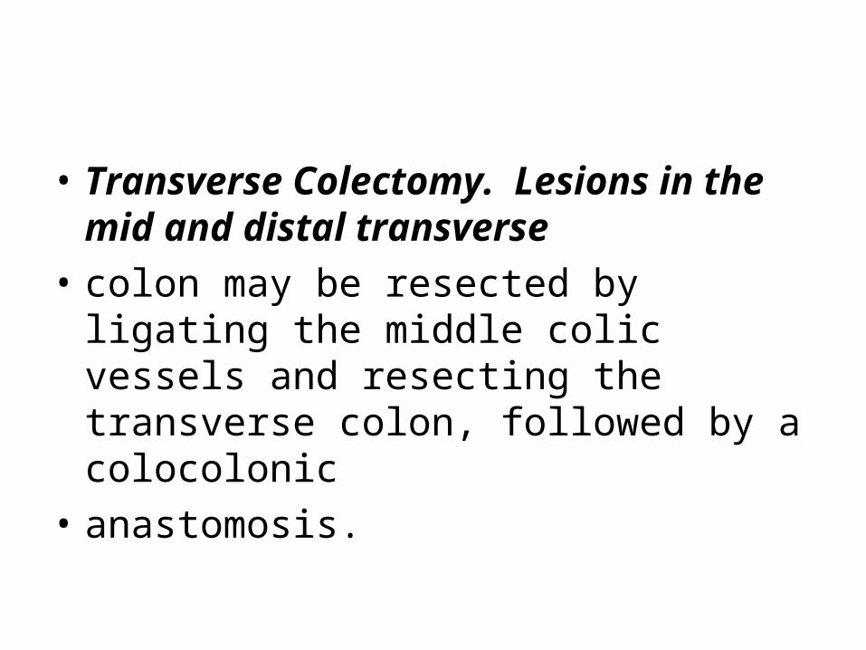
• Transverse Colectomy. Lesions in the mid and distal transverse
• colon may be resected by ligating the middle colic vessels and resecting the transverse colon, followed by a colocolonic
• anastomosis.
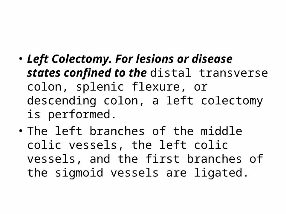
• Left Colectomy. For lesions or disease states confined to the distal transverse colon, splenic flexure, or descending colon, a left colectomy is performed.
• The left branches of the middle colic vessels, the left colic vessels, and the first branches of the sigmoid vessels are ligated.
• Extended Left Colectomy. An extended left colectomy is an option for removing lesions in the distal transverse colon.
• In this operation, the left colectomy is extended proximally to include the right branches of the middle colic vessels.
• Sigmoid Colectomy. Lesions in the sigmoid colon require
• ligation and division of the sigmoid branches of the inferior mesenteric artery. In general, the entire sigmoid colon should be resected to the level of the peritoneal reflection and an anastomosis created between the descending colon and upper rectum.
• .
• Full mobilization of the splenic flexure is often required to create a tension-free anastomosis.
• Total and Subtotal Colectomy. Total or subtotal colectomy is occasionally required for patients with fulminant colitis, attenuatedFAP, or synchronous colon carcinomas.
• In this procedure, the ileocolic vessels, right colic vessels, middle colic vessels,and left colic vessels are ligated and divided.
• The superior rectal vessels are preserved. If it is desired to preserve the sigmoid, the distal sigmoid vessels are left intact, and an anastomosis is created between the ileum and distal sigmoid colon (subtotal
• colectomy with ileosigmoid anastomosis). • .
• If the sigmoid is to be resected, the sigmoidal vessels are ligated and divided, and
• the ileum is anastomosed to the upper rectum (total abdominal colectomy with ileorectal anastomosis).
• If an anastomosis is contraindicated, an end ileostomy is created, and the remaining sigmoid or rectum is managed either as a mucus fistula or a Hartmann’s pouch
• Total Proctocolectomy. In this procedure, the entire colon, rectum, and anus are removed and the ileum is brought to the skin as a Brooke ileostomy

ADJUVANT THERAPYBASIS• Despite curative surgery half of these patients suffer
INCURABLE TUMOR RECURRENCE leading to cancer related death
• Therefore there is a need of adjuvant therapy to improve DFS and OS
• Establishment of adjuvant therapy as a standard treatment in stage III colon cancer based on improvement in overall survival
• In stage II colon cancer adjuvant treatment remains controversial
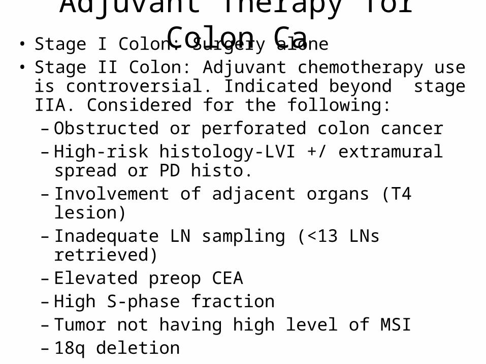
Adjuvant Therapy for Colon Ca• Stage I Colon: Surgery alone• Stage II Colon: Adjuvant chemotherapy use is
controversial. Indicated beyond stage IIA. Considered for the following:– Obstructed or perforated colon cancer– High-risk histology-LVI +/ extramural spread or PD
histo.– Involvement of adjacent organs (T4 lesion)– Inadequate LN sampling (<13 LNs retrieved)– Elevated preop CEA– High S-phase fraction– Tumor not having high level of MSI– 18q deletion
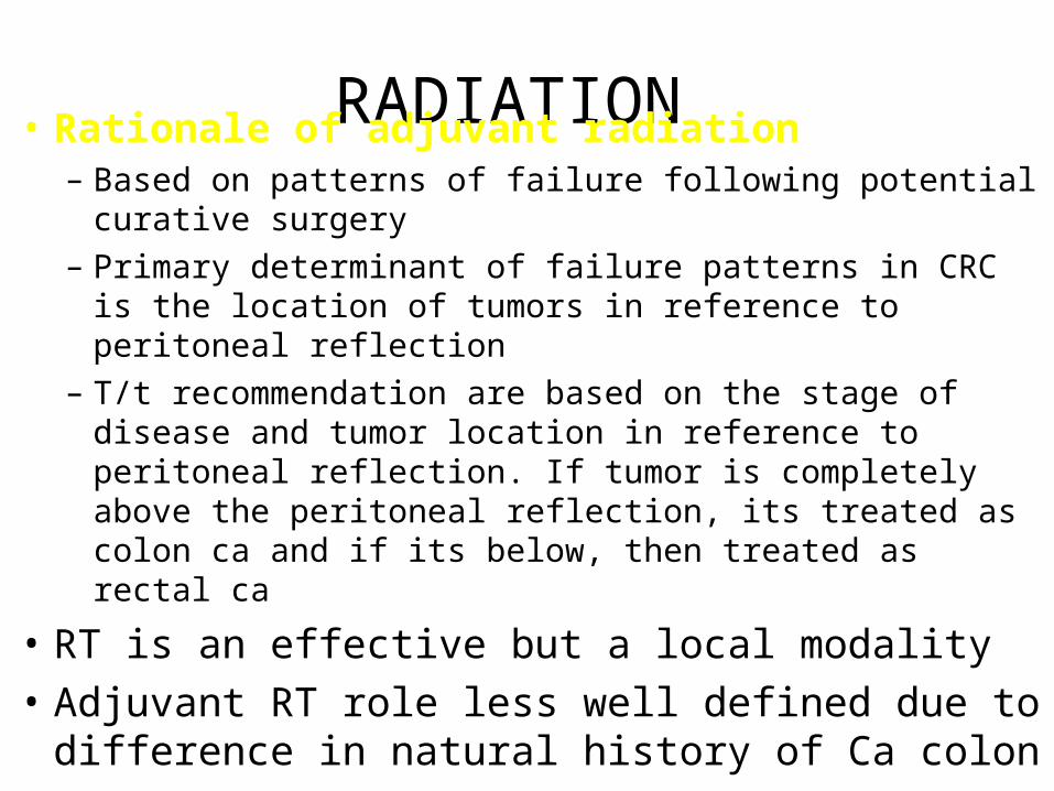
RADIATION • Rationale of adjuvant radiation – Based on patterns of failure following potential curative
surgery
– Primary determinant of failure patterns in CRC is the location of tumors in reference to peritoneal reflection
– T/t recommendation are based on the stage of disease and tumor location in reference to peritoneal reflection. If tumor is completely above the peritoneal reflection, its treated as colon ca and if its below, then treated as rectal ca
• RT is an effective but a local modality
• Adjuvant RT role less well defined due to difference in natural history of Ca colon
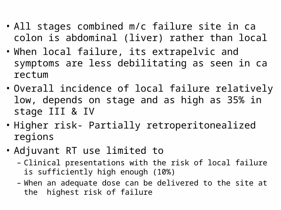
• All stages combined m/c failure site in ca colon is abdominal (liver) rather than local
• When local failure, its extrapelvic and symptoms are less debilitating as seen in ca rectum
• Overall incidence of local failure relatively low, depends on stage and as high as 35% in stage III & IV
• Higher risk- Partially retroperitonealized regions
• Adjuvant RT use limited to – Clinical presentations with the risk of local failure is sufficiently
high enough (10%)
– When an adequate dose can be delivered to the site at the highest risk of failure
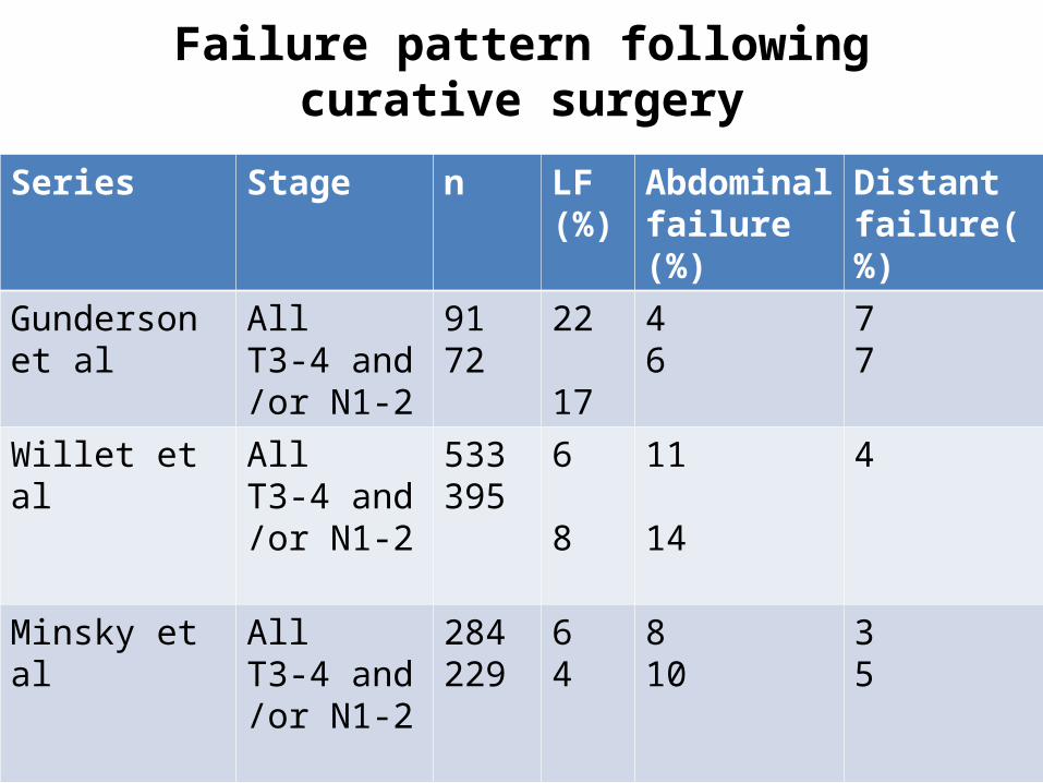
Failure pattern following curative surgery
Series Stage n LF (%)
Abdominal failure (%)
Distant failure(%)
Gunderson et al
All T3-4 and /or N1-2
9172
22
17
46
77
Willet et al All T3-4 and /or N1-2
533395
6
8
11
14
4
Minsky et al All T3-4 and /or N1-2
284229
64
810
35
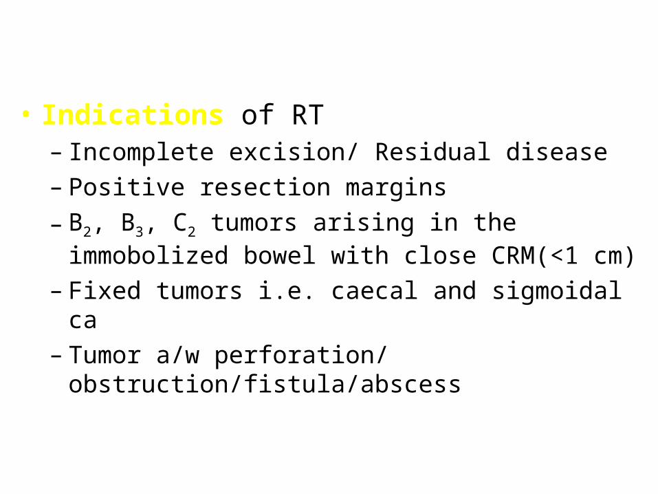
• Indications of RT– Incomplete excision/ Residual disease
– Positive resection margins
– B2, B3, C2 tumors arising in the immobolized bowel with close CRM(<1 cm)
– Fixed tumors i.e. caecal and sigmoidal ca
– Tumor a/w perforation/ obstruction/fistula/abscess
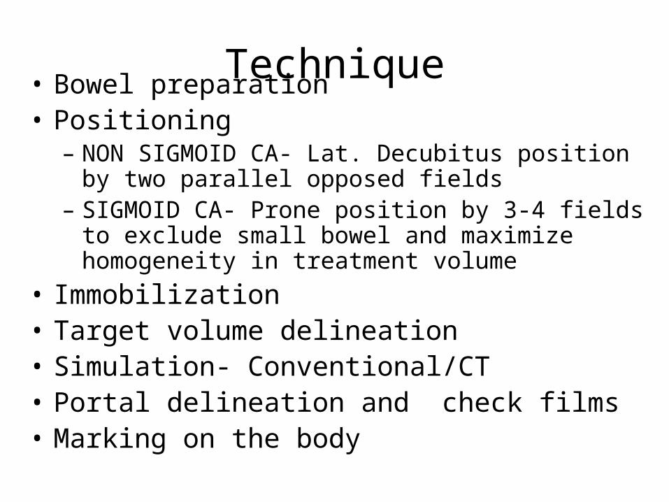
Technique• Bowel preparation• Positioning
– NON SIGMOID CA- Lat. Decubitus position by two parallel opposed fields
– SIGMOID CA- Prone position by 3-4 fields to exclude small bowel and maximize homogeneity in treatment volume
• Immobilization• Target volume delineation• Simulation- Conventional/CT• Portal delineation and check films • Marking on the body
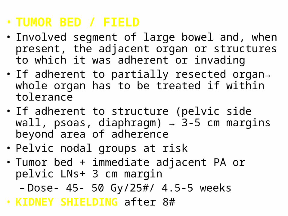
• TUMOR BED / FIELD• Involved segment of large bowel and, when present, the
adjacent organ or structures to which it was adherent or invading
• If adherent to partially resected organ→ whole organ has to be treated if within tolerance
• If adherent to structure (pelvic side wall, psoas, diaphragm) → 3-5 cm margins beyond area of adherence
• Pelvic nodal groups at risk• Tumor bed + immediate adjacent PA or pelvic LNs+ 3 cm
margin– Dose- 45- 50 Gy/25#/ 4.5-5 weeks
• KIDNEY SHIELDING after 8#
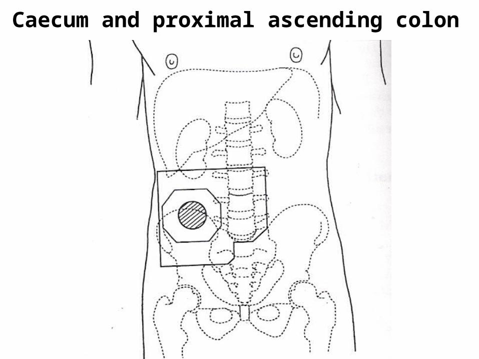
Caecum and proximal ascending colon
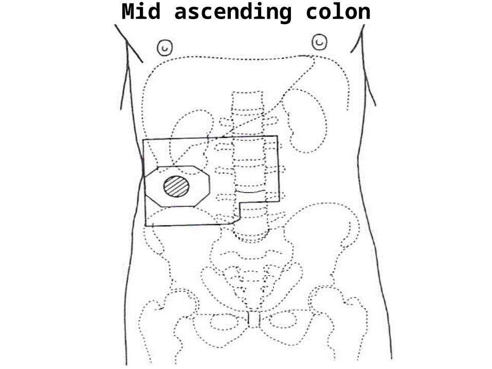
Mid ascending colon
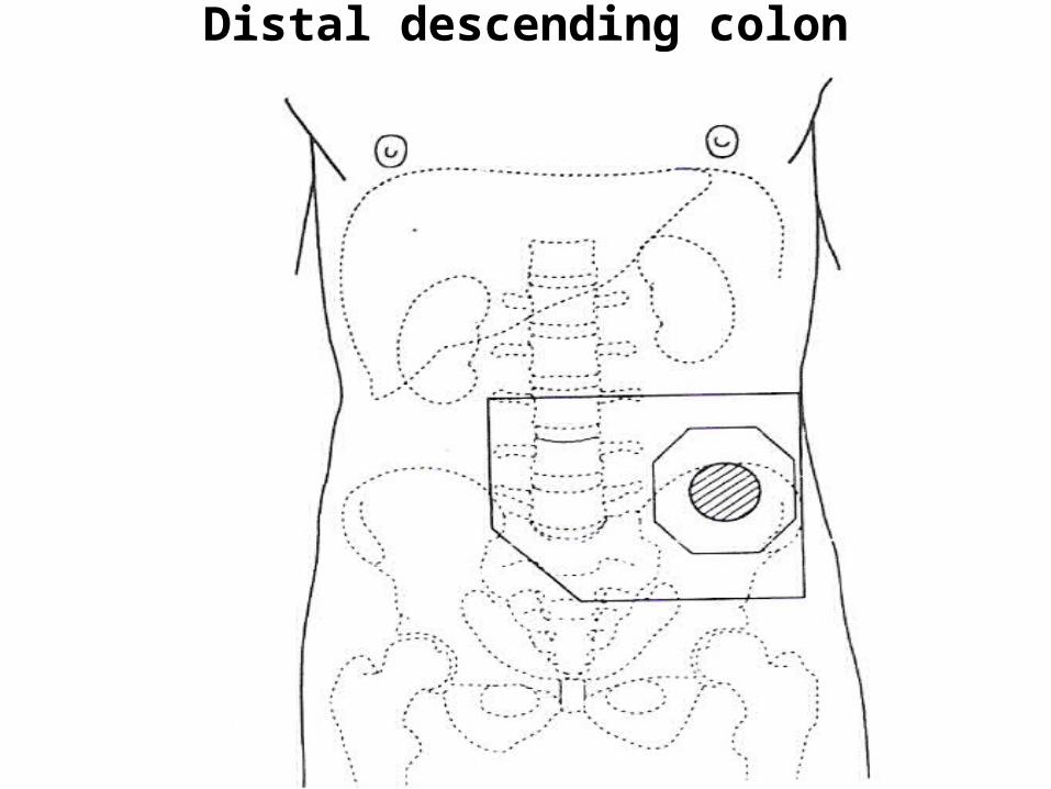
Distal descending colon
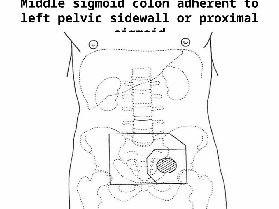
Middle sigmoid colon adherent to left pelvic sidewall or proximal sigmoid
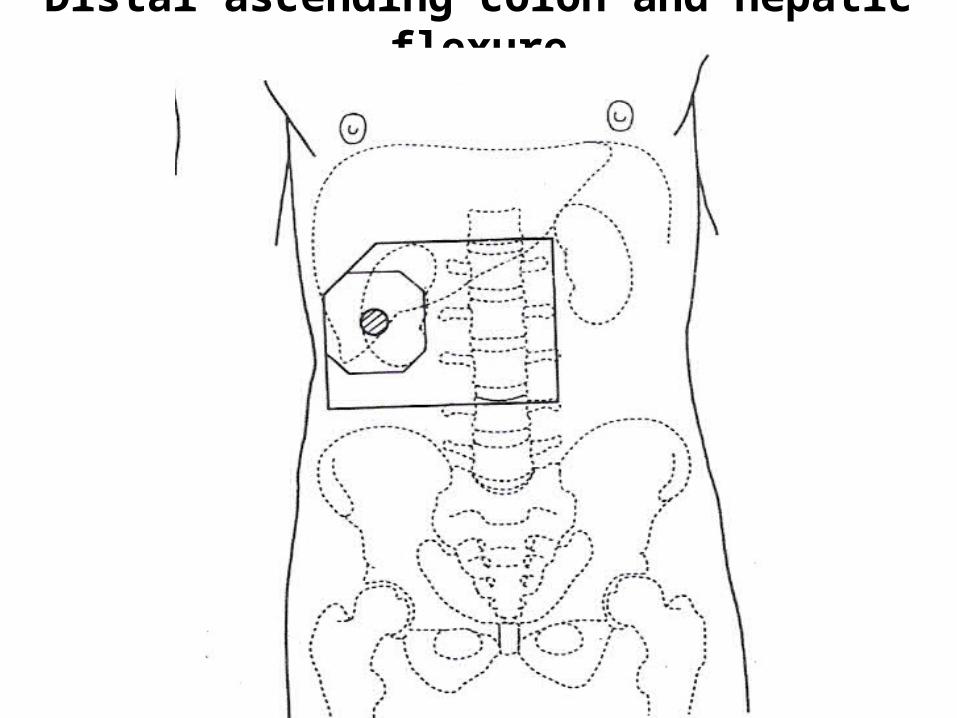
Distal ascending colon and hepatic flexure
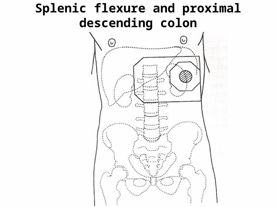
Splenic flexure and proximal descending colon
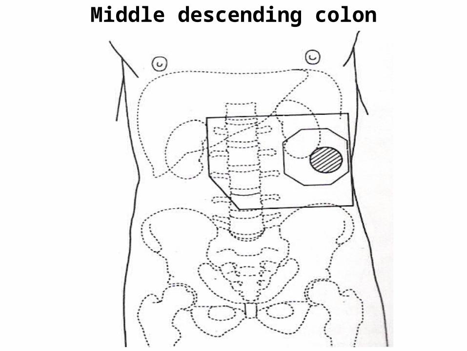
Middle descending colon
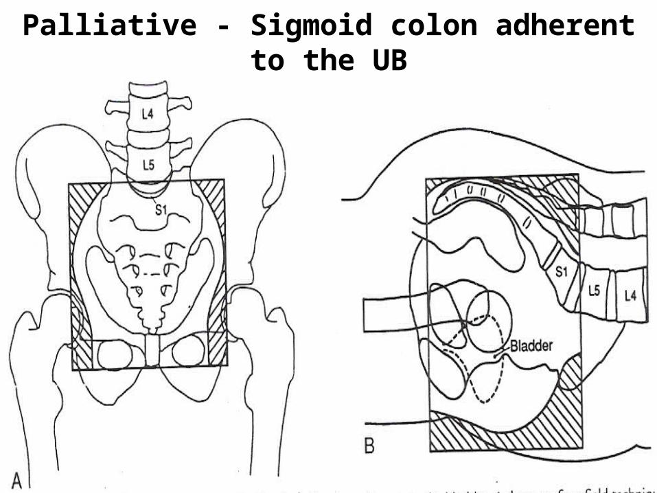
Palliative - Sigmoid colon adherent to the UB
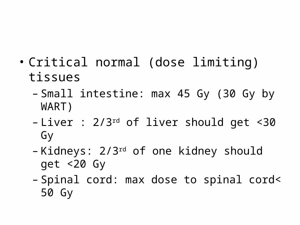
• Critical normal (dose limiting) tissues– Small intestine: max 45 Gy (30 Gy by WART)
– Liver : 2/3rd of liver should get <30 Gy
– Kidneys: 2/3rd of one kidney should get <20 Gy
– Spinal cord: max dose to spinal cord< 50 Gy
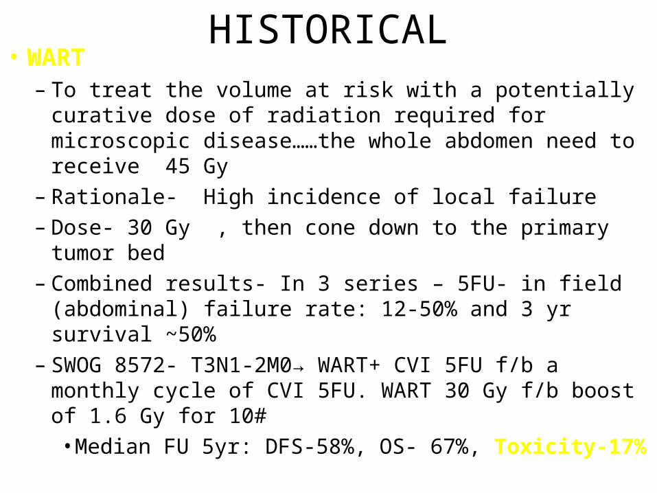
HISTORICAL• WART
– To treat the volume at risk with a potentially curative dose of radiation required for microscopic disease……the whole abdomen need to receive 45 Gy
– Rationale- High incidence of local failure
– Dose- 30 Gy , then cone down to the primary tumor bed
– Combined results- In 3 series – 5FU- in field (abdominal) failure rate: 12-50% and 3 yr survival ~50%
– SWOG 8572- T3N1-2M0→ WART+ CVI 5FU f/b a monthly cycle of CVI 5FU. WART 30 Gy f/b boost of 1.6 Gy for 10#
• Median FU 5yr: DFS-58%, OS- 67%, Toxicity-17%
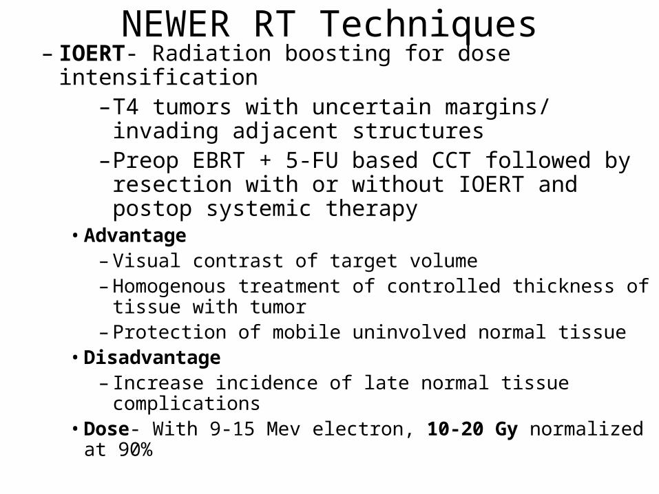
NEWER RT Techniques– IOERT- Radiation boosting for dose intensification
–T4 tumors with uncertain margins/ invading adjacent structures
–Preop EBRT + 5-FU based CCT followed by resection with or without IOERT and postop systemic therapy
• Advantage– Visual contrast of target volume– Homogenous treatment of controlled thickness of tissue
with tumor– Protection of mobile uninvolved normal tissue
• Disadvantage– Increase incidence of late normal tissue complications
• Dose- With 9-15 Mev electron, 10-20 Gy normalized at 90%
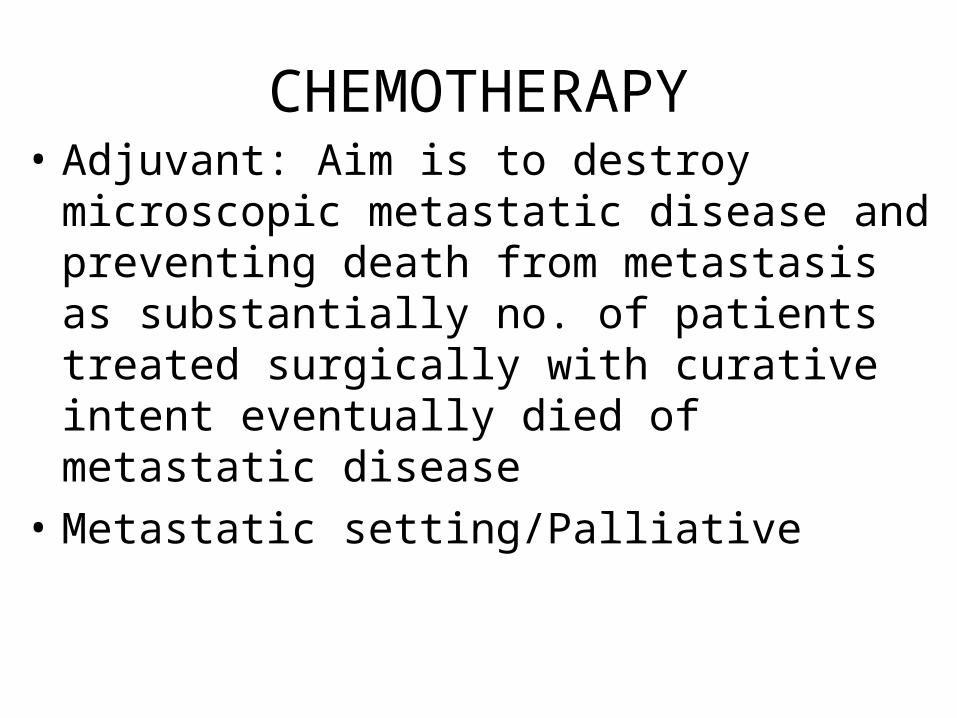
CHEMOTHERAPY• Adjuvant: Aim is to destroy microscopic
metastatic disease and preventing death from metastasis as substantially no. of patients treated surgically with curative intent eventually died of metastatic disease
• Metastatic setting/Palliative

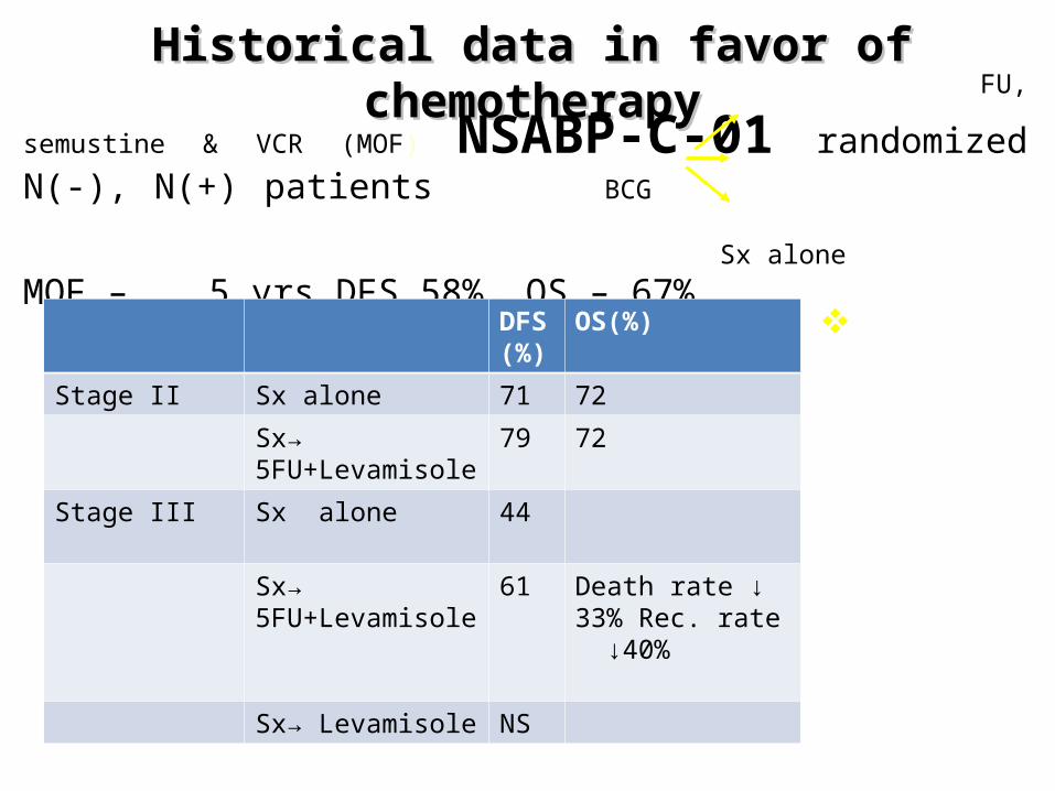
Historical data in favor of chemotherapyHistorical data in favor of chemotherapy FU, semustine & VCR (MOF)
NSABP-C-01 randomized N(-), N(+) patients BCG Sx alone
MOF – 5 yrs DFS 58%, OS – 67%
DFS(%)
OS(%)
Stage II Sx alone 71 72
Sx→ 5FU+Levamisole
79 72
Stage III Sx alone 44
Sx→ 5FU+Levamisole
61 Death rate ↓ 33% Rec. rate ↓40%
Sx→ Levamisole NS
NCI 1990 consensus established adjuvant CCT as standard of care of patients with node (+) resected ca colon
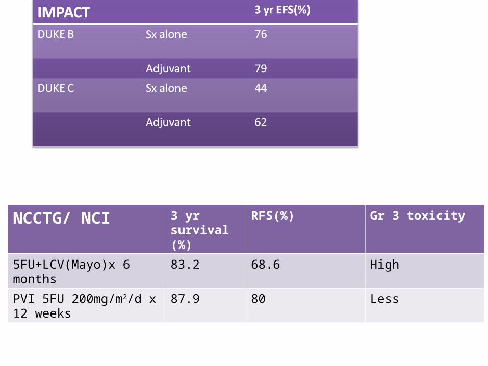
5FU 370-400mg/m2 + LCV 200mg/m2 D1-D5x4 weeklyx6 cycles
NCCTG/ NCI 3 yr survival (%)
RFS(%) Gr 3 toxicity
5FU+LCV(Mayo)x 6 months 83.2 68.6 High
PVI 5FU 200mg/m2/d x 12 weeks
87.9 80 Less
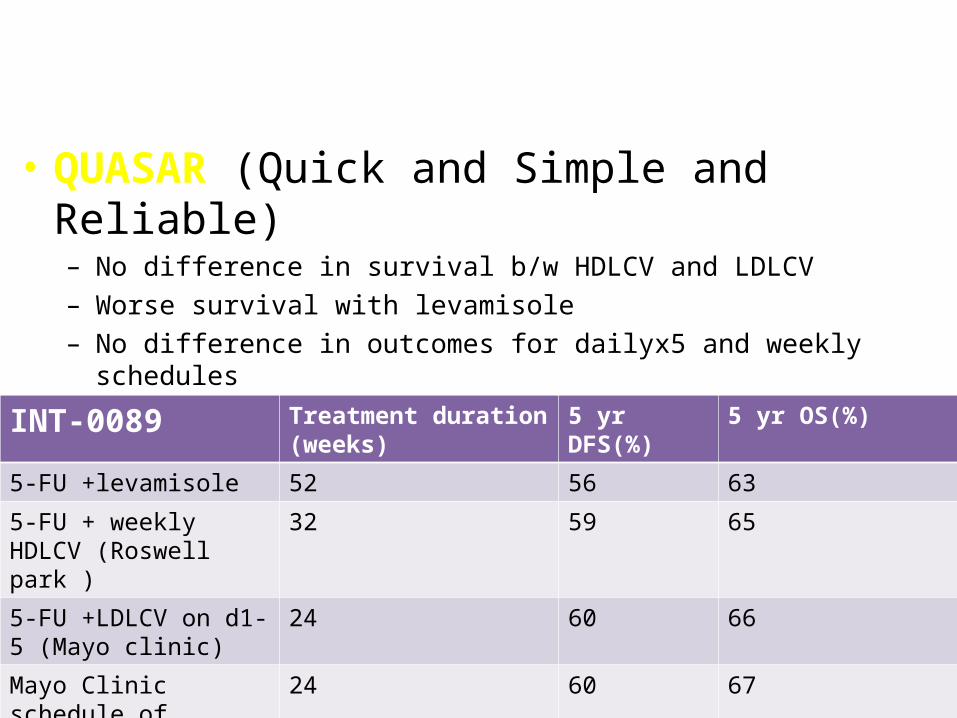
• QUASAR (Quick and Simple and Reliable)– No difference in survival b/w HDLCV and LDLCV– Worse survival with levamisole– No difference in outcomes for dailyx5 and weekly schedules– Once weekly regimen less toxic
INT-0089 Treatment duration (weeks)
5 yr DFS(%) 5 yr OS(%)
5-FU +levamisole 52 56 63
5-FU + weekly HDLCV (Roswell park )
32 59 65
5-FU +LDLCV on d1- 5 (Mayo clinic)
24 60 66
Mayo Clinic schedule of 5-FU/LCV + levamisole.
24 60 67
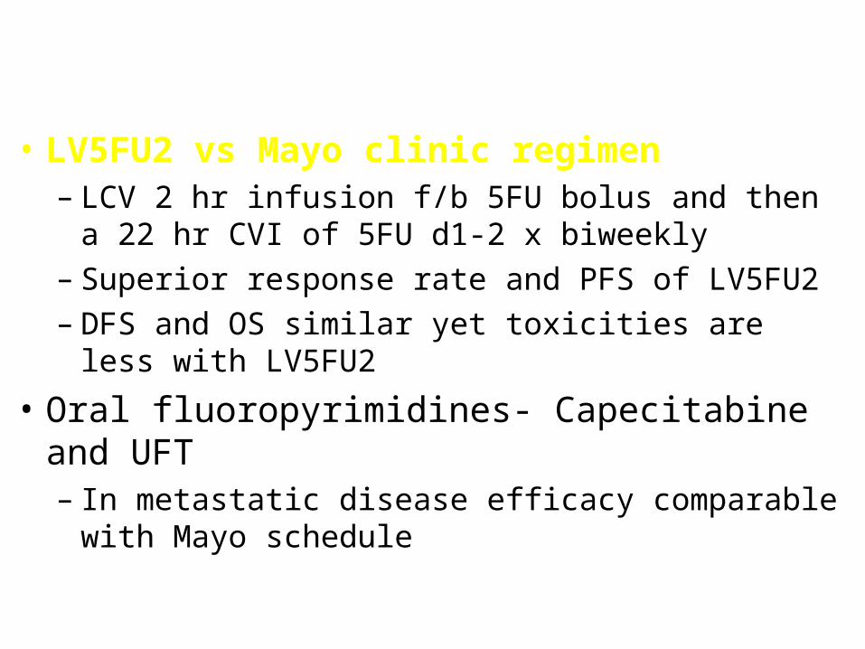
• LV5FU2 vs Mayo clinic regimen– LCV 2 hr infusion f/b 5FU bolus and then a 22 hr CVI of
5FU d1-2 x biweekly– Superior response rate and PFS of LV5FU2 – DFS and OS similar yet toxicities are less with LV5FU2
• Oral fluoropyrimidines- Capecitabine and UFT– In metastatic disease efficacy comparable with Mayo
schedule
MOSAIC Trial: LV5FU2 vs LV5FU2 + FOLFOX-4 LV5FU2 (n = 1,123) (%)
FOLFOX-4 (n = 1,123) (%)
3 yr DFS stage III 66 72
3 yr DFS stage II 84 87
OS NA NA
Grade 3-4 neutropenia 5 41
Neutropenic fever 0 1
Grade 3-4 diarrhea 0 1
Grade 3-4 vomiting 7 11
Neuropathy, any grade 0 92
Neuropathy, grade 3 0 12
Persistent neuropathy, grade 2-3, 1 year after t/t 0 5
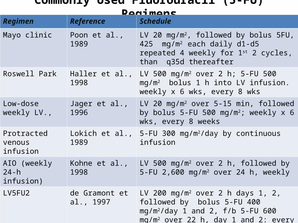
Commonly Used Fluorouracil (5-FU) RegimensRegimen Reference Schedule
Mayo clinic Poon et al., 1989 LV 20 mg/m2, followed by bolus 5FU, 425 mg/m2
each daily d1-d5 repeated 4 weekly for 1st 2 cycles, than q35d thereafter
Roswell Park Haller et al., 1998 LV 500 mg/m2 over 2 h; 5-FU 500 mg/m2 bolus 1 h into LV infusion. weekly x 6 wks, every 8 wks
Low-dose weekly LV.,
Jager et al., 1996 LV 20 mg/m2 over 5-15 min, followed by bolus 5-FU 500 mg/m2; weekly x 6 wks, every 8 weeks
Protracted venous infusion
Lokich et al., 1989 5-FU 300 mg/m2/day by continuous infusion
AIO (weekly 24-h infusion)
Kohne et al., 1998 LV 500 mg/m2 over 2 h, followed by 5-FU 2,600 mg/m2 over 24 h, weekly
LV5FU2 de Gramont et al., 1997
LV 200 mg/m2 over 2 h days 1, 2, followed by bolus 5-FU 400 mg/m2/day 1 and 2, f/b 5-FU 600 mg/m2 over 22 h, day 1 and 2: every 14 days
Simplified LV5FU2 Adapted from Andre et al., 1999
LV 400 mg/m2 over 2 h, followed by bolus 5-FU 400 mg/m2, followed by 5-FU 2,400-3,000 mg/m2 over 46-48 h; cycles repeated every 14 days
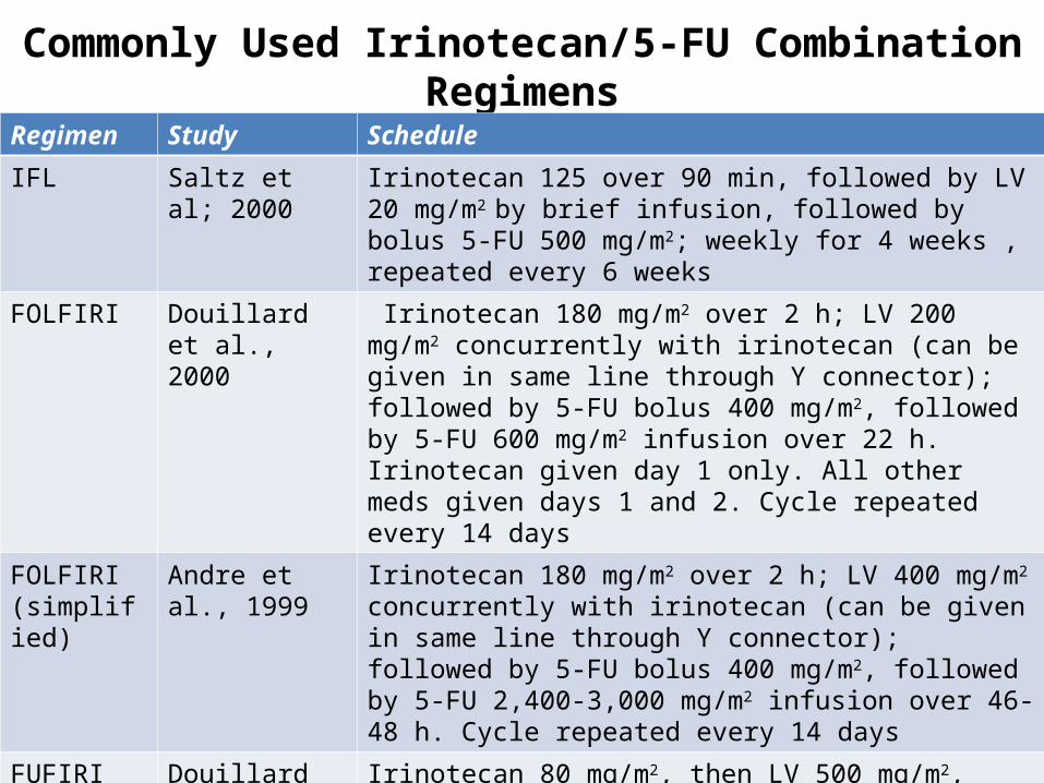
Commonly Used Irinotecan/5-FU Combination Regimens
Regimen Study Schedule
IFL Saltz et al; 2000 Irinotecan 125 over 90 min, followed by LV 20 mg/m2 by brief infusion, followed by bolus 5-FU 500 mg/m2; weekly for 4 weeks , repeated every 6 weeks
FOLFIRI Douillard et al., 2000
Irinotecan 180 mg/m2 over 2 h; LV 200 mg/m2 concurrently with irinotecan (can be given in same line through Y connector); followed by 5-FU bolus 400 mg/m2, followed by 5-FU 600 mg/m2 infusion over 22 h. Irinotecan given day 1 only. All other meds given days 1 and 2. Cycle repeated every 14 days
FOLFIRI (simplified)
Andre et al., 1999
Irinotecan 180 mg/m2 over 2 h; LV 400 mg/m2 concurrently with irinotecan (can be given in same line through Y connector); followed by 5-FU bolus 400 mg/m2, followed by 5-FU 2,400-3,000 mg/m2 infusion over 46-48 h. Cycle repeated every 14 days
FUFIRI Douillard et al., 2000
Irinotecan 80 mg/m2, then LV 500 mg/m2, followed by 5-FU 2,300 mg/m2; all drugs given weekly for 6 weeks, repeated every 7 weeks
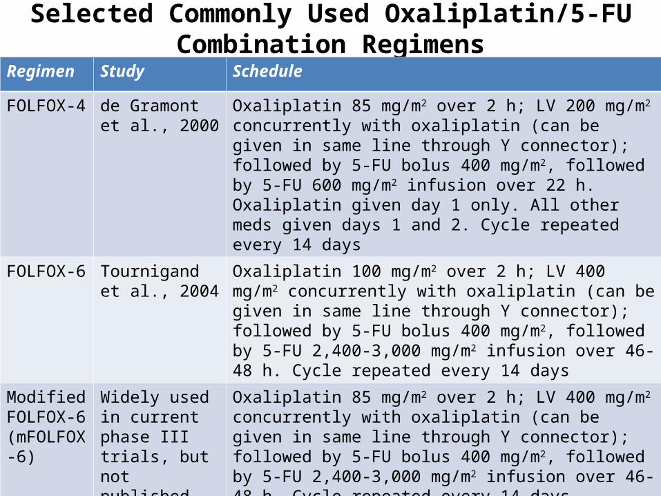
Selected Commonly Used Oxaliplatin/5-FU Combination Regimens
Regimen Study Schedule
FOLFOX-4 de Gramont et al., 2000
Oxaliplatin 85 mg/m2 over 2 h; LV 200 mg/m2 concurrently with oxaliplatin (can be given in same line through Y connector); followed by 5-FU bolus 400 mg/m2, followed by 5-FU 600 mg/m2 infusion over 22 h. Oxaliplatin given day 1 only. All other meds given days 1 and 2. Cycle repeated every 14 days
FOLFOX-6 Tournigand et al., 2004
Oxaliplatin 100 mg/m2 over 2 h; LV 400 mg/m2 concurrently with oxaliplatin (can be given in same line through Y connector); followed by 5-FU bolus 400 mg/m2, followed by 5-FU 2,400-3,000 mg/m2 infusion over 46-48 h. Cycle repeated every 14 days
Modified FOLFOX-6 (mFOLFOX-6)
Widely used in current phase III trials, but not published
Oxaliplatin 85 mg/m2 over 2 h; LV 400 mg/m2 concurrently with oxaliplatin (can be given in same line through Y connector); followed by 5-FU bolus 400 mg/m2, followed by 5-FU 2,400-3,000 mg/m2 infusion over 46-48 h. Cycle repeated every 14 days
FUFOX Grothey et al., 2002
Oxaliplatin 50 mg/m2 over 2 h, followed by LV 500 mg/m2, followed by 5-FU 2,000 mg/m2over 24 h, weekly for 5 weeks, repeated every 6 weeks.
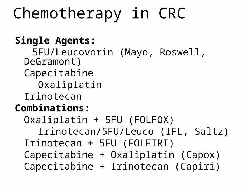
Single Agents: 5FU/Leucovorin (Mayo, Roswell, DeGramont)
Capecitabine Oxaliplatin
IrinotecanCombinations:
Oxaliplatin + 5FU (FOLFOX) Irinotecan/5FU/Leuco (IFL, Saltz)
Irinotecan + 5FU (FOLFIRI)Capecitabine + Oxaliplatin (Capox)Capecitabine + Irinotecan (Capiri)
Chemotherapy in CRC
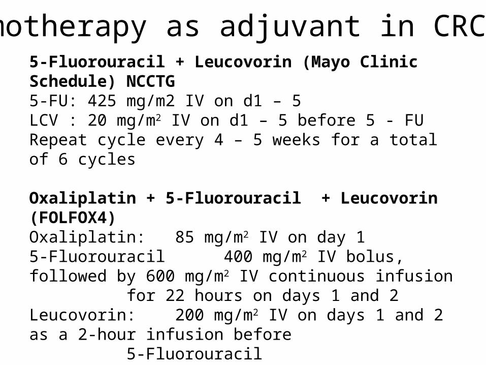
5-Fluorouracil + Leucovorin (Mayo Clinic Schedule) NCCTG5-FU: 425 mg/m2 IV on d1 – 5LCV : 20 mg/m2 IV on d1 – 5 before 5 - FURepeat cycle every 4 – 5 weeks for a total of 6 cycles
Oxaliplatin + 5-Fluorouracil + Leucovorin (FOLFOX4)Oxaliplatin: 85 mg/m2 IV on day 15-Fluorouracil 400 mg/m2 IV bolus, followed by 600 mg/m2 IV continuous infusion for 22 hours on days 1 and 2Leucovorin: 200 mg/m2 IV on days 1 and 2 as a 2-hour infusion before
5-FluorouracilRepeat cycle every 2 weeks
Chemotherapy as adjuvant in CRC
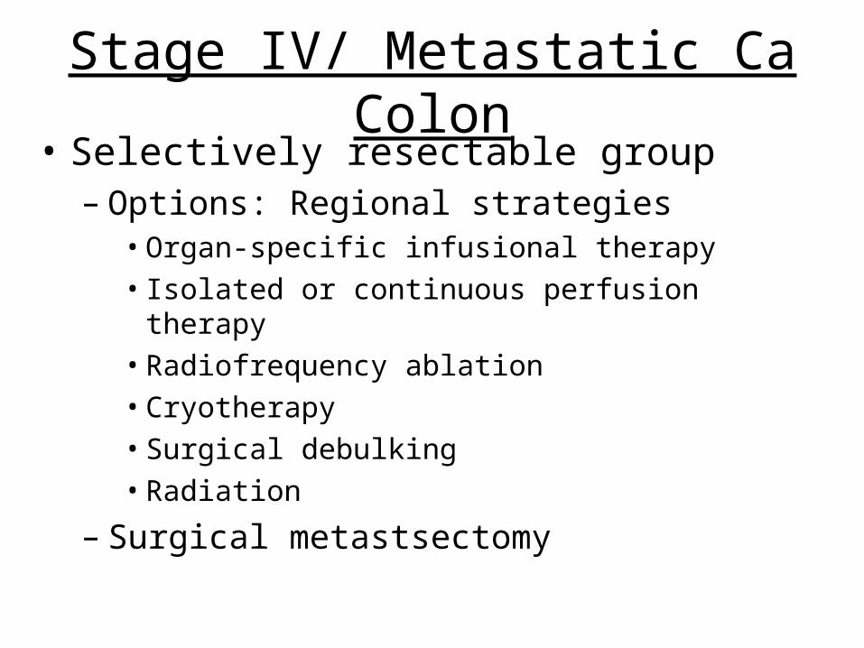
Stage IV/ Metastatic Ca Colon• Selectively resectable group
– Options: Regional strategies• Organ-specific infusional therapy• Isolated or continuous perfusion therapy• Radiofrequency ablation • Cryotherapy• Surgical debulking• Radiation
– Surgical metastsectomy
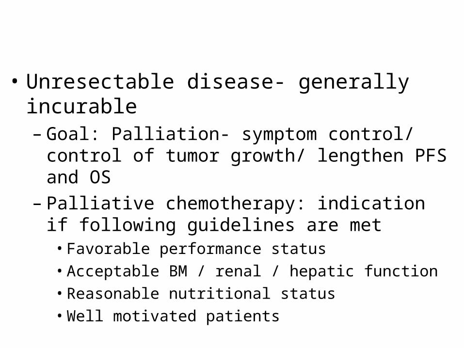
• Unresectable disease- generally incurable– Goal: Palliation- symptom control/ control of tumor
growth/ lengthen PFS and OS– Palliative chemotherapy: indication if following
guidelines are met• Favorable performance status• Acceptable BM / renal / hepatic function• Reasonable nutritional status• Well motivated patients
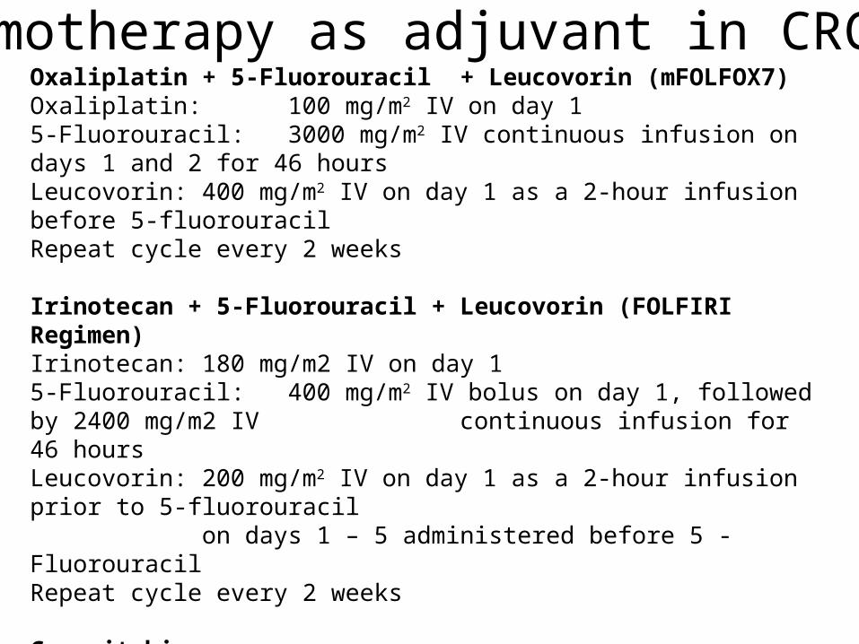
Oxaliplatin + 5-Fluorouracil + Leucovorin (mFOLFOX7)Oxaliplatin: 100 mg/m2 IV on day 15-Fluorouracil: 3000 mg/m2 IV continuous infusion on days 1 and 2 for 46 hoursLeucovorin: 400 mg/m2 IV on day 1 as a 2-hour infusion before 5-fluorouracilRepeat cycle every 2 weeks
Irinotecan + 5-Fluorouracil + Leucovorin (FOLFIRI Regimen)Irinotecan: 180 mg/m2 IV on day 15-Fluorouracil: 400 mg/m2 IV bolus on day 1, followed by 2400 mg/m2 IV
continuous infusion for 46 hoursLeucovorin: 200 mg/m2 IV on day 1 as a 2-hour infusion prior to 5-fluorouracil
on days 1 – 5 administered before 5 - Fluorouracil Repeat cycle every 2 weeks
CapecitabineCapecitabine: 1250 mg/m2 PO bid on days 1 – 14Repeat cycle every 21 days for a total of 8 cycles. Dose may be decreased to 850-1000 mg/m2 PO bid on days 1-14 to reduce the risk of toxicity without compromising clinical efficacy
Chemotherapy as adjuvant in CRC
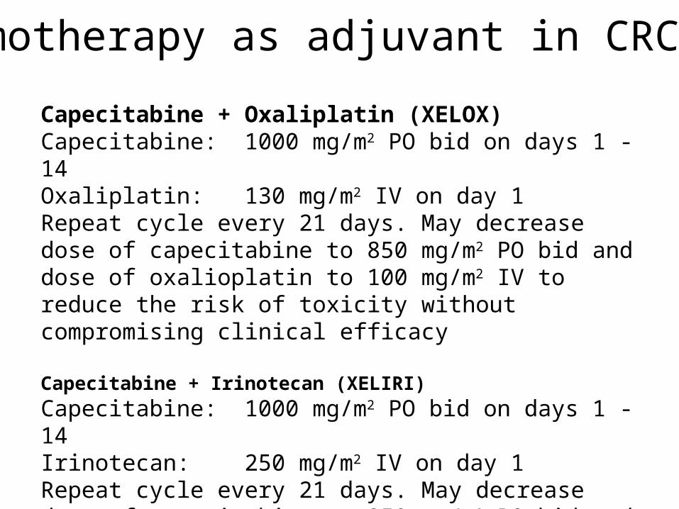
Capecitabine + Oxaliplatin (XELOX)Capecitabine: 1000 mg/m2 PO bid on days 1 - 14Oxaliplatin: 130 mg/m2 IV on day 1Repeat cycle every 21 days. May decrease dose of capecitabine to 850 mg/m2 PO bid and dose of oxalioplatin to 100 mg/m2 IV to reduce the risk of toxicity without compromising clinical efficacy
Capecitabine + Irinotecan (XELIRI)
Capecitabine: 1000 mg/m2 PO bid on days 1 - 14Irinotecan: 250 mg/m2 IV on day 1 Repeat cycle every 21 days. May decrease dose of capecitabine to 850 mg/m2 PO bid and dose of irinotecan to 200 mg/m2 IV to reduce the risk of toxicity without compromising clinical efficacy
Chemotherapy as adjuvant in CRC
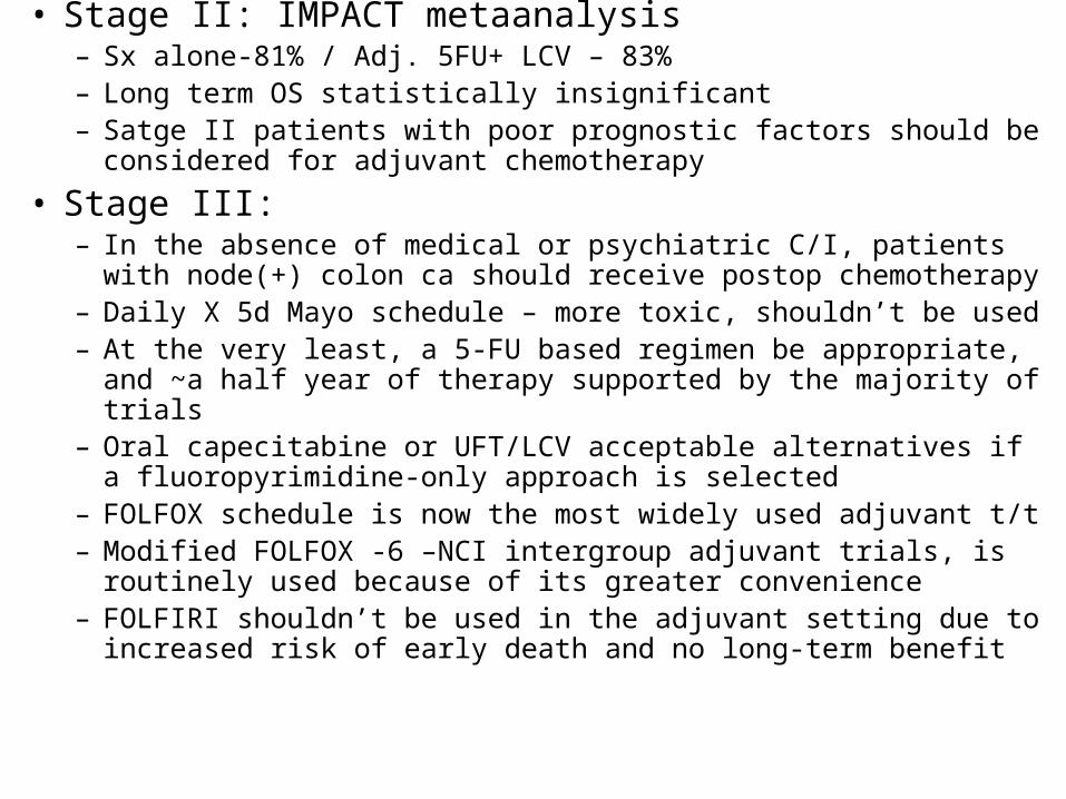
• Stage II: IMPACT metaanalysis– Sx alone-81% / Adj. 5FU+ LCV – 83%– Long term OS statistically insignificant– Satge II patients with poor prognostic factors should be considered for
adjuvant chemotherapy
• Stage III: – In the absence of medical or psychiatric C/I, patients with node(+)
colon ca should receive postop chemotherapy– Daily X 5d Mayo schedule – more toxic, shouldn’t be used– At the very least, a 5-FU based regimen be appropriate, and ~a half
year of therapy supported by the majority of trials– Oral capecitabine or UFT/LCV acceptable alternatives if a
fluoropyrimidine-only approach is selected– FOLFOX schedule is now the most widely used adjuvant t/t– Modified FOLFOX -6 –NCI intergroup adjuvant trials, is routinely used
because of its greater convenience– FOLFIRI shouldn’t be used in the adjuvant setting due to increased risk
of early death and no long-term benefit
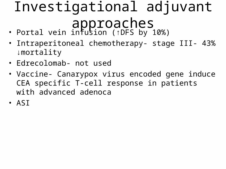
Investigational adjuvant approaches• Portal vein infusion (↑DFS by 10%)• Intraperitoneal chemotherapy- stage III- 43% ↓mortality• Edrecolomab- not used• Vaccine- Canarypox virus encoded gene induce CEA specific T-cell
response in patients with advanced adenoca • ASI
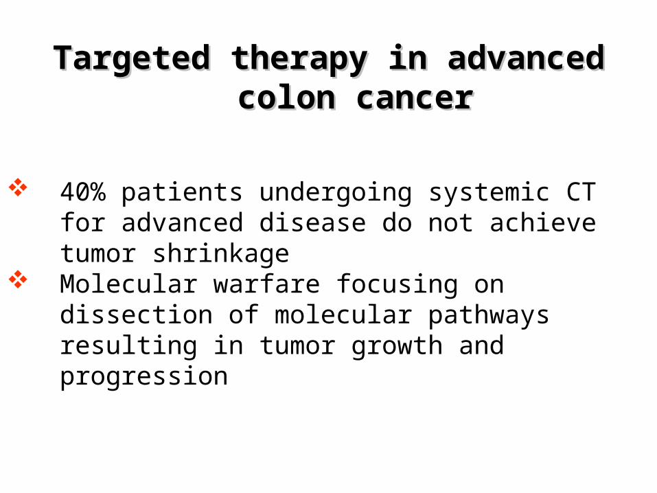
Targeted therapy in advanced colon cancerTargeted therapy in advanced colon cancer
40% patients undergoing systemic CT for advanced disease do not achieve tumor shrinkage
Molecular warfare focusing on dissection of molecular pathways resulting in tumor growth and progression
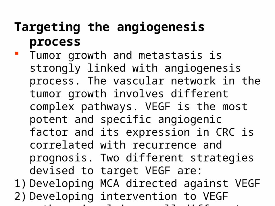
Targeting the angiogenesis process Tumor growth and metastasis is strongly linked with
angiogenesis process. The vascular network in the tumor growth involves different complex pathways. VEGF is the most potent and specific angiogenic factor and its expression in CRC is correlated with recurrence and prognosis. Two different strategies devised to target VEGF are:
1) Developing MCA directed against VEGF2) Developing intervention to VEGF pathway involving
small different molecules with tyrosine kinase inhibition activity directed towards the VEGF receptors

Bevacizumab
Anti – VEGF monoclonal antibody
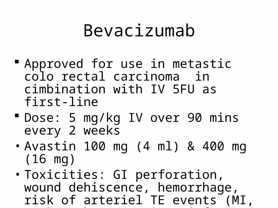
Bevacizumab
Approved for use in metastic colo rectal carcinoma in cimbination with IV 5FU as first-line
Dose: 5 mg/kg IV over 90 mins every 2 weeks• Avastin 100 mg (4 ml) & 400 mg (16 mg)• Toxicities: GI perforation, wound dehiscence,
hemorrhage, risk of arteriel TE events (MI, stoke), hypertension, infusion-related toxicity, proteinuria and nehprotic syndrome
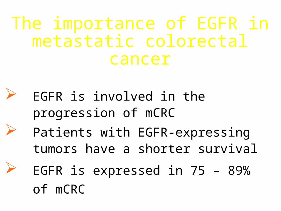
The importance of EGFR in metastatic colorectal cancer
EGFR is involved in the progression of mCRC
Patients with EGFR-expressing tumors have a shorter survival
EGFR is expressed in 75 – 89% of mCRC
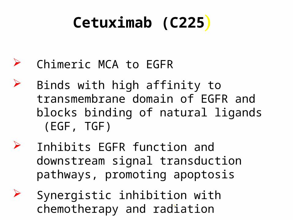
Cetuximab (C225)
Chimeric MCA to EGFR
Binds with high affinity to transmembrane domain of EGFR and blocks binding of natural ligands (EGF, TGF)
Inhibits EGFR function and downstream signal transduction pathways, promoting apoptosis
Synergistic inhibition with chemotherapy and radiation
.
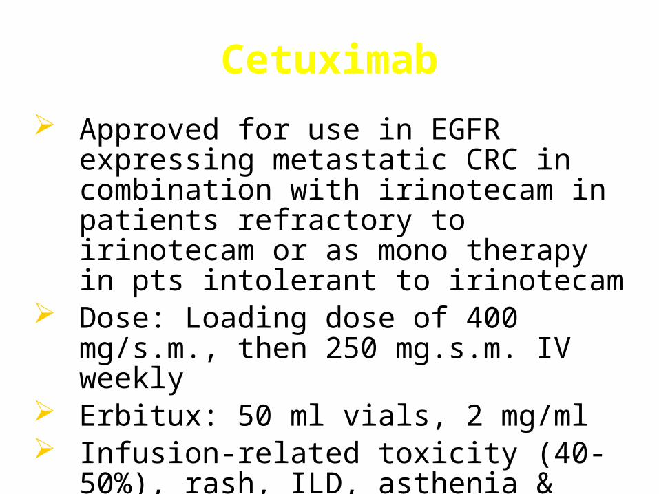
Cetuximab
Approved for use in EGFR expressing metastatic CRC in combination with irinotecam in patients refractory to irinotecam or as mono therapy in pts intolerant to irinotecam
Dose: Loading dose of 400 mg/s.m., then 250 mg.s.m. IV weekly
Erbitux: 50 ml vials, 2 mg/ml Infusion-related toxicity (40-50%), rash, ILD,
asthenia & fatigue, paronychia
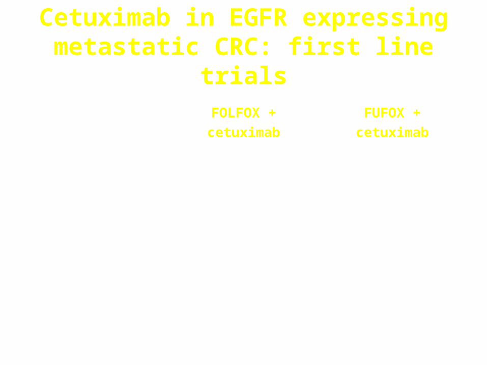
Cetuximab in EGFR expressing metastatic CRC: first line trials
Hoehler T. et al.
Proc ESMO 2004
Van Cutsem E. et al.
Proc ESMO 2004
Reference
21% resection of
livermetastases
remarks
21 %2%PD
24 %17%SD
55 %81% (74% conf.)
(61-88)
CR + PR
(95% CI)
3843Total patients
FUFOX +
cetuximab
FOLFOX +
cetuximab
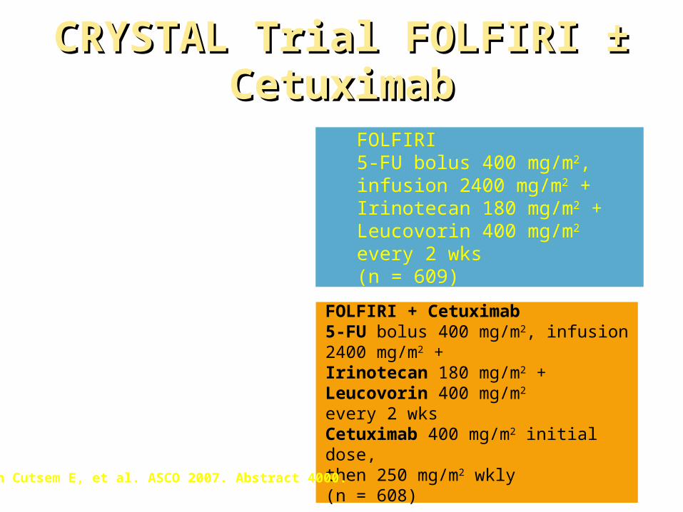
FOLFIRI5-FU bolus 400 mg/m2, infusion 2400 mg/m2 + Irinotecan 180 mg/m2 +Leucovorin 400 mg/m2
every 2 wks(n = 609)
Patients with previously untreated EGFR-expressing metastatic colorectal cancer, stratified by geographical region, ECOG PS
(N = 1217)
FOLFIRI + Cetuximab5-FU bolus 400 mg/m2, infusion 2400 mg/m2 + Irinotecan 180 mg/m2 +Leucovorin 400 mg/m2 every 2 wksCetuximab 400 mg/m2 initial dose, then 250 mg/m2 wkly(n = 608)Van Cutsem E, et al. ASCO 2007. Abstract 4000.
CRYSTAL Trial FOLFIRI ± CetuximabCRYSTAL Trial FOLFIRI ± Cetuximab
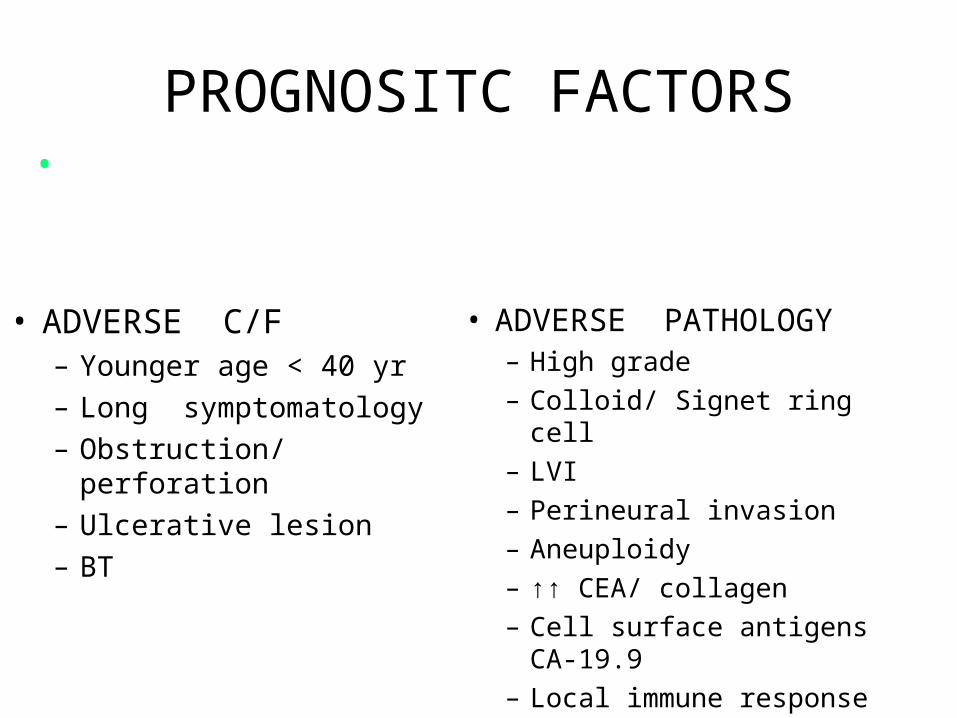
PROGNOSITC FACTORS
• ADVERSE C/F– Younger age < 40 yr
– Long symptomatology
– Obstruction/ perforation
– Ulcerative lesion
– BT
• ADVERSE PATHOLOGY– High grade
– Colloid/ Signet ring cell
– LVI
– Perineural invasion
– Aneuploidy
– ↑↑ CEA/ collagen
– Cell surface antigens CA-19.9
– Local immune response
•Most important guide to prognosis is STAGE of the disease i.e. depth of penetration and number of LNs involved
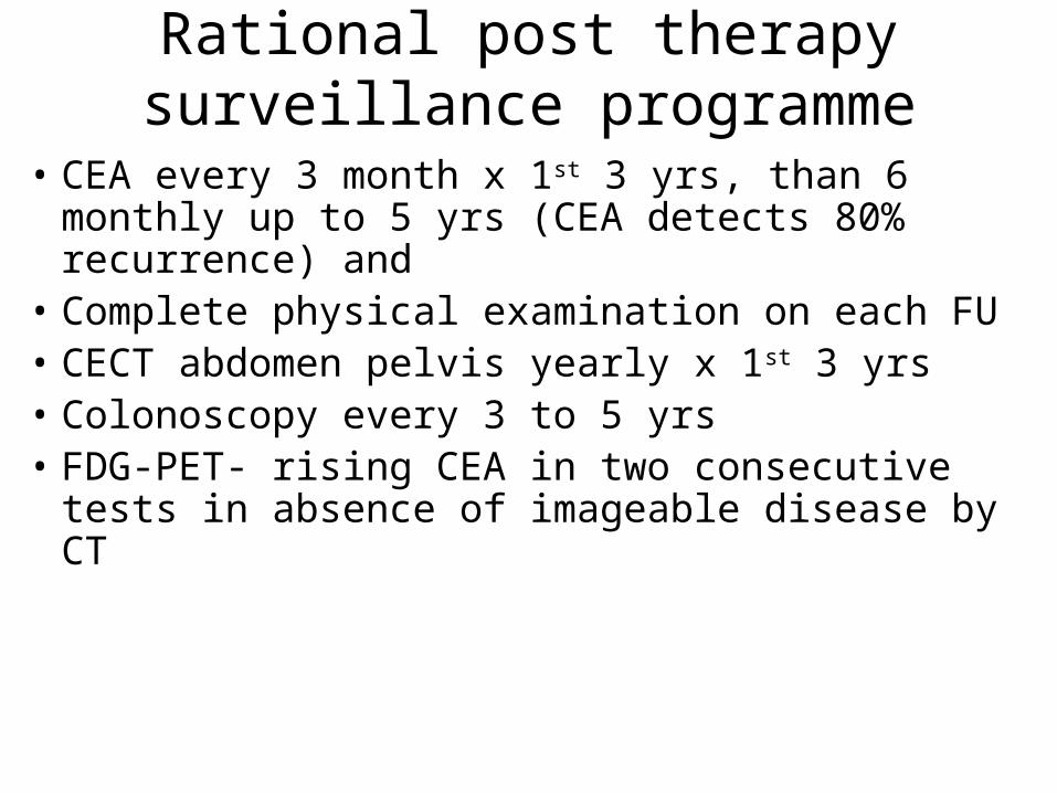
Rational post therapy surveillance programme
• CEA every 3 month x 1st 3 yrs, than 6 monthly up to 5 yrs (CEA detects 80% recurrence) and
• Complete physical examination on each FU• CECT abdomen pelvis yearly x 1st 3 yrs• Colonoscopy every 3 to 5 yrs• FDG-PET- rising CEA in two consecutive tests in
absence of imageable disease by CT
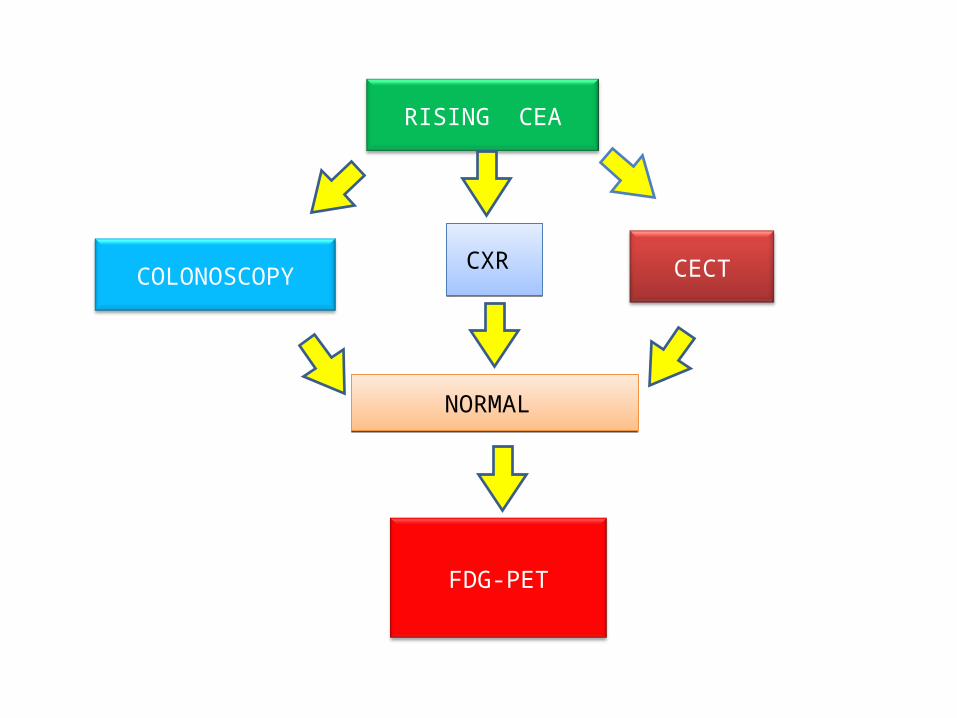
RISING CEA
COLONOSCOPY
FDG-PET
NORMAL NORMAL
CXR CXR CECT
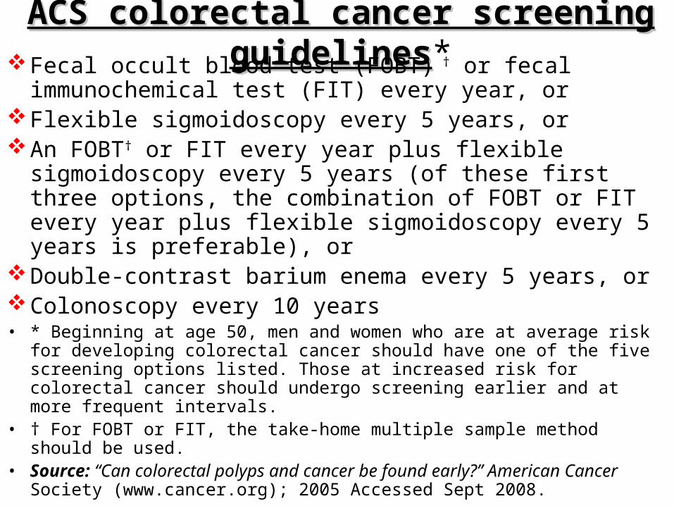
ACS colorectal cancer screening guidelinesACS colorectal cancer screening guidelines*Fecal occult blood test (FOBT) † or fecal immunochemical test
(FIT) every year, orFlexible sigmoidoscopy every 5 years, orAn FOBT† or FIT every year plus flexible sigmoidoscopy every
5 years (of these first three options, the combination of FOBT or FIT every year plus flexible sigmoidoscopy every 5 years is preferable), or
Double-contrast barium enema every 5 years, orColonoscopy every 10 years• * Beginning at age 50, men and women who are at average risk for developing
colorectal cancer should have one of the five screening options listed. Those at increased risk for colorectal cancer should undergo screening earlier and at more frequent intervals.
• † For FOBT or FIT, the take-home multiple sample method should be used.• Source: “Can colorectal polyps and cancer be found early?” American Cancer
Society (www.cancer.org); 2005 Accessed Sept 2008.

THANK YOU
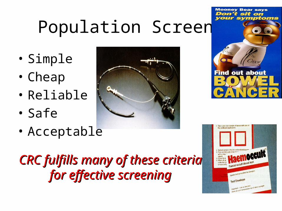
Population Screening
• Simple• Cheap• Reliable• Safe• Acceptable
CRC fulfills many of these criteria CRC fulfills many of these criteria for effective screeningfor effective screening