Embed Size (px)
Citation preview
1053
James Renwick BA MD FRCPC, Charles Kerr MD FRCPC, Rodney McTaggart asc MD FRCPC, John Yeung MB FRCPC
Review article
Cardiac electrophysiology and conduction pathway ablation
Invasive cardiac electrophysiological (EP) testing and transcath- eter ablation are new methods available for the diagnosis and
treatment o f complex dysrhythmias. The purpose o f this review is to familiarize anaesthetists with these procedures. The #l-
formation presented combines a literature review with the au- thors' experience. This article reviews normal cardiac conduc-
tion, tachycardia pathogenesis, principles o f cardiac EP study
and techniques o f conduction pathway ablation. The anaes- thetic considerations, including the choice o f anaesthetic agent,
monitoring problems, drug interactions, special methods o f dys- rhythmia termination in the EP lab, and complications specific to these procedures, are detailed. Balanced general anaesthesia
or monitored anaesthesia care (MAC) sedation with benzodi- azepines, propofol and narcotics are acceptable. Several con- clusions can be drawn: transcatheter ablation is an effective treatment for many reentry tachycardias; anaesthetic assistance for this procedure will increasingly be needed; anaesthesia can easily be provided without influencing accurate EP testing; over- drive pacing is the method o f choice for terminating tachy-
dysrhythmias in the EP lab.
Les tests d~lectrophysiologie (EP) invasive et I'ablation trans-
cathdtdrisme cardiaque reprdsentent des nouvelles mdthodes de diagnostic et de traitement des arythmies complexes. Cet article vise ~ familiariser les anesthdsistes avec ces interventions par l'association d'une revue de la litt&ature ~ l'expdrience des
auteurs. On y passe en revue la conduction cardiaque normale, la pathogdnkse des tachycardies, les principes d~nvestigation EP et les techniques d'ablation des voies de conduction. Les consi-
Key words ANAESTHESIA; cardiovascular; HEART: arrhythmia, electrocardiography.
From the Departments of Anaesthesiology and Cardiology, University Hospital, University of British Columbia, Vancouver, Canada.
Accepted for publication 26th July, 1993.
d~rations anesth~siques dont le choix des agents, le monitorage et ses probl~mes, les interactions pharmacologiques, les prin-
cipales m~thodes d'arr~t des arythmie en laboratoire d'EP et les complications sp~cifiques ~ ces interventions sont abord$s.
L'anesth~sie g~n&ale $quilibr~e ou la s~dation monitor~e avec les benzodiaz~pines, propofol et opiac~s sont approprides. On
peut tirer plusieurs conclusions: l'ablation transcath$t&isme est
une traitement efficace pour plusieurs tachyarythmies de r~en- tr$e; on aura de plus en besoin de l'assistance des anesth$sistes
pour cette intervention; l'anesth~sie elle-m~me n'inter~re pas n~cessairement avec les ~preuves EP; l'entrafnement ~lectro-
systolique rapide est la mdthode de choix pour mettre f in aux tachydysrythmies en laboratoire d'EP.
Contents Introduction Normal cardiac conduction Tachycardia pathogenesis Cardiac electrophysiologieal study (EPS) Influence of anaesthetic agents on cardiac conduction Antidysrhythmic drugs Percutaneous catheter ablation Management of anaesthesia Dysrhythmia termination Complications Summary
Invasive cardiac electrophysiologicai (EP) testing and transcatheter ablation techniques for treatment of reen- trant dysrhythmias are in a rapid phase of development. An increasing number of centres now offers this tech- nology to patients, and many thousands have been suc- cessfully treated. 1.2 As cardiac electrophysiologists gain experience in diagnosing and managing more seriously compromised patients, anaesthetists will be asked for as- sistance more frequently. Anaesthetists are asked to at- tend the EP lab for several purposes: to provide anaes- thesia and airway management for cardioversion, to
CAN J ANAESTH 1993 / 40: I 1 / pp 1053-64
1054 CANADIAN JOURNAL OF ANAESTHESIA
supervise sedation and haemodynamic monitoring, and to provide general anaesthesia and invasive monitoring for selected patients undergoing transvascular ablative procedures or intemal defibrillator checks.
Cardiac EP study and conduction ablation therapy present the anaesthetist with unique problems. These in- chide the underlying cardiac disease, interaction between anaesthetic and antidysrhythmic drugs, interference by anaesthetic drugs on cardiac conduction pathway map- ping, distinct monitoring requirements and special meth- ods of dysrhythmia termination unique to the EP lab.
The purpose of this review is to provide an overview of the role of cardiac electrophysiological testing and transcatheter ablation therapy, as well as background in- formation helpful to the safe conduct of anaesthesia in the EP lab. Normal cardiac conduction and mechanisms of tachycardia generation are reviewed. The principles of EP study are discussed and the effects of anaesthetic drugs on cardiac conduction examined. Antidysrhyth- mics, especially those with notable side effects, are dis- cussed. The transcatheter ablation technique is detailed. This is followed by recommendations on the management of anaesthesia. Special methods of dysrhythmia termi- nation in the EP lab are discussed. Important compli- cations specific to these procedures are highlighted.
Normal cardiac conduction The embryological and histopathological understanding of the cardiac conduction system is incomplete and un- dergoing continued investigation. Action potentials initiat- ing cardiac depolarization begin at the sino-atrial (SA) node which is a non-discrete area of specialized cells pos- sessing automaticity located near the junction of the su- perior vena cava and the right atrium. Primary pace- maker function of the heart is provided by a pacemaker complex which includes the SA node and secondary atrial pacemakers (SAP) and is referred to as a multicentric atrial pacemaker complex. 3,4 From the SA node, de- polarization spreads through atrial muscle to the atrio- ventricular (AV) node. Cardiac muscle fibres branch and are interconnected by intercalated discs. Current is able to pass rapidly because of low resistance tight gap june- tions in the regions of the intercalated discs. Preferred internodal conduction exists via atrial muscle bundles rather than via specialized tracts. These preferential routes of depolarization appear to be a product of the geometric configuration of the atrium around the openings of the superior and inferior vena cavae, foramen ovale and cor- onary sinus. Widespread depolarization of the ventricles from the atria is prevented by the interposed, electrically inert fibrous skeleton of the heart.
The AV node is located in the endocardium of the right atrium between the coronary sinus and the medial
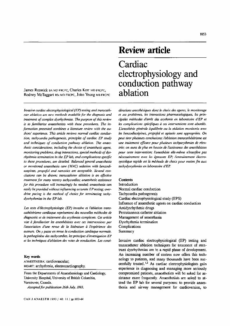
FIGURE 1 Cardiac conduction system. Possible accessory connections include: (1) atrioventricular bypass; (2) AV nodal bypass; (3) nodal-ventricular bypass; (4) His-ventricular bypass tracts.
tricuspid valve leaflet. It contains a graduated hetero- geneous group of specialized conducting cells, which are designated atrionodal (AN), nodal (N), nodal-His (Nil) and His (H) bundle cells. These cells slow conduction allowing ventricular fdling to occur. The histological basis for slowing of conduction in the AV node is related to the sparse intercellular connections, and thus few inter- calated discs and tight gap junctions. This slowing is dem- onstrated on the surface ECG by the P-R interval. The atrioventricular (His) bundle emerges from the AV node and descends along the posterior border of the mem- branous ventricular septum. Depolarization accelerates in the His bundle. It divides almost immediately into left and right bundle branches located on each side of the septum. The left bundle divides further into anterior and posterior fascicles. Interindividual anatomical variation exists in the configuration of the bundle branches. De- polarization then proceeds swiftly to the ventricular mus- cle mass via the Purkinje fibres. The rapid, almost simul- taneous, depolarization of ventricular muscle produces synchronized contraction and systolic ejection (Figure 1).
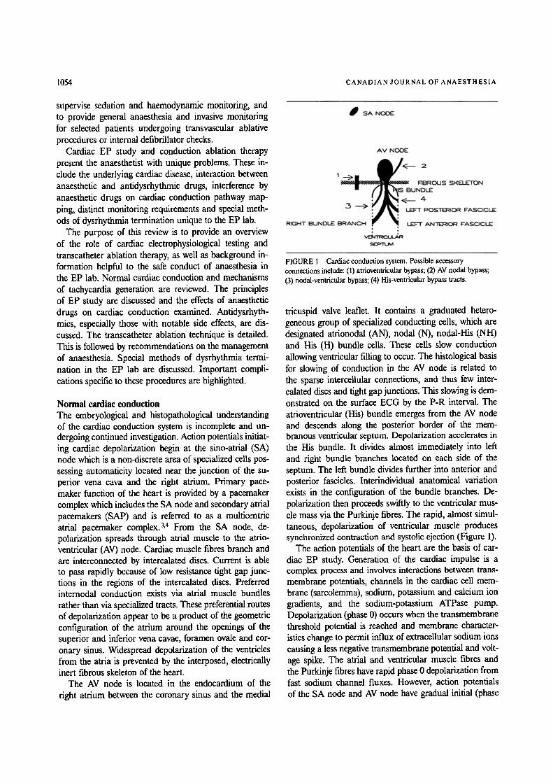
The action potentials of the heart are the basis of car- diac EP study. Generation of the cardiac impulse is a complex process and involves interactions between tmns- membrane potentials, channels in the cardiac cell mem- brane (sarcolemma), sodium, potassium and calcium ion gradients, and the sodium-potassium ATPase pump. Depolarization (phase 0) occurs when the transmembrane threshold potential is reached and membrane character- istics change to permit influx of extracellular sodium ions causing a less negative transmembrane potential and volt- age spike. The atrial and ventricular muscle fibres and the Purkinje fibres have rapid phase 0 depolarization from fast sodium channel fluxes. However, action potentials of the SA node and AV node have gradual initial (phase
Renwick et al.: CARDIAC ELECTROPHYS1OLOGY 1055
1 1
o 3
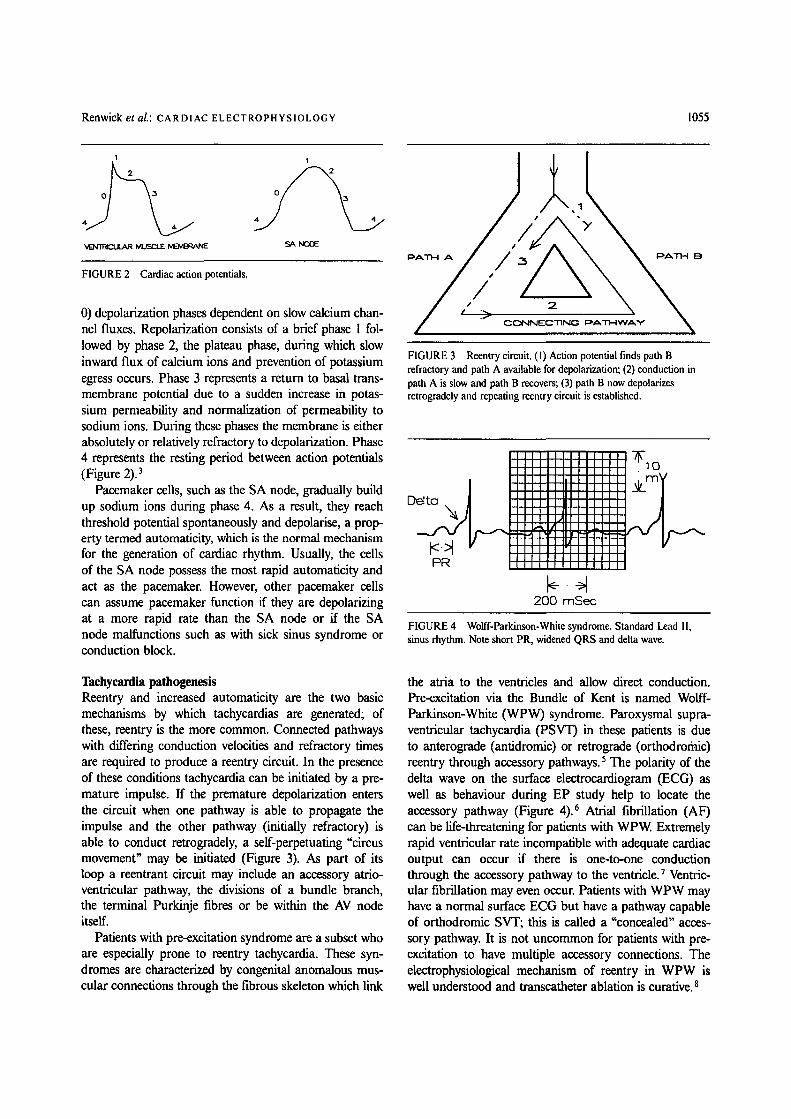
FIGURE 2 Cardiac action potentials.
0) depolarization phases dependent on slow calcium chan- nel fluxes. Repolarization consists of a brief phase 1 fol- lowed by phase 2, the plateau phase, during which slow inward flux of calcium ions and prevention of potassium egress occurs. Phase 3 represents a return to basal trans- membrane potential due to a sudden increase in potas- sium permeability and normalization of permeability to sodium ions. During these phases the membrane is either absolutely or relatively refractory to depolarization. Phase 4 represents the resting period between action potentials (Figure 2). 3
Pacemaker cells, such as the SA node, gradually build up sodium ions during phase 4. As a result, they reach threshold potential spontaneously and depolarise, a prop- erty termed automaticity, which is the normal mechanism for the generation of cardiac rhythm. Usually, the cells of the SA node possess the most rapid automaticity and act as the pacemaker. However, other pacemaker cells can assume pacemaker function if they are depolarizing at a more rapid rate than the SA node or if the SA node malfunctions such as with sick sinus syndrome or conduction block.
p A - r l - i A / i j / / / I N E t " T I N r':- P A T H B
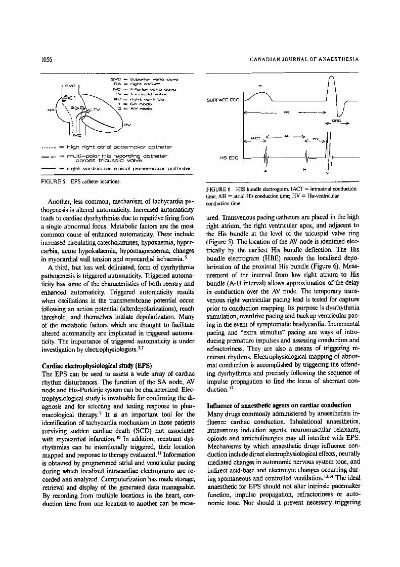
FIGURE 3 Reentry circuit. ( I ) Action potential finds path B refractory and path A available for depolarization; (2) conduction in path A is slow and path B recovers; (3) path B now depolarizes retrogradely and repeating reentry circuit is established.
Delta
PR
I.,41 . . . . I.,~ I / [ II
' I I I I I I 1 I I /
200 mSec
Jlq I t l l l i l t J l
ErA
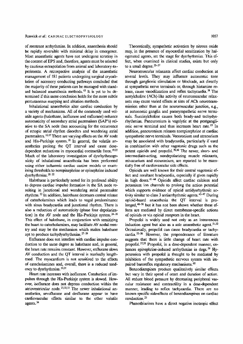
FIGURE 4 Wolff-Parkinson-White syndrome. Standard Lead I1, sinus rhythm. Note short PR, widened QRS and delta wave.
Tachycardia pathogenesis Reentry and increased automaticity are the two basic mechanisms by which tachycardias are generated; of these, reentry is the more common. Connected pathways with differing conduction velocities and refractory times are required to produce a reentry circuit. In the presence of these conditions tachycardia can be initiated by a pre- mature impulse. If the premature depolarization enters the circuit when one pathway is able to propagate the impulse and the other pathway (initially refractory) is able to conduct retrogradely, a self-perpetuating "circus movement" may be initiated (Figure 3). As part of its loop a reentrant circuit may include an accessory atrio- ventricular pathway, the divisions of a bundle branch, the terminal Purkinje fibres or be within the AV node itself.
Patients with pre-excitation syndrome are a subset who are especially prone to reentry tachycardia. These syn- dromes are characterized by congenital anomalous mus- cular connections through the fibrous skeleton which link
the atria to the ventricles and allow direct conduction. Pre-excitation via the Bundle of Kent is named Wolff- Parkinson-White (WPW) syndrome. Paroxysmal supra- ventricular tachycardia (PSVT) in these patients is due to anterograde (antidromic) or retrograde (orthodroriaic) reentry through accessory pathways, s The polarity of the delta wave on the surface electrocardiogram (ECG) as well as behaviour during EP study help to locate the accessory pathway (Figure 4). 6 Atrial fibrillation (AF) can be life-threatening for patients with WPW. Extremely rapid ventricular rate incompatible with adequate cardiac output can occur if there is one-to-one conduction through the accessory pathway to the ventricle. 7 Ventric- ular fibrillation may even occur. Patients with WPW may have a normal surface ECG but have a pathway capable of orthodromic SVT; this is called a "concealed" acces- sory pathway. It is not uncommon for patients with pre- excitation to have multiple accessory connections. The electrophysiological mechanism of reentry in WPW is well understood and transcatheter ablation is curative. 8
1056 C A N A D I A N J O U R N A L OF A N A E S T H E S I A
r R A ~ r i c J h t o b - ; ~ r ' ~
- ~ = "b ' i (=u=p |d v<~ lve I ~ R V ~ r~cJFl~ v~mfa- ; ( : l~
1 = S A n o c l ~ R A ~' ~ A V r - . ~ d e
�9 R V
. . . . . . . . h lgh right otriol N e e m Q k e r ~---x3NetE
- - m u f t i - - p o l a r H i s r~con=ling ~ t h e t e r o c r o s s t r i c u s p i d v o l v e
- - = r i g h t v e n t r i c u l o r a p i c e l ICX~ce rnoke r c a t h e t e r
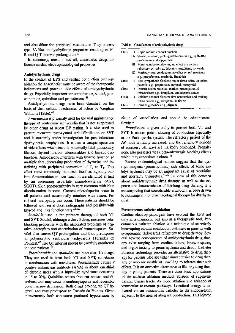
FIGURE 5 EPS catheter locations.
Another, less common, mechanism of tachycardia pa- thogenesis is altered automaticity. Increased automaticity leads to cardiac dysrhythmias due to repetitive firing from a single abnormal focus. Metabolic factors are the most common cause of enhanced automaticity. These include increased circulating catecholamines, hypoxaemia, hyper- carbia, acute hypokalaemia, hypomagnesaemia, changes in myocardial wall tension and myocardial ischaemia. 5
A third, but less well deliniated, form of dysrhythmia pathogenesis is triggered automaticity. Triggered automa- ticity has some of the characteristics of both reentry and enhanced automaticity. Triggered automaticity results when oscillations in the transmembrane potential occur following an action potential (afterdepolarizations), reach threshold, and themselves initiate depolarization. Many of the metabolic factors which are thought to facilitate altered automaticity are implicated in triggered automa- ticity. The importance of triggered automaticity is under investigation by electrophysiologists. 5,7
Cardiac electrophysiologieal study (EPS) The EPS can be used to assess a wide array of cardiac rhythm disturbances. The function of the SA node, AV node and His-Purkinje system can be characterized. Elec- trophysiological study is invaluable for confu'rning the di- agnosis and for selecting and testing response to phar- macological therapy. 9 It is an important tool for the identification of tachycardia mechanism in those patients surviving sudden cardiac death (SCD) not associated with myocardial infarction, io In addition, reentrant dys- rhythmias can be intentionally triggered, their location mapped and response to therapy evaluated. I i Information is obtained by programmed atrial and ventricular pacing during which localized intracardiac electrograms are re- corded and analyzed. Computerization has made storage, retrieval and display of the generated data manageable. By recording from multiple locations in the heart, con- duction time from one location to another can be meas-
SUPJ:ACE ECG
HIS ECG
P
<
, A C T , t( _ _ .-->
--4 A
<._. QRS._._~
V
FIGURE 6 HIS bundle electrogram. IACT = intraatrial conduction time; AH = atrial-His conduction time; HV = His-ventricular conduction time.
ured. Transvenous pacing catheters are placed in the high right atrium, the right ventricular apex, and adjacent to the His bundle at the level of the tricuspid valve ring (Figure 5). The location of the AV node is identified elec- trically by the earliest His bundle deflection. The His bundle electrogram (HBE) records the localized depo- larization of the proximal His bundle (Figure 6). Meas- urement of the interval from low right atrium to His bundle (A-H interval) allows approximation of the delay in conduction over the AV node. The temporary trans- venous right ventricular pacing lead is tested for capture prior to conduction mapping. Its purpose is dysrhythmia stimulation, overdrive pacing and backup ventricular pac- ing in the event of symptomatic bradycardia. Incremental pacing and "extra stimulus" pacing are ways of intro- ducing premature impulses and assessing conduction and refractoriness. They are also a means of triggering re- entrant rhythms. Electrophysiological mapping of abnor- mal conduction is accomplished by triggering the offend- ing dysrhythmia and precisely following the sequence of impulse propagation to find the locus of aberrant con- duction. ~2
Influence of anaesthetic agents on cardiac conduction Many drugs commonly administered by anaesthetists in- fluence cardiac conduction. Inhalational anaesthetics, intravenous induction agents, neuromuscular relaxants, opioids and anticholinergics may all interfere with EPS. Mechanisms by which anaesthetic drugs influence con- duction include direct electrophysiological effects, neurally mediated changes in autonomic nervous system tone, and indirect acid-base and electrolyte changes occurring dur- ing spontaneous and controlled ventilation. ~3,~4 The ideal anaesthetic for EPS should not alter intrinsic pacemaker function, impulse propagation, refractoriness or auto- nomic tone. Nor should it prevent necessary triggering
Renwick et al.: CARDIAC ELECTROPHYSIOLOGY 1057
of reentrant arrhythmias. In addition, anaesthesia should be rapidly reversible with minimal delay in emergence. Most anaesthetic agents have not undergone scrutiny in the context of EPS and, therefore, agents must be selected by cautious extrapolation from animal and laboratory ex- periments. A retrospective analysis of the anaesthetic management of 181 patients undergoing surgical cryoab- lation of accessory conducting pathways concluded that the majority of these patients can be managed with stand- ard balanced anaesthesia methods.IS It is yet to be de- termined if this same conclusion holds for the more subtle percutaneous mapping and ablation methods.
Inhalational anaesthetics alter cardiac conduction by a variety of mechanisms. All of the commonly used vol- atile agents (halothane, isoflurane and enflurane) enhance automaticity of secondary atrial pacemakers (SAP's) rel- ative to the SA node thus accounting for the occurrence of ectopic atrial rhythm disorders and wandering atrial pacemakers. 16,17 There are varying effects on the AV node and His-Purkinje system, is In general, the volatile an- aesthetics prolong the QT interval and cause dose- dependent reductions in myocardial contractile force. ~9.23 Much of the laboratory investigation of dysrhythmoge- nicity of inhalational anaesthesia has been performed using either ischaemic cardiac canine models or exam- ining thresholds to norepinephrine or epinephrine induced dysrhythmias. 2o-22
Halothane is particularly noted for its profound ability to depress cardiac impulse formation in the SA node re- sulting in junctional and wandering atrial pacemaker rhythms. 23 In addition, halothane decreases central release of catecholamines which leads to vagal predominance with sinus bradycardia and junctional rhythm. There is also a reduction of automaticity (phase four depolariza- tion) in the AV node and the His-Purkinje system. 24-26 This effect of halothane, in conjunction with sensitizing the heart to catecholamines, may facilitate AV nodal reen- try and may be the mechanism which makes halothane apt to produce tachydysrhythmias. 27-29
Enflurane does not interfere with cardiac impulse con- duction to the same degree as halothane and, in general, the heart rate remains constant. However, enflurane slows AV conduction and the QT interval is markedly length- ened. The myocardium is not sensitized to the effects of catecholamines and, overall, there is a reduced tend- ency to dysrhythmias. 3~
Heart rate increases with isoflurane. Conduction of im- pulses through the His-Purkinje system is slowed. How- ever, isoflurane does not depress conduction within the atrioventricular node.23,32,33 The newer inhalational an- aesthetics, sevoflurane and desflurane appear to have cardiovascular effects similar to the other volatile agents. 34
Theoretically, sympathetic activation by nitrous oxide may, in the presence of myocardial sensitization by hal- ogenated agents, set the stage for dysrhythmias. This ef- fect, when examined in clinical studies, exists but only to a small degree. 35-37
Neuromuscular relaxants affect cardiac conduction at several levels. They may influence autonomic tone through ganglionic stimulation or blockade, act directly at sympathetic nerve terminals or, through histamine re- lease, cause vasodilatation and reflex tachycardia. 38 The acetylcholine (ACh)-like activity of neuromuscular relax- ants may cause varied effects at sites of ACh neurotrans- mission other than at the neuromuscular junction, e.g., at autonomic ganglia and parasympathetic nerve termi- nals. Succinylcholine causes both brady-and tachydys- rhythmias. Pancuronium is vagolytic at the postgangli- onic nerve terminal and thus increases heart rate. 23 In addition, pancuronium releases norepinephrine at cardiac sympathetic nerve terminals. Vecuronium and atracurium may be associated with bradycardia, particularly if used in combination with other vagotonic drugs such as the potent opioids and propofol. 39,4~ The newer, short- and intermediate-acting, nondepolarizing muscle relaxants, mivacurium and rocuronium, are reported to be essen- tially free of cardiovascular side effects.
Opioids are well known for their central vagotonic ef- fect and resultant bradycardia, especially if given rapidly in high doses. 41-44 Opioids affect cardiac calcium and potassium ion channels to prolong the action potential which supports evidence of opioid antidysrhythmic ac- tivity similar to class 3 antiarrhythmic agents. 45-47 During opioid-based anaesthesia the QT interval is pro- longed, 4s-5~ but it has not been shown whether these ef- fects are mediated by direct membrane-specific actions of opioids or via opioid receptors in the heart.
Propofol is widely used not only as an intravenous induction agent but also as a sole anaesthetic agent. 51,52 Occasionally, propofol can cause bradycardia or tachy- cardia. 53-56 However, the preponderance of literature suggests that there is little change of heart rate with propofol. 57,5s Propofol, in a dose-dependent manner, en- hances epinephrine-induced arrhythmias in dogs. 59 ny- potension with propofol is thought to be mediated by inhibition of the sympathetic nervous system with im- paired baroreflex regulatory mechanisms.6~
Benzodiazepines produce qualitatively similar effects but vary in their speed of onset and duration of action. All reduce blood pressure by decreasing peripheral vas- cular resistance and contractility in a dose-dependent manner, leading to reflex tachycardia. There are no known specific side-effects of benzodiazepines on cardiac conduction. 23
Phenothiazines have a direct negative inotropic effect
1058 CANADIAN JOURNAL OF ANAESTHESIA
and also dilate the peripheral vasculature. They possess type I A-like antidysrhythmic properties resulting in P- R and Q-T interval prolongation. 23
In summary, most, if not all, anaesthetic drugs in- fluence cardiac electrophysiological properties.
Antidysrhythmie drugs In the context of EPS and cardiac conduction pathway ablation the anaesthetist must be aware of the therapeutic indications and potential side effects of antidysrhythmic drugs. Especially important are amiodarone, sotalol, pro- cainamide, quinidine and propafenone. 61
Antidysrhythmic drugs have been classified on the basis of their cellular mechanism of action by Vaughan Williams (Table). 62
Amiodarone is primarily used for the oral maintenance therapy of ventricular tachycardia that is not suppressed by other drugs at repeat EP testing. It is also used to prevent recurrent paroxysmal atrial fibrillation or SVT and is currently under investigation for post-infarction dysrhythmia prophylaxis. It causes a unique spectrum of side effects which include potentially fatal pulmonary fibrosis, thyroid function abnormalities and hepatic dys- function. Amiodarone interferes with thyroid function at multiple sites, decreasing production of thyroxine and in- terfering with peripheral conversion of T3 t o "1"4. This effect most commonly manifests itself as hypothyroid- ism. Abnormalities in liver function are identified at fast by an increasing aspartate aminotransferase (AST, SGOT). Skin photosensitivity is very common with blue discolouration in some. Corneal microdeposits occur in all patients and occasionally interfere with vision. Pe- ripheral neuropathy can occur. These patients should be followed with serial chest radiographs and possibly with thyroid and fiver function tests.63-~
Sotalol is used in the primary therapy of both VT and SVT. Sotalol, although a class 3 drug, possesses beta- blocking properties with the associated problems of neg- ative inotropism and exacerbation of bronchospasm. So- talol also causes QT prolongation and thus predisposes to polymorphic ventricular tachycardia (Torsades de Pointes).67 The QT interval should be carefully monitored in these patients. 68
Procainamide and quinidine are both class IA drugs. They are used to treat both VT and SVT, sometimes in combination with mexilitene. Procainamide causes a positive antinuclear antibody (ANA) in about one-third of chronic users with a lupus-like syndrome occurring in 15 to 20%. Quinidine causes frequent nausea and di- arrhoea and may cause thrombocytopenia and reversible bone marrow depression. Both drugs prolong the QT in- terval and may predispose to Torsade de Pointes. Given intravenously both can cause profound hypotension by
TABLE Classification of antidysrhythmic drugs
Class 1 Rapid sodium channel blockers IA Slow conduction, prolong refractoriness e.g., quinidine,
procainamide, disopyramide 1B Minor conduction slowing, no effect or shortens
refractory period e.g., lidoeaine, mexilitene, tocainide IC Markedly slow conduction, no effect on refractoriness
e.g., propafenone, encainide, flecainide Class 2 Beta sympathetic blockers; major direct affect on action
potential e.g., propranolol, esmolol, metoprolol Class 3 Prolong action potential, marked prolongation of
refractoriness e.g., bretylium, amiodarone, sotalol Class 4 Calcium channel blockers slow conduction and prolong
refractoriness e.g., verapamil, diltiazem Class 5 Cardiac glycosides e.g., digoxin
virtue of vasodilation and should be administered slowly. 69
Propafenone is given orally to prevent both VT and SVT. It causes potent slowing of conduction especially in the Purkinje-His system. The refractory period of the AV node is mildly increased, and the refractory periods of accessory pathways are markedly prolonged. Propafe- none also possesses weak beta-adrenergic blocking effects which may exacerbate asthma. 7~
Recent epidemiological studies suggest that the dys- rhythmogenic (proarrhythmic) side effects of some an- tidysrhythmics may be an important cause of morbidity and mortality themselves. 71-74 In view of this concern about antidysrhythmic drug therapy, as well as the ex- pense and inconvenience of life-long drug therapy, it is not surprising that considerable attention has been drawn to nonsurgical, nonpharmacological therapy for dysrhyth- mias.
Percutaneous catheter ablation Cardiac electrophysiologists have evolved the EPS not only as a diagnostic but also as a therapeutic tool. Per- cutaneous catheter ablation is a technique of selectively interrupting cardiac conduction pathways in patients with symptomatic tachycardia refractory to drug therapy. Sev- eral adverse consequences of antidysrhythmic drug ther- apy exist ranging from cardiac failure, bronchospasm, and organ toxicity to proarrhythmia and death. Catheter ablation technology provides an alternative to drug ther- apy for patients who are either unresponsive to drug ther- apy or who are unable or unwilling to tolerate their side effects. It is an attractive alternative to life-long drug ther- apy in young patients. There are three basic applications of the catheter ablation method: ablation of supraven- tricular bypass tracts, AV node ablation and ablation of ventficular re-entrant pathways. Localized energy is de- livered via an intracardiac catheter to the endocardium adjacent to the area of aberrant conduction. This injured
Renwick et aL: CARDIAC ELECTROPHYSIOLOGY 1059
tissue becomes electrophysiologically inactive, scars and prevents recurrence. The technique has superseded open cardiac procedures for most cases. 75,76
Transcatheter ablation for control of tachydysrhyth- mias was first used in 1982.77,78 The initial energy source used DC current generated by a defibrillator and passed through a catheter electrode. The high level of uncon- trolled energy caused frequent complications, including perforation and proarrhythmia. Ventricular fibrillation may occur immediately after or up to seven days after DC ablation. 79,80 Since 1986, radiofrequency (RF) energy has replaced DC shock as a more common energy source and is a safer energy source for ablation of the AV node and accessory pathways, s~ The RF energy is low-power, high-frequency alternating current which causes injury by generating heat at the electrode/tissue interface. The advantages include control of energy delivery, the creation of a smaller area of injury, and the ability to be used safely in thin-walled structures such as the coronary sinus. It is usually painless and seldom triggers malignant dys- rhythmias. When DC current is used general anaesthesia is required because of the severity of pain. 82-84
Supraventricular tachycardias amenable to catheter ab- lation technique include PSVT, paroxysmal atrial fibril- lation (PAl7), chronic AF and atrial fib-flutter. 85 In pa- tients with pre-excitation, SVT can be abolished either by ablation of the AV node or ideally by creating a lesion in the accessory pathway itself. Injury to the AV node or His bundle, intentional or accidental, may cause com- plete heart block. If this occurs, a permanent ventricular pacemaker is required. When it is possible to ablate the bypass tract specifically and prevent re-entry the patient will be cured.
Another application is the specific destruction of an identifiable ventricular reentrant focus. This subset of pa- tients presents with paroxysmal ventricular tachycardia (PVT) or paroxysmal ventricular fibrillation (PVF).
Delivering a small, precisely located, energy surge to the moving endocardium is technically difficult and often requires multiple attempts. In the patient with pre- excitation and PSVT, successful ablation is recognized by the disappearance of delta waves and lengthening of the P-R interval. In patients with AF and rapid ven- tricular response the goal is AV dissociation. Inability to re-trigger VT identifies success in the patient with PVT.
Effective ablation is further tested by attempting to re- trigger the offending dysrhythmia after administration of atropine (0.6-1.2 mg) and isoproterenol (0.5-71~g ' min -l) by infusion. A period of observation in the EP laboratory helps to exclude delayed recovery of an ablated area. We have commonly observed arousal and awakening dur- ing light levels of anaesthesia when isoproterenol is in- fused and we deepen anaesthesia at this time. After suc-
cessful RF destruction of the proximal AV node or of- fending accessory pathway the patient is monitored by telemetry for 24 hr. If the pacemaker function of the AV node is damaged a well-secured temporary pacemaker is left in situ before leaving the EP laboratory and the patient monitored in the cardiac care unit (CCU) until a permanent pacemaker can be inserted.
Management of anaesthesia Preoperative preparation includes thorough patient his- tory, physical examination, laboratory testing and optim- ization of medical therapy. The anaesthetist should en- quire about the frequency and duration of rhythm disturbances. It is of critical importance to the safe con- duct of anaesthesia that patients who decompensate dur- ing tachycardia, with symptoms of syncope, severe angina or congestive heart failure, be identified before EPS. Rapid identification of these symptoms during EPS will facilitate rapid termination of the causative rhythm. Anx- iolysis by reassurance and benzodiazepine premedication minimizes excessive sympathetic tone. All non-essential drugs, especially antidysrhythmic agents are discontinued several days before EPS in order to promote SVT or VT. 86 Optimization of medical therapy generally consti- tutes adequate treatment of angina and congestive heart failure. Patients with sustained PVT often have severe previous myocardial damage, are at high risk for any procedure, and may also be under consideration for in- sertion of an automatic defibrillator. ~ Minimal preoper- ative laboratory tests include complete blood count (CBC), platelet count, prothombin time (PT, INR), elec- trolytes, urea, creatinine and ECG. Abnormalities of elec- trolytes, PT and platelet numbers should be identified and corrected beforehand. Patients are fasted preoper- atively; those with increased risk for aspiration are given appropriate prophylaxis. Anaesthesia options are dis- cussed with the patient. 6,87-89
These procedures are exacting for the cardiac electro- physiologist to perform. A motionless patient is required, who, if sedated, is still able to communicate cardiopul- monary and neurological symptoms. Apart from vascular access and the RF bursts the procedure is generally not painful. Analgesia for line insertion is easily accomplished with local anaesthesia. During the 5 to 45 sec periods of RF ablation, repeated up to 15 or more times during the course of the procedure, some patients may experience brief retrostemal angina-like chest pain of mild to mod- erate intensity.
The EP suite is a difficult environment for the an- aesthetist. It is remote from the operating room, dark, and there is limited access to the patient. There is in- creased potential for electrical hazard. We maintain a fully operational anaesthesia machine with all ancillary equip-
1060 CANADIAN JOURNAL OF ANAESTHESIA
ment and drugs in the laboratory. We employ continuous ECG, combined pulse oximetry/plethysmography and noninvasive blood pressure monitoring as minimum mon- itors. Arterial monitoring is used for patients with haem- odynamically unstable rhythm disturbances. Mapping catheters may partially occlude arterial catheter sheaths; thus a separate radial line is preferred. A defibrillator with leads attached and with the ECG displayed on the screen is employed throughout. Patients with VT or PVF are connected to a defibrillator by hard wired gel pads placed over the sternum and back.
The patient is protected from radiation exposure with thyroid and gonadal lead shields. In anticipation of a long procedure the patient may have a urinary catheter inserted and measures are instituted to prevent the in- sidious development of hypothermia. Patients may receive antibiotic prophylaxis with a cephalosporin and are anti- coagulated with heparin if arterial or transeptal heart catheters are introduced.
We have employed both intravenous sedation and gen- eral anaesthesia. Our experience is that propofol by in- fusion, in the sedative to total anaesthesia dose range of (25-200 ~tg- kg -I- min -I ), seems to have little influence on EPS and the ability to trigger either SVT or VT. 9~ In those patients unable to tolerate propofol-induced hy- potension we have been successful using propofol in an attenuated dose supplemented with 70% nitrous oxide. We have observed no difficulty with EPS using alfentanil for sedation or as part of total intravenous anaesthesia (TIVA) in doses of 0.25-1.5 ~tg- kg -m. min -I , sometimes combined with midazolam in 0.5-2 mg increments. Intra- venous sedation has the advantage of allowing the patient to communicate symptoms. The anaesthetist must keep in close verbal contact with patients who are being se- dated while haemodynamically unstable rhythms are trig- gered so that impending loss of consciousness can be identified quickly and measures instituted rapidly to pro- vide airway and vasopressor support while the offending rhythm is terminated. Angina or presyncope signals the need for prompt retum to sinus rhythm. Nitroglycerin (NTG) spray or /v NTG (1-3 ~.g" kg -I) by infusion is used to treat persisting angina. Coronary spasm may re- spond to sublingual nifedipine 10-20 rag. We prefer phe- nylephrine (25-I00 I~g bolus), because of its pure alpha agonisfic properties, for initial vasopressor support.
In patients with severe haemodynamic instability or who are at increased risk for aspiration, the advantages of general anaesthesia with a secured airway outweigh the identification of symptoms. General anaesthesia can be satisfactorily accomplished using propofol 100-200 vtg-kg -m -min -I , or isoflurane 0.5-1% and supple- mented with nitrous oxide if necessary, with minimal in- terference on EPS. In patients requiring general anaes-
thesia, but who are at low risk for aspiration, we prefer airway management with Brain's laryngeal mask airway. Prior to transfer to the CCU or telemetry unit, patients are monitored in the post-anaesthetic care unit (PACU).
Dysrhythmia termination All anaesthetists are familiar with the guidelines and pro- tocols for cardiopulmonary resuscitation (CPR) and emergency cardiac care (ECC) outlined by the American Heart Association (AHA) and taught in the Advanced Cardiac Life Support (ACLS) programme. 91 In addition, there are special techniques of dysrhythmia termination available in the EP laboratory. 92,93
Patients being investigated for haemodynamically un- stable rhythms such as VT or VF are provided with hard wired defibrillator gel pads over the right sternum and back so that cardioversion or defibrillation can be per- formed immediately without having to move fluoroscopic equipment and place defibrillator paddles manually. A machine check, to ensure that current can be delivered, is performed. In anaesthetised patients undergoing au- tomatic implanted cardiac defibrillator (AICD) check, this circuit must be tested on the patient with low energy (e.g., 2 joules) before inducing VF to ensure that the pa- tient can be "rescued"! 94 The right ventricular apical pac- ing catheter must also be checked at the beginning of EPS to ensure capture in the event of symptomatic bra- dycardia.
Electrical pacing is available for dysrhythmia termi- nation and is the method of choice for terminating both SVT and VT in the EP lab. In haemodynamically stable patients, introduced ventricular premature beats (VPBs) or atrial premature beats (APBs) will often convert the rhythm to sinus. If introduced APBs or VPBs fail to terminate the taehycardia then "burst" pacing is em- ployed. In "burst" pacing a train of three to ten asyn- chronous paced beats is introduced at a rate faster than the tachycardia and then stopped abruptly. In either case the goal is to terminate reentry by making the circuit refractory to conduction, obviating the need for time- consuming and deleterious effects of drug therapy. For patients not responsive to these measures or who are haemodynamically unstable direct DC cardioversion is employed.
Patients being investigated and treated for PSVT, par- ticularly those with accessory pathways, may inadvert- ently be triggered into AE Atrial fibrillation is often a difficult rhythm to terminate. It may sometimes be con- verted to sinus rhythm with/v procainamide, but usually synchronized electrical cardioversion at 100-200 joules is required. Thiopentone, methohexitone and etomidate have all been used successfully for cardioversion. 95 We prefer to use propofol in a hypnotic dose of 1-1.5
Renwick et al.: CARDIAC ELECTROPHYSIOLOGY 1061
mg. kg- i for cardioversion. It has the advantages of short duration of action and salutary effects on the muscles of the airway making ventilation by bag and mask easier than if thiopentone or methohexitone are used. Propofol has become the agent of choice for cardioversion in some centres. 96 Midazolam, with flumazenil reversal, has also been reported to provide anaesthesia safely for cardio- version. 97
vide an anaesthetic safely which takes into account the electrophysiologist's requirements for minimal cardiac conduction interference yet provides a motionless patient who is painfree and recovers rapidly. In addition, the anaesthetist must be alert to procedural complications and their management. There is the potential for col- laborative work between cardiac electrophysiologists and anaesthetists in this new and exciting field.
Complications In general EPS and cardiac transvascular ablation are safe. Complications of the procedure are those of heart catheterization plus those of the ablation technique. Line insertion related problem include haematomas, bleeding, phlebitis, thromboembolism, pneumothorax, pericardial tamponade, pacemaker failure, and rarely endocarditis. A large pneumothorax can be diagnosed most rapidly by fluoroscopy. The Percutaneous Cardiac Mapping and Ablation Registry (PCMAR) reported, in 1988, two deaths in 522 patients who underwent attempted AV junc- tional ablation with DC shock. 9s The PCMAR reported a 6.7% occurrence of procedural related deaths in those patients undergoing VT ablation in a group of patients with severe pre-existing cardiac disease. Although as yet undocumented, RF ablation appears to be considerably safer, and equally effective as DC current ablation.
The EPS study may cause cardiac ischaemia or failure during mapping of haemodynamically unstable rhythms. This may result in cardiac infarction, pulmonary oedema, hypotension, new dysrhythmia or syncope. Patients ex- periencing angina are rapidly converted to sinus rhythm and given nitroglycerin if angina persists. All patients have blood samples taken at the end of the procedure to measure cardiac isoenzyme levels. Catheter-induced brady- and tachydysrhythmias can degenerate into un- stable rhythms leading to death. Coronary spasm has also been reported after attempted ablation. 99 Direct cur- rent, though not commonly used at our centre, is par- ticularly noted for its proarrhythmic potential. Perforation and acute pericardial tamponade have been caused by DC shock delivered across the coronary sinus. J00
Summary Cardiac electrophysiological testing and conduction path- way ablation are in an era of rapid evolution with ex- panding indications and improving technology. The an- aesthetist has a special role to play in the management of these challenging patients. The anaesthetist must have a working knowledge of normal and abnormal cardiac conduction and be familiar with antidysrhythmic drugs and their complications. The environment of the EP lab, the patient's disease process and the procedure all present unique problems. The anaesthetist is challenged to pro-
References 1 Scheinman MM, Laks MM, DiMarco J, Plumb V.
Current role of catheter ablative procedures in patients with cardiac arrhythmias: a report for health professionals from the subcommittee on electrocardiography and electrophysi- ology, American Heart Association. Circulation 1991; 83: 2146-53.
2 Schienman MM. Interventional electrophysiology: catheter ablation for patients with cardiac arrhythmias. Cardiol Clin 1986; 4: 543-9.
3 Boineau JP,, Canavan TE, Schuessler RB, Cain ME, Corr PB, Cox JL. Demonstration of a widely distributed atrial pacemaker complex in the human heart. Circulation 1988; 77: 1221-37.
4 Boineau JP, Schuessler RB, Canavan TE, Corr PB, Cain ME, Cox JL. The human atrial pacemaker complex. J Electrocardiol 1989; 22: 189-97.
5 Becker AE, Anderson RH, Durrer D, Wellens HJJ. The anatomical substrates of Wolff-Parkinson-White syndrome. A cfinicopathologic correlation in seven patients. Circula- tion 1978; 57: 870-9.
6 Wellens HI,, Atie J, Penn OC, Gorgels AP,, Brugada P, Smeets JL. Diagnosis and treatment of patients with ac- cessory pathways. Cardiol Clin 1990; 8: 503-21.
7 Wellens H J, Brugada P, Penn OC. The management of pre-excitation syndromes. JAMA 1987; 257: 2325-33.
8 Fisch C. Clinical electrophysiological studies and the Wolff-Parkinson-White pattern. Circulation 1990; 82: 1872-3.
9 Schuger CD, Steinman RT, Meissner MD, Mosteller RD, Lehmann MH. Clinical management of patients with atrioventricular nodal reentrant taehyeardia. Cardiol Clin 1990; 8: 491-501.
10 Kasanuki H, Ohnishi S, Tanaka E, Hirosawa K. Mechanism and prediction of sudden cardiac death in ar- rhythmia patients using electrophysiologieal studies. Jpn Cire J 1989; 53: 1565-70.
11 Gomes JA, Hariman R J, Kang PS, Showdry IH. Sustained symptomatic sinus node reentrant tachycardia: incidence, clinical significance, eleetrophysiologic observa- tions and the effects of antiarrhythmie agents. J Am Coil Cardiol 1985; 5: 45-57.
12 Cambell RF. Mapping of ventricular arrhythmias. Cardiol Clin 1986; 4: 497-505.
1062 CANADIAN JOURNAL OF ANAESTHESIA
13 Atlee JL, Bosnjak ZJ Mechanisms for cardiac dysrhyth- mias during anesthesia. Anesthesiology 1990; 72: 347-74.
14 Pratila MG, Pratilas V. Anesthetic agents and cardiac elec- tromechanical activity. Anesthesiology 1978; 49: 338-60.
15 Irish CL, Murkin JM, Guiraudon GM. Anaesthetic man- agement for surgical cryoablation of accessory conducting pathways: a review and report of 181 cases. Can J Anaesth 1988; 35: 634-40.
16 Bosnjak Z J, Kampine JP. Effects of halothane, enflurane, and isoflurane on the SA node. Anesthesiology 1983; 58: 314-21.
17 Marshall BE, Longnecher DE. General Anesthetics. In: Gilman AG, Rail TW, Nies AS, Taylor R The Pharmaco- logical Basis of Therapeutics. 8th cd. New York, Permagon, 1990: 285-310.
18 Lazlo A, Polic S, Atlee JL, Kampine JP, Bosnjak ZJ. Anesthetics and automaticity in latent pacemaker fibers: I. Effects of halothane, enflurane, and isoflurane on automa- ticity and recovery of automaticity from overdrive suppres- sion in Purkinje fibers derived from canine hearts. Anesthe- siology 1991; 75: 98-105.
19 Riley DC, Schmeling WT, AI-Wathiqui MH, Kampine JP,, Warltier DC. Prolongation of the QT interval by volatile anesthetics in chronically instrumented dog. Anesth Analg 1988; 67: 741-9.
20 Denniss AR, Richards DA, Taylor AT, Uther JB. Halothane anesthesia reduces inducibility of ventricular ta- ehyarrhythmias in chronic canine myocardial infarction. Basic Res Cardiol 1989; 84: 5-12.
21 Deutsch N, Hantler CB, Tait AR, Uprichard A, Schork MA, Knight PR. Suppression of ventricular arrhythmias by volatile anesthetics in a canine model of chronic myocar- dial infarction. Anesthesiology 1990; 72: 1012-21.
22 Atlee JL II1, Yeager TS. Electrophysiologic assessment of the effects of enflurane, halothane, and isoflurane on prop- erties affecting supraventricular re-entry in chronically in- strumented dogs. Anesthesiology 1989; 71: 941-52.
23 Atlee JL IIL Alexander SC. Halothane effects on conduc- tivity of the AV node and His-Purkinje system in the dog. Anesth Analg 1977; 56: 378-86.
24 Eger E1 II, Smith NT, Stoelting RK, Cullen D J, Kadis LB, Whitcher CE. Cardiovascular effects of halothane in man. Anesthesiology 1970; 2: 396-409.
25 Lynch C IlL Vogel S, Sperelakis N. Halothane depression of myocardial slow action potentials. Anesthesiology 1981; 55: 360-8.
26 Polic S, Atlee JL III, Laslo A, Kampine JP, Bosnjak ZJ Anesthetics and automaticity in latent pacemaker fibers: II. Effects of halothane and epinephrine or norepinephrine on automaticity of dominant and subsidiary atrial pacemakers in the canine heart. Anesthesiology 1991; 75: 298-304.
27 Reynolds AK. On the mechanism of myocardial sensitiza-
tion to catecholamines by hydrocarbon anesthetics. Can J Physiol Pharmacol 1984; 62: 183-98.
28 Maze M, Smith CM. Identification of receptor mechanism mediating epinephrine-induced arrhythmias during halo- thane anesthesia in the dog. Anesthesiology 1983; 59: 322-6.
29 Gallagher JD, McClernan CA. The effects of halothane on ventricular tachycardia in intact dogs. Anesthesiology 1991; 75: 866-75.
30 Calverley RK, Smith NT, Prys-Roberts C, Eger EI IL Jones CW. Cardiovascular effects of enflurane anesthesia during controlled ventilation in man. Anesth Analg 1978; 57: 619-28.
31 Hief C, Borggrefe M, Chen X, et al. Effects of enflurane on inducibility of ventricular tachycardia. Am J Cardiol 1991; 68: 609-13.
32 Blitt CD, Raessler KL Wightman MA, Groves BM, Wall CL, Geha DG. Atrioventricular conduction in dogs during anesthesia with isoflurane. Anesthesiology 1979; 50: 210-2.
33 Skovsted P, Sapthavichaikul S. The effects of isoflurane on arterial pressure, pulse rate, autonomic nervous system activity, and barostatic reflexes. Can Anaesth Soc J 1977; 24: 304-14.
34 Weiskopf RB, Holmes MA, Rampil IJ,, et al. Cardiovascular safety and actions of high concentrations of 1-653 and isoflurane in swine. Anesthesiology 1989; 70:
793-8. 35 Eisele JH, Smith NT. Cardiovascular effects of 40 percent
nitrous oxide in man. Anesth Analg 1972; 51: 956--63. 36 Smith NT, Eger E1 II, Stoelting RK, Whayne TF,, Cullen
D, Kadis LB. The cardiovascular and sympathomimetic responses to the addition of nitrous oxide to halothane in man. Anesthesiology 1970; 32: 410-21.
37 Lampe GH, Donegan JD, Rupp SM, et al. Nitrous oxide and epinephrine-induced arrhythmias. Anesth Analg 1990; 71: 602-5.
38 Harrah MD, Way WL, Katzung BG. The interaction of d- tubocurarine with antiarrhythmic drugs. Anesthesiology 1970; 33: 406-10.
39 Hardy PAJ. Atracurium and bradycardia (Letter). Anaes- thesia 1985; 40: 504-5.
40 Cozanitis DA, Lindgren L, Rosenberg PH. Bradyeardia in patients receiving atracurium or vecuronium in conditions of low vagal stimulation. Anaesthesia 1989; 44: 303-5.
41 Gautret B, Schmitt H. Cardiac slowing induced by periph- eral K opiate receptors in rats. Eur J Pharmacol 1984; 102: 159-63.
42 Maryniak JK, Bishop VA. Sinus arrest after alfentanil (Letter). Br J Anaesth 1987; 59: 390-1.
43 Sebel PS, Bovili JG, van der Haven A. Cardiovascular ef- fects of alfentanil anaesthesia. Br J Anaesth 1982; 54: 1185-90.
Renwiek et aL: CARDIAC ELECTROPHYSIOLOGY 1063
44 Schmeling WT, Bernstein JS, Vucins EL. Persistent brady- cardia with episodic sinus arrest after sufentanil and vecuro- nium administration - successful treatment with isoprote- renol. Journal of Cardiothoracic Anesthesia 1990; 4: 89-94.
45 Pruett JK, Blair JR, Adams RJ Cellular and subeellular actions of opioids in the heart. In: Estafanous FG. Opioids in Anesthesia. Butterworth-Heinemann, Boston. 1991.
46 Blair JR, Pruett JK, Introna RPS, Adams R J, Balser JS. Cardiac electrophysiologic effects of fentanyl and sufentanil in canine cardiac Purkinje fibers. Anesthesiology 1989; 71: 565-70.
47 Dashwood MR, Spyer KM. Autoradiographic localization of alpha-adrenoreceptors, muscarinic aeetylcholine recep- tors and opiate receptors in the heart. Eur J Pharmacol 1986; 127: 279-82.
48 Blair JR, Pruett JK, Crumrine RS. Prolongation of QT interval in association with the administration of large doses of opiates (Letter). Anesthesiology 1987; 67: 442-3.
49 Helgesen KG, Refsum H. Arrhythmogenic, antiarrhythmic and inotropic properties of opioids. Effects of piritramide, pethidine and morphine compared on heart muscle isolated from rats. Pharmacology 1987; 35: 121-9.
50 Gtmez-Arnau J, Mdrquez-Montes J, Avello F. Fentanyl and droperidol effects on the refractoriness of the accessory pathway in the Wolff-Parkinson-White syndrome. Anesthe- siology 1983; 58: 307-13.
51 Sebel PS, Lowdon JD. Propofol: a new intravenous anes- thetic. Anesthesiology 1989; 71: 260-77.
52 Turtle MZ Cullen R Prys-Roberts C, Coates D, Monk CR, Faroqui MH. Dose requirements for propofol by infusion during nitrous oxide anaesthesia in man. II. Patients pre- medicated with lorazepam. Br J Anaesth 1987; 59: 283-7.
53 Thompson S J, }rate PM. Bradycardia after propofol infu- sion (Letter). Anaesthesia 1987; 42: 430.
54 Colson R Barlet H, Roquefeuill B, Eledjam JJ. Mechanism of propofol bradycardia (Letter). Anesth Analg 1988; 67: 906-7.
55 Guise PA. Asystole following propofol and fentanyl in an anxious patient. Anaesth Intensive Care 1991; 19:116-7.
56 Dorrington KL. Asystole with convulsion following a sub- anaesthetic dose of propofol plus fentanyl. Anaesthesia. 1989; 44: 658-9.
57 Ebert TJ,, Muzi M, Berens R, Goff D, Kampine JP. Sympathetic responses to induction of anesthesia in hu- mans with propofol or etomidate. Anesthesiology 1992; 76: 725-33.
58 Munoz R, Goldberg ME, Cantillo s Subramoni J, Nemi- roffMS. Perioperative arrhythmias with a propofol-based anesthetic. J Cfin Anesth 1991; 3: 149-52.
59 Kamibayashi T, Hayashi Y, Sumikawa K, Yamatodani A, Kawabata K, Yoshiya L Enhancement by propofol of epinephrine-induced arrhythmias in dogs. Anesthesiology 1991; 75: 1035-40.
60 Cullen PM, Turtle M, Prys-Roberts C, Way WL, Dye J.. Effect of propofol anesthesia on baroreflex activity in hu- mans. Anesth Analg 1987; 66:1115-20.
61 Lucas WJ,, Maccioli GA, Mueller RA. Advances in oral anti-arrhythmic therapy: implications for the anaesthetist. Can J Anaesth 1990; 37: 94-101.
62 Vaughn Williams EM. A classification of antiarrhythmic actions reassessed after a decade of new drugs. J Clin Phar- macol 1984; 24: 129-47.
63 Liberman BA, Teasdale SJ Anaesthesia and amiodarone. Can Anaesth Soc J 1985; 32: 629-38.
64 Satz AK, Stoelting RK, Gibbs PS. Intraoperative pulmo- nary edema in a patient being treated with amiodarone. Anesth Analg 1991; 73: 821-3.
65 Kay GN, Epstine AE, Kirklin JK, Diethelm AS, Graybar G, Plumb VJ Fatal postoperative amiodarone pulmonary toxicity. Am J Cardiol 1988; 62: 490-2.
66 Gallagher JD. The electrophysiologic effects of amioda- rone and halothane on canine Purldnje fibers. Anesthesiol- ogy 1991; 75: 106-12.
67 Singh SN, Lazin A, Cohen A, Johnson M, Fletcher RD Sotalol-induced Torsades des pointes successfully treated with hemodialysis after failure of conventional therapy. Am Heart J 1991; 121: 601-2.
68 Trappe H J, Klein H, Lichtlen R Sotalol in patients with life-threatening ventricular taehyarrhythmias. Cardiovasc Drugs Ther 1990; 4: 1425-32.
69 Gallagher JD, Gessman I.s Moura R Klerns D Electrophysiologic effects of halothane and quinidine on ca- nine Purkinje fibers: evidence for a synergistic interaction. Anesthesiology 1986; 65: 278-85.
70 Hernandez M, Reder RF, Marinchak RA, Rials S J,, Kowey PR. Propafenone for malignant ventricular arrhyth- mia: an analysis of the literature. Am Heart J 1991; 121: 1178-84.
71 Podrid P J,, Lampert S, Graboys TB, Blatt CM, Lown B. Aggravation of arrhythmias by antiarrhythmic drugs - in- cidence and predictors. Am J Cardiol 1987; 59: 38E-44E.
72 Ruskin JN, McGovern B, Garan H, DiMarco JP, Kelly E. Antiarrhythmic drugs: a possible cause of out-of-hospital cardiac arrest. N Engl J Med 1983; 309: 1302-6.
73 Levine JH, Morganroth J, Kadish AH. Mechanisms and risk factors for proarrhythmia with type I a compared with lc antiarrhythmic drug therapy. Circulation 1989; 80: 1063-9.
74 Pratt CM, Moye LA. The cardiac arrhythmia suppression trial: background, interim results and impfications. Am J Cardiol 1990; 65: 20B-29B.
75 Scheinman MM. Nonpharmacological treatment of life- threatening cardiac arrhythmias. Am Heart J 1987; 114: 1291-8.
76 Davis M J, Mews GC, Cope GD Transvenous ablation of atrioventricular conduction for refractory or malignant su-
1064 CANADIAN JOURNAL OF ANAESTHESIA
praventricular arrhythmias. Aust N Z J Med 1984; 14: 479-86.
77 Scheinman MM, Morady F, Hess DS, Gonzalez R. Catheter-induced ablation of the atrioventricular junction to control refractory supraventricular arrhythmia. JAMA 1982; 248: 851-5.
78 Gallagher JJ, Svenson RH, Kasell JH, et al. Catheter technique for closed-chest ablation of the atrioventricular conduction system: a therapeutic alternative for the treat- ment of refractory supraventricular taehycardia. N Engl J Med 1982; 306: 194-200.
79 Westveer DC, Nelson T, Stewart JR, Thornton EP,, Gor- don S, Timmis GC. Sequelae of lett ventrieular electrical endocardial ablation. J Am Coil Cardiol 1985; 5: 956-62.
80 Chapman PD, Klopfenstein S, Troup P J, Brooks HL Evaluation of a percutaneous catheter technique for abla- tion of ventricular tachycardia in a canine model. Am Heart J 1985; I10: 1-8.
81 Langberg JJ, Chin MC, Rosenqvist M, et al. Catheter ab- lation of the atrioventricular junction with radiofrequency energy. Circulation 1989; 80: 1527-35.
82 Scheinman MM. Interventional electrophysiology: catheter ablation for patients with cardiac arrhythmias. Cardiol Clinics 1986; 4: 543-50.
83 Scheimnan MM. Catheter techniques for ablation of su- praventricular taehyeardia (Editorial). N Engl J Met 1989; 320: 460-1.
84 Warin J-F, Haissaguerre M, Lemetayer P, Guillem J-P, Blanchot P. Catheter ablation of accessory pathways with a direct approach: results in 35 patients. Circulation 1988; 78: 800-15.
85 Jackman WM, Beckman K J, McClelland JH, et al. Treatment of supraventricular tachycardia due to aWioven- tricular nodal reentry by radiofrequency catheter ablation of slow-pathway conduction. N Engl J Med 1992; 327: 313-9.
86 Poser RF, Podrid P J, Lombardi F,, Lown B. Aggravation of arrhythmia induced with antiarrhythmic drugs during electrophysiologic testing. Am Heart J 1985; 110: 9-16.
87 Jones RM, Broadbent MP, Adams A P Anaesthetic con- siderations in patients with paroxysmal supraventrieular ta- chycardia. A review and report of cases. Anaesthesia 1984; 39: 307-13.
88 Sadowsky AR, Moyers J R Anesthetic management of Wolff-Parkinson-White syndrome. Anesthesiology 1979; 51: 553-6.
89 Van der Starre PJA. Wolff-Parkinson-White syndrome during anesthesia. Anesthesiology 1978; 48: 369-72.
90 Galletly DC, Short TG. Total intravenous anaesthesia using propofol infusion: 50 consecutive cases. Anaesth In- tensive Care 1988; 16: 150-7.
91 Guidelines for cardiopulmonary resuscitation and emer- gency cardiac care. JAMA 1992; 268: 2171-302.
92 Akhtar. Management of ventricular tachyarrhythmias. Part I JAMA 1982; 24: 671-4.
93 Platia Elf. Management of Cardiac Arrhythmias. Phila- delphia, JB Lippincott, 1987.
94 Lehmann MH, Saksena S. Implantable cardioverter deft- brillators in cardiovascular practice: report of the Policy Conference of the North American Society of Pacing and Electrophysiology. NASPE Policy Conference Committee. PACE Pacing Clin Electrophysiol 1991; 14: 969-79.
95 Canessa R, Lema G, Urzua J, Dagnino J,, Concha M. Anesthesia for elective cardioversion: a comparison of four anesthetic agents. J Cardiothorac Vase Anesth 1991; 5: 566-8.
96 Lechleimer P, Genser N, Mitterschiffihaler G, Dienstl E Propofol for direct current cardioversion in cardiac risk patients. Eur Heart J 1991; 12: 813-7.
97 Fennelly ME, Powell H, Galletly DC, Whitwam JG. Midazolam sedation reversed with flumazenil for cardiov- ersion. Br J Anaesth 1992; 68: 303-5.
98 Evans GT Jr, Scheinman MM, Zipes DP, et al. The Per- cutaneous Cardiac Mapping and Ablation Registry: final summary of results. PACE Pacing Clin Electrophysiol 1988; 11: 1621-6.
99 Hartzler GO, Giorgi LV,, Diehl AM, Hamaker WR. Right coronary spasm complicating electrode catheter ab- lation of a right lateral accessory pathway. J Am Coil Cardiol 1985; 6: 250-3.
100 Fisher JD, Brodman R, Kim SG, et al. Attempted non- surgical electrical ablation of accessory pathways via the coronary sinus in the Wolfe-Parkinson-White syndrome. J Am Coil Cardiol 1984; 4: 685-94.