Embed Size (px)
Citation preview
Cardiovascular Imaging in MiceColin K.L. Phoon1 and Daniel H. Turnbull2,3
1Division of Pediatric Cardiology, Department of Pediatrics, New York University Schoolof Medicine, New York, New York
2Departments of Radiology and Pathology, New York University School of Medicine, NewYork, New York
3Skirball Institute of Biomolecular Medicine, New York University School of Medicine,New York, New York
The mouse is the mammalian model of choice for investigating cardiovascularbiology, given our ability to manipulate it by genetic, pharmacologic, mechani-cal, and environmental means. Imaging is an important approach to phenotypingboth function and structure of cardiac and vascular components. This reviewdetails commonly used imaging approaches, with a focus on echocardiographyand magnetic resonance imaging and brief overviews of other imaging modal-ities. We also briefly outline emerging imaging approaches but caution thatreliability and validity data may be lacking. C© 2016 by John Wiley & Sons,Inc.
Keywords: Doppler � echocardiography � magnetic resonance imaging �
mouse models � ultrasound biomicroscopy
How to cite this article:Phoon, C.K.L. and Turnbull, D.H. 2016. Cardiovascular imaging in
mice. Curr. Protoc. Mouse Biol. 6:15-38.doi: 10.1002/9780470942390.mo150122
INTRODUCTION
Cardiovascular diseases remain a leading cause of morbidity and mortality around theworld, but particularly in developed societies (Mozaffarian et al., 2015). Much of the re-search into their causes and treatments relies on animal models. The mouse has emerged asthe mammalian model of choice, due to its parallels to human cardiovascular physiologyand human development, as well as our ability to modify mouse genetics. Cardiovascularimaging plays an important role in studying adult mouse cardiovascular physiology andpathophysiological processes, where important questions focus on myocardial infarction,atherosclerosis, pathologic hypertrophy, and heart failure. In addition, investigations intocardiovascular development are important for several reasons. First, congenital heart de-fects are the most common birth defect, occurring in approximately 1% of all live births;prenatal incidence is approximately 10-fold higher than postnatal incidence (Hoffman,1995; Hoffman and Kaplan, 2002). Second, the concerted effort that is the InternationalMouse Phenotyping Consortium aims to knock out every single gene in the mousegenome, and it is estimated this will lead to prenatal lethality in some 30% of knockouts;thus, there will be a wealth of information on how gene function affects embryonic devel-opment and how loss of many genes will affect cardiovascular processes (Adams et al.,2013; Norris et al., 2013). Finally, an understanding of heart development and particu-larly myocardial development forms the basis for much of regenerative cardiovascularmedicine.
Current Protocols in Mouse Biology 6:15-38, March 2016Published online March 2016 in Wiley Online Library (wileyonlinelibrary.com).doi: 10.1002/9780470942390.mo150122Copyright C© 2016 John Wiley & Sons, Inc.
CardiovascularImaging in Mice
15
Volume 6

Imaging of mouse cardiovascular biology focuses primarily on myocardial and vascularfunction. Cardiac and vascular imaging in mice is challenging due to the small size ofthese structures, high heart rates, and the need, in most cases, for sedation. Most invivo cardiovascular imaging is accomplished using ultrasound (echocardiography; high-resolution ultrasound is also called ultrasound biomicroscopy [UBM]) and to a lesserextent, magnetic resonance imaging (MRI). Imaging systems must be optimized foradequate spatial and temporal resolution, all the while maintaining as close to normala physiological state as possible. Trade-offs in structural visualization and high framerates, however, may be acceptable depending on the question being addressed. Formost mouse biologists, echocardiography is readily available, although local expertisein hands-on imaging may be variable; therefore, this review focuses in more detail oncardiovascular ultrasound imaging. MRI is a more specialized approach and complementscardiovascular ultrasound in specific circumstances. In this article, we detail experimentalimaging protocols commonly used in our laboratories in both the prenatal and postnatalmouse, while also touching on additional imaging options in more specialized areas.
IMAGING CONSCIOUS VERSUS ANESTHETIZED MICE:CONSIDERATIONS FOR ANESTHESIA
Concerns about anesthesia include depression of myocardial function and heart rate, aswell as autonomic reflex control. Echocardiography can be performed in either consciousor anesthetized mice. (Note: MRI cannot be done in conscious mice.) While someinvestigators have advocated for echocardiography of conscious mice due to a morephysiological state, this approach requires training the mice over several days prior toimaging—which may not be feasible for studies of cardiovascular development—and canalso result in stress in the mouse during imaging, resulting in physiological variability.Different anesthetics exhibit different effects on cardiovascular physiology; none areperfect. A recent study from the Vatner lab suggests that intraperitoneal avertin alonemay exert the least effects on cardiovascular parameters (Pachon et al., 2015). Moreover,at least one study has suggested that deep sedation with morphine/midazolam exertedless of a depressive effect on cardiac function than general anesthesia with isofluranein mice undergoing MRI imaging (Berry et al., 2009). Ketamine/xylazine fared worsethan isoflurane, however (Kober et al., 2004), and it should be noted that one needs toconsider the respiratory artifacts induced by different concentrations of isoflurane (Koberet al., 2004). Most labs use inhalational isoflurane, which had been touted by Roth et al.(2002) as the optimal anesthetic agent. Our labs use primarily isoflurane; importantly, weuse a consistent approach to anesthesia for all mice, to reduce variability between miceand experimental groups. We also believe isoflurane is the preferred anesthetic whenserial (e.g., daily) imaging is required—as we have performed in certain experimentalprotocols (Phoon et al., 2007; Nomura-Kitabayashi et al., 2009)—as we have concernsabout the pharmacokinetics and half-lives of injectable anesthetics when administereddaily. Detailed data on effects of anesthesia are beyond the scope of this article and canbe found elsewhere (Roth et al., 2002; Gao et al., 2011; Pachon et al., 2015).
CARDIAC IMAGING: ULTRASOUND (ECHOCARDIOGRAPHY)
Postnatal Echocardiography: Neonatal, Juvenile, and Adult
Mouse cardiac imaging (echocardiography) has been in use for �2 decades, at firstemploying clinical imaging systems coupled to transducers meant for human clinicaluse (Manning et al., 1994; Gardin et al., 1995; Collis et al., 2007). More recently—forapproximately the past 15 years—high-frequency systems have been available that permitadequate spatial resolution for true mouse cardiac work. Modern, phased array imagingsystems offer not only high spatial resolution but also the temporal resolution neededfor the very rapid mouse heart rates. In general, 40 MHz center frequency transducers
CardiovascularImaging in Mice
16
Volume 6 Current Protocols in Mouse Biology

will achieve an axial resolution of 40 to 70 μm, which is more than adequate for a leftventricle (LV) that is typically 2 to 4 mm internal diameter. The temporal resolution ofcurrent imaging systems is now in the range of 200 to 500 frames/sec, depending onthe field of view. A history of the technical innovations leading to “micro-ultrasound”(ultrasound biomicroscopy) was published by Foster et al. (2011).
The goals of mouse cardiac imaging include: (1) functional analysis of the myocardium,typically of the LV; (2) functional analysis of valves, mostly the left-sided valves (mitraland aortic); (3) analysis of flow, for example in transverse aortic constriction; and (4)miscellaneous applications, including pulmonary artery flow and right ventricle analysis.Most of the literature on mouse echocardiography focuses on effects of genetic, pharma-cologic, and mechanical manipulation (coronary ligation, transverse aortic constriction)of the LV, reflecting the highest-priority heart diseases of coronary artery disease, heartfailure, and cardiomyopathy.
NOTE: All protocols using live animals must first be reviewed and approved by an Insti-tutional Animal Care and Use Committee (IACUC) and must conform to governmentalregulations regarding the care and use of laboratory animals.
Materials
Mouse of interestIsofluraneMedical oxygen (optional)70% ethanolDepilatory cream (if fur clippers are used for shaving)Ultrasound (acoustic coupling) gel
Scale (to weigh mouse)Anesthesia setup
Anesthetic induction chamberIsoflurane distributorMouse anesthesia nosecone delivery system
High-resolution ultrasound imaging system coupled to the anesthesia setup and to awarming pad that doubles as an imaging platform
Electrocardiography (EKG) system including EKG leads and electrodes (usuallyincorporated into imaging platform)
Strips of tape, cut to size (for paws)Hair dryer (for warm air convection) or warming lampRazor blade (not safety razor) or fur clippersGauze pads or KimwipesThermistor
1. Weigh mouse.
Mouse can be weighed after falling asleep as well.
2. Place the mouse in the anesthetic induction chamber.
3. Mix isoflurane with medical oxygen, typically 2% to 3% at 1 liter/min flow, toinduce sedation in the induction chamber.
Room air will also work in place of medical oxygen, but one must be consistent in allexperiments.
Sedation typically occurs within 1 to 2 min, when the mouse is still and breathingslowly.
CardiovascularImaging in Mice
17
Current Protocols in Mouse Biology Volume 6

4. Quickly transfer the mouse from the induction chamber to the imaging platform, andplace its nose in the anesthetic nosecone. Re-route the isoflurane/oxygen mixtureto the warming pad/imaging platform but maintain at the higher level until afterremoving fur. Determine the level of sedation by paw pinch, corneal reflex, andlevel of respirations (both rate and amplitude).
5. Tape the paws to the EKG leads on the warming platform.
The tips of the paws should be uncovered so the EKG electrode gel can make contactwith both the paws and the EKG pads on the platform.
In addition to monitoring the anesthetized mouse, the EKG can detect cardiac rhythmdisturbances that may lead to further scientific questions in a particular mouse model(Danielson et al., 2013).
6. Remove fur from the chest as follows:
a. Apply a generous amount of 70% ethanol to both the left chest and most of theright chest, including the superior (cranial) and inferior (caudal) margins of thethoracic cage.
Use ethanol for shaving lubricant as water does not work as well, and creams will bedifficult to wipe off. However, this should not be so much as to have excessive ethanolrun off onto the imaging platform and tape at the paws.
b. Use the razor blade to carefully shave the chest.
The shaved fur can be wiped off with a gauze pad or Kimwipe.
Alternatively, fur clippers can be used, followed by depilatory cream. Be careful in thisinstance not to leave the cream on the chest for too long, which will irritate the skin.Less than 1 min should be adequate. Once the cream is wiped off, clean the skin with awater-soaked gauze pad to remove traces of the depilatory cream.
7. Reduce the isoflurane to 1%. Determine the level of sedation by paw pinch, cornealreflex, and level of respirations (both rate and amplitude).
8. Begin warm air convection.
9. Insert the thermistor probe into the rectum.
The probe should be well in, and taped securely in place, to accurately reflect the coretemperature of the mouse
10. Apply EKG gel to paws and EKG leads, and adjust the EKG tracing on the imagingsystem.
Acoustic coupling gel can double as EKG gel.
Many investigators call for warming the gel. In our experience, the gel cools quickly uponapplication to the chest anyway, so we find this unnecessary. However, this will not hurt.
11. Once the core temperature is physiological and stable (takes several minutes) with astable heart rate (see Roth et al., 2002), apply the acoustic coupling gel to the chest.
In our experience, it will take several minutes for the core temperature to rise to the 37°Cto 38°C range. Acoustic coupling gel may decrease the temperature slightly, but whenapplied to the mouse chest, generally does not affect this much.
Heart rates of sedated or anesthetized mice are typically 400 to 500 bpm. This will behigher for conscious mice (600 to 700 bpm), which we do not discuss in detail here.
12. Optimize the acoustic windows; make sure the fur is completely removed.
Sometimes, lung can interfere with imaging, and tilting the imaging platform to theleft may help. Conversely, one should be flexible in imaging from the right parasternal
CardiovascularImaging in Mice
18
Volume 6 Current Protocols in Mouse Biology
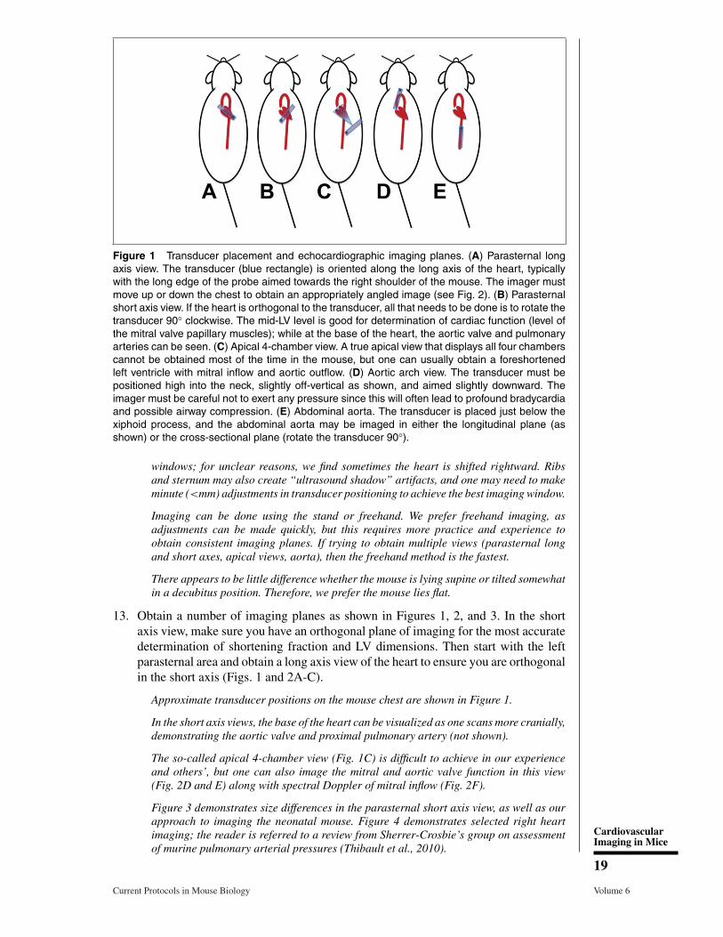
Figure 1 Transducer placement and echocardiographic imaging planes. (A) Parasternal longaxis view. The transducer (blue rectangle) is oriented along the long axis of the heart, typicallywith the long edge of the probe aimed towards the right shoulder of the mouse. The imager mustmove up or down the chest to obtain an appropriately angled image (see Fig. 2). (B) Parasternalshort axis view. If the heart is orthogonal to the transducer, all that needs to be done is to rotate thetransducer 90° clockwise. The mid-LV level is good for determination of cardiac function (level ofthe mitral valve papillary muscles); while at the base of the heart, the aortic valve and pulmonaryarteries can be seen. (C) Apical 4-chamber view. A true apical view that displays all four chamberscannot be obtained most of the time in the mouse, but one can usually obtain a foreshortenedleft ventricle with mitral inflow and aortic outflow. (D) Aortic arch view. The transducer must bepositioned high into the neck, slightly off-vertical as shown, and aimed slightly downward. Theimager must be careful not to exert any pressure since this will often lead to profound bradycardiaand possible airway compression. (E) Abdominal aorta. The transducer is placed just below thexiphoid process, and the abdominal aorta may be imaged in either the longitudinal plane (asshown) or the cross-sectional plane (rotate the transducer 90°).
windows; for unclear reasons, we find sometimes the heart is shifted rightward. Ribsand sternum may also create “ultrasound shadow” artifacts, and one may need to makeminute (<mm) adjustments in transducer positioning to achieve the best imaging window.
Imaging can be done using the stand or freehand. We prefer freehand imaging, asadjustments can be made quickly, but this requires more practice and experience toobtain consistent imaging planes. If trying to obtain multiple views (parasternal longand short axes, apical views, aorta), then the freehand method is the fastest.
There appears to be little difference whether the mouse is lying supine or tilted somewhatin a decubitus position. Therefore, we prefer the mouse lies flat.
13. Obtain a number of imaging planes as shown in Figures 1, 2, and 3. In the shortaxis view, make sure you have an orthogonal plane of imaging for the most accuratedetermination of shortening fraction and LV dimensions. Then start with the leftparasternal area and obtain a long axis view of the heart to ensure you are orthogonalin the short axis (Figs. 1 and 2A-C).
Approximate transducer positions on the mouse chest are shown in Figure 1.
In the short axis views, the base of the heart can be visualized as one scans more cranially,demonstrating the aortic valve and proximal pulmonary artery (not shown).
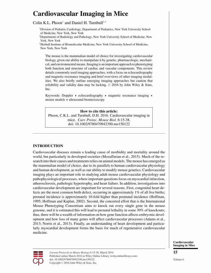
The so-called apical 4-chamber view (Fig. 1C) is difficult to achieve in our experienceand others’, but one can also image the mitral and aortic valve function in this view(Fig. 2D and E) along with spectral Doppler of mitral inflow (Fig. 2F).
Figure 3 demonstrates size differences in the parasternal short axis view, as well as ourapproach to imaging the neonatal mouse. Figure 4 demonstrates selected right heartimaging; the reader is referred to a review from Sherrer-Crosbie’s group on assessmentof murine pulmonary arterial pressures (Thibault et al., 2010).
CardiovascularImaging in Mice
19
Current Protocols in Mouse Biology Volume 6
Figure 2 Heart images from different planes. (A) Parasternal long axis view. The left ventricularapex is to our left, with the mitral valve, aortic valve, and aortic root shown. Note how the heartlies “flat” and is orthogonal to the transducer, and the normal left ventricle is shaped like a prolateellipsoid ("bullet shaped"). (B) Parasternal long axis view, with superimposed color Doppler flowmap, showing mitral inflow and aortic outflow. (C) Parasternal short axis view. The normal leftventricle is round, and systolic function is normally determined at the level of the papillary muscles.The right ventricle is often poorly seen. (D) Apical 4-chamber view, with superimposed colorDoppler flow map showing mitral inflow (red jet). (E) Apical 4-chamber view, with superimposedcolor Doppler flow map showing aortic outflow (blue jet). (F) Pulsed-wave Doppler signal of mitralinflow plus off-angle aortic outflow.
Figure 3 Parasternal short axis imaging of the left ventricle through the ages. (A) Adult heart.(B) Corresponding M-mode echocardiogram. (C) Juvenile (few weeks old, post-weaning). (D)Newborn pup. Note the far grainier image. (E) Newborn pup being imaged. The pup is held verygently with the thumb and forefinger, and ultrasound gel is applied all over the chest, taking care notto cover the snout. Parasternal long and short axis imaging can be accomplished in this manner.Scale bars (A, C, D): 2 mm.
14. Perform 2D (or B-mode) imaging.
Typically, 2D imaging alone can be done by one person who can use the foot pedalto freeze the cine loop. However, Doppler interrogation of blood flow will require anadditional observer who can place the Doppler sample volume and work the controls.
A typical echocardiogram of a mouse will take under 10 min; several minutes are requiredfor sedation, preparation, and warming to an appropriate core temperature. The imagingitself, in experienced hands, will take only 1 to 2 min.
15. When imaging is completed, wipe off the gel, remove the mouse from the imagingplatform, and place the mouse back in its cage.
CardiovascularImaging in Mice
20
Volume 6 Current Protocols in Mouse Biology
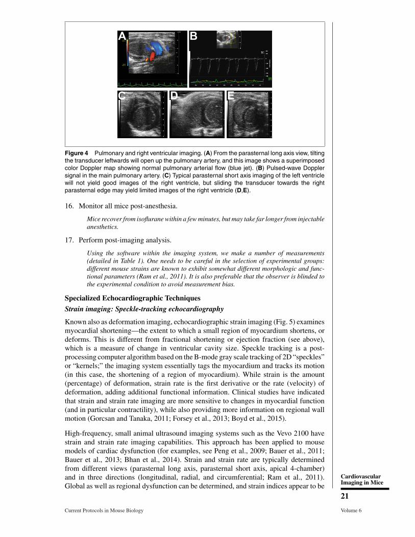
Figure 4 Pulmonary and right ventricular imaging. (A) From the parasternal long axis view, tiltingthe transducer leftwards will open up the pulmonary artery, and this image shows a superimposedcolor Doppler map showing normal pulmonary arterial flow (blue jet). (B) Pulsed-wave Dopplersignal in the main pulmonary artery. (C) Typical parasternal short axis imaging of the left ventriclewill not yield good images of the right ventricle, but sliding the transducer towards the rightparasternal edge may yield limited images of the right ventricle (D,E).
16. Monitor all mice post-anesthesia.
Mice recover from isoflurane within a few minutes, but may take far longer from injectableanesthetics.
17. Perform post-imaging analysis.
Using the software within the imaging system, we make a number of measurements(detailed in Table 1). One needs to be careful in the selection of experimental groups:different mouse strains are known to exhibit somewhat different morphologic and func-tional parameters (Ram et al., 2011). It is also preferable that the observer is blinded tothe experimental condition to avoid measurement bias.
Specialized Echocardiographic Techniques
Strain imaging: Speckle-tracking echocardiography
Known also as deformation imaging, echocardiographic strain imaging (Fig. 5) examinesmyocardial shortening—the extent to which a small region of myocardium shortens, ordeforms. This is different from fractional shortening or ejection fraction (see above),which is a measure of change in ventricular cavity size. Speckle tracking is a post-processing computer algorithm based on the B-mode gray scale tracking of 2D “speckles”or “kernels;” the imaging system essentially tags the myocardium and tracks its motion(in this case, the shortening of a region of myocardium). While strain is the amount(percentage) of deformation, strain rate is the first derivative or the rate (velocity) ofdeformation, adding additional functional information. Clinical studies have indicatedthat strain and strain rate imaging are more sensitive to changes in myocardial function(and in particular contractility), while also providing more information on regional wallmotion (Gorcsan and Tanaka, 2011; Forsey et al., 2013; Boyd et al., 2015).
High-frequency, small animal ultrasound imaging systems such as the Vevo 2100 havestrain and strain rate imaging capabilities. This approach has been applied to mousemodels of cardiac dysfunction (for examples, see Peng et al., 2009; Bauer et al., 2011;Bauer et al., 2013; Bhan et al., 2014). Strain and strain rate are typically determinedfrom different views (parasternal long axis, parasternal short axis, apical 4-chamber)and in three directions (longitudinal, radial, and circumferential; Ram et al., 2011).Global as well as regional dysfunction can be determined, and strain indices appear to be
CardiovascularImaging in Mice
21
Current Protocols in Mouse Biology Volume 6
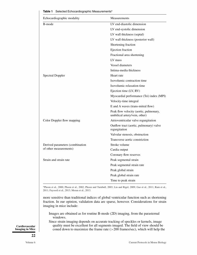
Table 1 Selected Echocardiographic Measurementsa
Echocardiographic modality Measurements
B-mode LV end-diastolic dimension
LV end-systolic dimension
LV wall thickness (septal)
LV wall thickness (posterior wall)
Shortening fraction
Ejection fraction
Fractional area shortening
LV mass
Vessel diameters
Intima-media thickness
Spectral Doppler Heart rate
Isovolumic contraction time
Isovolumic relaxation time
Ejection time (LV, RV)
Myocardial performance (Tei) index (MPI)
Velocity-time integral
E and A waves (trans-mitral flow)
Peak flow velocity (aortic, pulmonary,umbilical artery/vein, other)
Color Doppler flow mapping Atrioventricular valve regurgitation
Outflow tract (aortic, pulmonary) valveregurgitation
Valvular stenosis, obstruction
Transverse aortic constriction
Derived parameters (combinationof other measurements)
Stroke volume
Cardia output
Coronary flow reserves
Strain and strain rate Peak segmental strain
Peak segmental strain rate
Peak global strain
Peak global strain rate
Time to peak strain
aPhoon et al., 2000; Phoon et al., 2002; Phoon and Turnbull, 2003; Liu and Rigel, 2009; Gao et al., 2011; Ram et al.,2011; Fayssoil et al., 2013; Moran et al., 2013.
more sensitive than traditional indices of global ventricular function such as shorteningfraction. In our opinion, validation data are sparse, however. Considerations for strainimaging in mice include:
Images are obtained as for routine B-mode (2D) imaging, from the parasternalwindows.
Since strain imaging depends on accurate tracking of speckles or kernels, imagequality must be excellent for all segments imaged. The field of view should beconed down to maximize the frame rate (>200 frames/sec), which will help the
CardiovascularImaging in Mice
22
Volume 6 Current Protocols in Mouse Biology
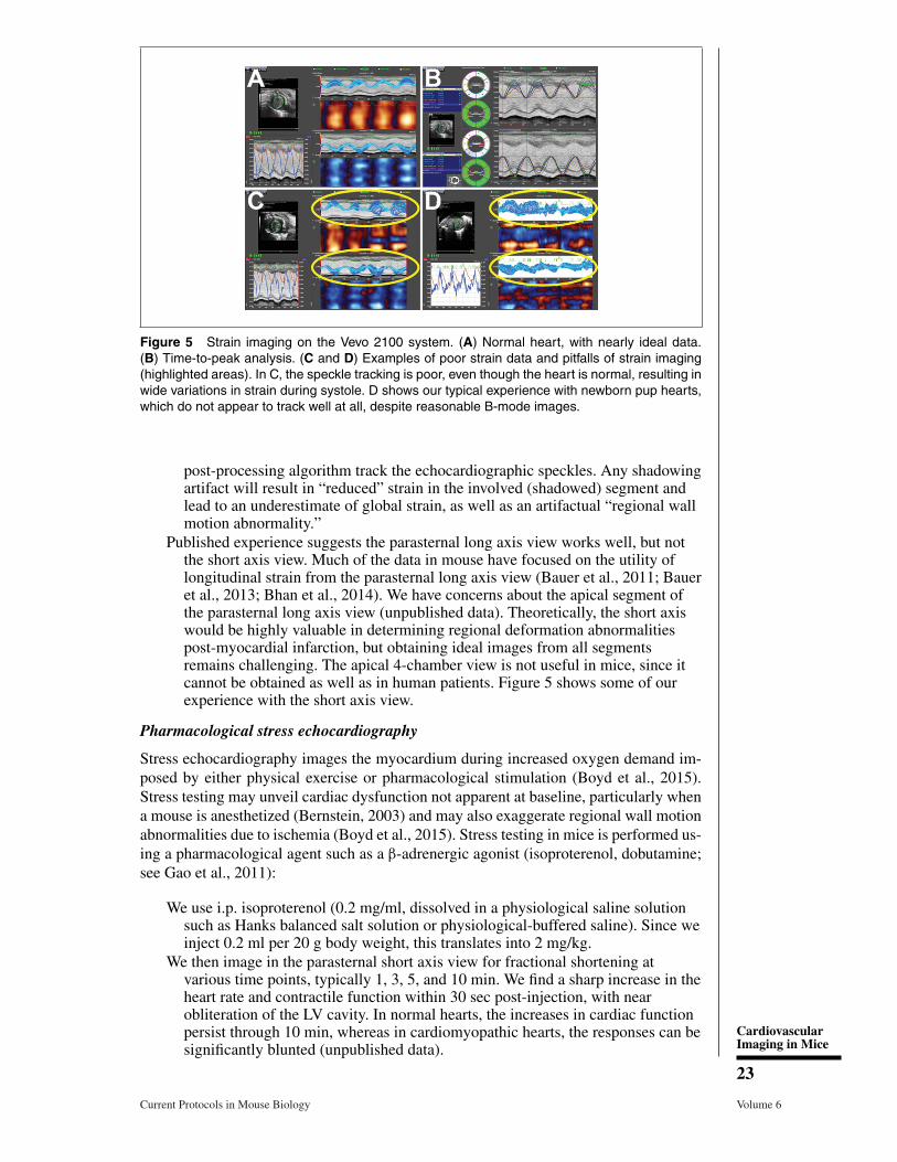
Figure 5 Strain imaging on the Vevo 2100 system. (A) Normal heart, with nearly ideal data.(B) Time-to-peak analysis. (C and D) Examples of poor strain data and pitfalls of strain imaging(highlighted areas). In C, the speckle tracking is poor, even though the heart is normal, resulting inwide variations in strain during systole. D shows our typical experience with newborn pup hearts,which do not appear to track well at all, despite reasonable B-mode images.
post-processing algorithm track the echocardiographic speckles. Any shadowingartifact will result in “reduced” strain in the involved (shadowed) segment andlead to an underestimate of global strain, as well as an artifactual “regional wallmotion abnormality.”
Published experience suggests the parasternal long axis view works well, but notthe short axis view. Much of the data in mouse have focused on the utility oflongitudinal strain from the parasternal long axis view (Bauer et al., 2011; Baueret al., 2013; Bhan et al., 2014). We have concerns about the apical segment ofthe parasternal long axis view (unpublished data). Theoretically, the short axiswould be highly valuable in determining regional deformation abnormalitiespost-myocardial infarction, but obtaining ideal images from all segmentsremains challenging. The apical 4-chamber view is not useful in mice, since itcannot be obtained as well as in human patients. Figure 5 shows some of ourexperience with the short axis view.
Pharmacological stress echocardiography
Stress echocardiography images the myocardium during increased oxygen demand im-posed by either physical exercise or pharmacological stimulation (Boyd et al., 2015).Stress testing may unveil cardiac dysfunction not apparent at baseline, particularly whena mouse is anesthetized (Bernstein, 2003) and may also exaggerate regional wall motionabnormalities due to ischemia (Boyd et al., 2015). Stress testing in mice is performed us-ing a pharmacological agent such as a β-adrenergic agonist (isoproterenol, dobutamine;see Gao et al., 2011):
We use i.p. isoproterenol (0.2 mg/ml, dissolved in a physiological saline solutionsuch as Hanks balanced salt solution or physiological-buffered saline). Since weinject 0.2 ml per 20 g body weight, this translates into 2 mg/kg.
We then image in the parasternal short axis view for fractional shortening atvarious time points, typically 1, 3, 5, and 10 min. We find a sharp increase in theheart rate and contractile function within 30 sec post-injection, with nearobliteration of the LV cavity. In normal hearts, the increases in cardiac functionpersist through 10 min, whereas in cardiomyopathic hearts, the responses can besignificantly blunted (unpublished data).
CardiovascularImaging in Mice
23
Current Protocols in Mouse Biology Volume 6
Miscellaneous approaches to studying myocardial function
Additional approaches include myocardial contrast echocardiography to measure my-ocardial perfusion (Scherrer-Crosbie and Kurtz, 2010; Foster et al., 2011; Ram et al.,2011; Moran et al., 2013), elastography to measure regional myocardial function (Luoand Konofagou, 2008), and coronary flow reserve, usually of the left coronary arteryunder conditions of myocardial damage or stress (Ram et al., 2011; Mercier et al., 2012).The reader is referred to these papers and reviews for further details.
Prenatal Assessment of the Embryonic/Fetal Mouse Cardiovascular System
The advantages of ultrasound/ultrasound biomicroscopy include rapid acquisition timesand in vivo imaging with good spatial resolution. However, image contrast tends tobe poor relative to other imaging modalities (Norris et al., 2013). Clearly, prenatalcardiovascular imaging of the mouse is far more challenging than postnatal imaging.Gui et al. (1996) first described their technique using a clinical 7 MHz ultrasoundsystem, followed by our lab’s approach with a prototype 40 MHz scanner (Srinivasanet al., 1998). With the advent of high-frequency ultrasound imaging systems and probes,arose innovative imaging approaches. Challenges include the far smaller size of thetarget (mouse embryos or fetuses), the number of mice within the uterine sacs, randomorientation of the organism, and movement within the uterus. Although mouse andhuman heart development are nearly parallel, important differences exist in their overalltimelines (Phoon, 2001; Krishnan et al., 2014). Nevertheless, such imaging has been ableto address important questions in developmental cardiology and hematology (Phoon,2006). Although cardiac structures are less often emphasized, our lab (Phoon et al., 2007)and others (Liu et al., 2014) have been able to detect congenital cardiac malformationsprenatally.
Materials
Mouse of interestIsofluraneMedical oxygen (optional)Ultrasound (acoustic coupling) gel
Scale (to weigh mouse)Anesthesia setup
Anesthetic induction chamberIsoflurane distributorMouse anesthesia nosecone delivery system
High-resolution ultrasound imaging system coupled to the anesthesia setup andwarming pad that doubles as an imaging platform
Electrocardiography (EKG) system including EKG leads and electrodes (usuallyincorporated into the imaging platform)
Strips of tape, cut to size (for paws)Hair dryer (for warm air convection) or warming lamp
1. Perform steps 1 through 10 of Cardiac imaging: Postnatal ultrasound (echocardiog-raphy) for neonatal, juveniles, and adults.
2. Once the core temperature is physiological and stable (takes several minutes) witha stable heart rate (Roth et al., 2002), apply the acoustic coupling gel to theabdomen.
In our experience, acoustic coupling gel on the gravid (pregnant) abdomen tends to coolthe core temperature far more than when applied to the chest. Therefore, we tend to allowthe core temperature to rise to 38°C before application of gel. When gel is applied, thecore temperature tends to fall by 1°C.
CardiovascularImaging in Mice
24
Volume 6 Current Protocols in Mouse Biology
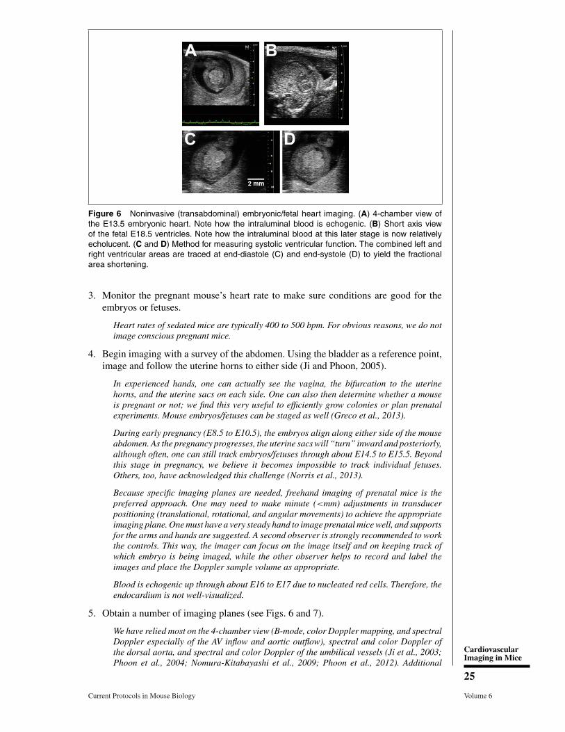
Figure 6 Noninvasive (transabdominal) embryonic/fetal heart imaging. (A) 4-chamber view ofthe E13.5 embryonic heart. Note how the intraluminal blood is echogenic. (B) Short axis viewof the fetal E18.5 ventricles. Note how the intraluminal blood at this later stage is now relativelyecholucent. (C and D) Method for measuring systolic ventricular function. The combined left andright ventricular areas are traced at end-diastole (C) and end-systole (D) to yield the fractionalarea shortening.
3. Monitor the pregnant mouse’s heart rate to make sure conditions are good for theembryos or fetuses.
Heart rates of sedated mice are typically 400 to 500 bpm. For obvious reasons, we do notimage conscious pregnant mice.
4. Begin imaging with a survey of the abdomen. Using the bladder as a reference point,image and follow the uterine horns to either side (Ji and Phoon, 2005).
In experienced hands, one can actually see the vagina, the bifurcation to the uterinehorns, and the uterine sacs on each side. One can also then determine whether a mouseis pregnant or not; we find this very useful to efficiently grow colonies or plan prenatalexperiments. Mouse embryos/fetuses can be staged as well (Greco et al., 2013).
During early pregnancy (E8.5 to E10.5), the embryos align along either side of the mouseabdomen. As the pregnancy progresses, the uterine sacs will “turn” inward and posteriorly,although often, one can still track embryos/fetuses through about E14.5 to E15.5. Beyondthis stage in pregnancy, we believe it becomes impossible to track individual fetuses.Others, too, have acknowledged this challenge (Norris et al., 2013).
Because specific imaging planes are needed, freehand imaging of prenatal mice is thepreferred approach. One may need to make minute (<mm) adjustments in transducerpositioning (translational, rotational, and angular movements) to achieve the appropriateimaging plane. One must have a very steady hand to image prenatal mice well, and supportsfor the arms and hands are suggested. A second observer is strongly recommended to workthe controls. This way, the imager can focus on the image itself and on keeping track ofwhich embryo is being imaged, while the other observer helps to record and label theimages and place the Doppler sample volume as appropriate.
Blood is echogenic up through about E16 to E17 due to nucleated red cells. Therefore, theendocardium is not well-visualized.
5. Obtain a number of imaging planes (see Figs. 6 and 7).
We have relied most on the 4-chamber view (B-mode, color Doppler mapping, and spectralDoppler especially of the AV inflow and aortic outflow), spectral and color Doppler ofthe dorsal aorta, and spectral and color Doppler of the umbilical vessels (Ji et al., 2003;Phoon et al., 2004; Nomura-Kitabayashi et al., 2009; Phoon et al., 2012). Additional
CardiovascularImaging in Mice
25
Current Protocols in Mouse Biology Volume 6

Figure 7 Examples of arterial flow in the prenatal mouse. (A) Sagittal view of an E12.5 mouseembryo, with superimposed color Doppler flow map showing the dorsal aorta. (B) Color Dopplerflow map of the umbilical vessels (red-blue “stripes” to the right of center; placenta is the homo-geneous gray region below the embryo). (C) Pulsed-wave Doppler signal in the dorsal aorta (notthe same embryo as in A). (D) Pulsed-wave Doppler signal in the umbilical cord (not the sameembryo as in B). The pulsatile positive deflection is the umbilical artery, while the more continuousnegative deflection is the umbilical vein. In C and D, the maternal EKG and respiratory waveformsare shown at the bottom, which help monitor the pregnant mouse during embryonic/fetal imaging.
information can be obtained from the vitelline circulation (Phoon and Turnbull, 2003;Phoon, 2006).
A typical echocardiogram of a pregnant mouse will take 30 to 60 min, depending on thenumber of parameters measured. Several minutes are required for sedation, preparation,and warming to an appropriate core temperature.
6. When imaging is completed, wipe off the gel, remove the mouse from the imagingplatform, and place it back in the cage.
7. Monitor all mice post-anesthesia.
8. Perform post-imaging analysis.
Using the software within the imaging system, we make a number of measurements (Table1). Preferably, the observer is blinded to the experimental condition in order to avoidmeasurement bias.
The Lo lab group has employed a 2-tier approach to screening for cardiac anomalies in aforward mutagenesis screen, using a lower-frequency, clinical ultrasound system for thefirst level of screening then moving to the Vevo systems for improved resolution. The readeris referred to their papers for details (Liu et al., 2013; Liu et al., 2014).
Newer Technologies: Annular Array Transducers for Fetal MouseEchocardiography
The technology for in vivo echocardiography in mice has improved significantly in re-cent years. The commercial scanners now available offer high frequency (30 to 50 MHz)linear array transducers with high spatial and temporal resolution for improved dynamicand functional analyses. Despite these advances, the current linear array transducers stillpresent limitations in spatial resolution, especially in the elevation (out-of-plane) direc-tion, where the width of the ultrasound beam is significantly larger than in the azimuthal(in-plane) direction. This can be problematic for analyses of very small cardiovascularstructures in mouse embryos, especially at earlier stages (E10.5 to E14.5) when manymorphological and functional changes occur and when many cardiac-specific phenotypesare first manifested in mutant embryos. These considerations have led us investigate the
CardiovascularImaging in Mice
26
Volume 6 Current Protocols in Mouse Biology
utility of annular array transducers that provide axi-symmetric focusing, integrated intoscanners with 3D image acquisition more suitable for in utero studies of developingmouse embryos (Aristizabal et al., 2006; Aristizabal et al., 2013).
Quantitative comparisons of imaging performance in phantoms designed to detect thesedifferences have clearly shown that axi-symmetrically focused annular array transducershave superior spatial resolution compared to commercially available linear arrays (Filouxet al., 2011; Filoux et al., 2012). These gains in resolution have translated into improvedcapability for 3D in utero analyses of brain development (Aristizabal et al., 2013).Since the annular array transducers cannot be used to steer the beam off axis, theymust be mechanically scanned to acquire 2D and 3D image data, which necessitatesmotion-gated acquisition, similar to most MRI methods. Respiratory-gated ultrasoundbiomicroscopy (UBM) was shown to be sufficient for high-throughput 3D fetal brainimaging (Aristizabal et al., 2013), but 3D imaging of the beating mouse heart willrequire combined respiratory and cardiac gating (Ketterling and Aristizabal, 2009). Inthe case of in utero echocardiography, where an EKG signal is not available, it is possibleto employ a Doppler ultrasound signal as the trigger for cardiac gating (Aristizabal et al.,2009).
VASCULAR IMAGING: ULTRASOUND
Vascular imaging is valuable in mouse models of atherosclerosis, vascular interventions,or to determine the success (degree) of transverse aortic constriction (Hartley et al.,2011; Ram et al., 2011). Atherosclerotic lesions, an abnormally thickened intima-medialayer, and vascular thromboses may be directly imaged with high resolution ultrasound(Choudhury et al., 2004; Chatterjee et al., 2014; Zhang et al., 2015) and also enhancedwith specific contrast agents (Wang et al., 2012; Foss et al., 2015). Arterial stiffness maybe determined by the pulse wave velocity (Hartley et al., 2011; Chatterjee et al., 2014)or a combination of aortic diameter and invasive blood pressure measurements (Kuoet al., 2014); for a more precise determination of localized arterial wall characteristics,pulse wave imaging has more recently been developed (Fujikura et al., 2007; Nandlallet al., 2014). Similarly, arterial wall strain may now be measured, as well (Favreauet al., 2014). To achieve the highest B-mode resolution images in general, the long axisof the vessel should be approximately orthogonal to the ultrasound beam; in contrast,Doppler beams must be placed as close to parallel to the artery as possible. Representativeimages from our lab of the normal aorta, aortic arch, and carotid artery are shown inFigure 8.
CARDIOVASCULAR MRI
MRI is now used for cardiac and vascular imaging in many human patients, althoughgenerally as a secondary modality following an initial exam with echocardiography.Over the past decade, dedicated high-field (�7 Tesla) MRI systems have become morecommon for small animal imaging, and many biologists now consider incorporatingMRI into studies of the mouse cardiovascular system. Traditional MRI images derivetissue contrast from differences in proton density, T1 or T2 relaxation times, but othermethods also are available to show differences in diffusion, perfusion, or blood flow.This makes MRI a very flexible imaging modality compared to echocardiography, withmany options for manipulating image contrast. MRI offers additional advantages overechocardiography including intrinsic three-dimensional image acquisition along image-defined projections so that assumptions about LV geometry do not need to be made (as isoften the case with echocardiography) and the ability to study many pathophysiologicalparameters in a single session, including energy metabolism (Akki et al., 2013). Despitethese apparent advantages, it must be recognized that MRI in mice, especially at fetaland neonatal stages, is very challenging due to the small size of the cardiac chambers Cardiovascular
Imaging in Mice
27
Current Protocols in Mouse Biology Volume 6
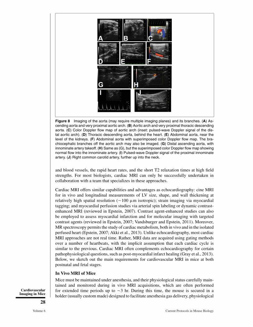
Figure 8 Imaging of the aorta (may require multiple imaging planes) and its branches. (A) As-cending aorta and very proximal aortic arch. (B) Aortic arch and very proximal thoracic descendingaorta. (C) Color Doppler flow map of aortic arch (inset: pulsed-wave Doppler signal of the dis-tal aortic arch). (D) Thoracic descending aorta, behind the heart. (E) Abdominal aorta, near thelevel of the kidneys. (F) Abdominal aorta with superimposed color Doppler flow map. The bra-chiocephalic branches off the aortic arch may also be imaged. (G) Distal ascending aorta, withinnominate artery takeoff. (H) Same as (G), but the superimposed color Doppler flow map showingnormal flow into the innominate artery. (I) Pulsed-wave Doppler signal of the proximal innominateartery. (J) Right common carotid artery, further up into the neck.
and blood vessels, the rapid heart rates, and the short T2 relaxation times at high fieldstrengths. For most biologists, cardiac MRI can only be successfully undertaken incollaboration with a team that specializes in these approaches.
Cardiac MRI offers similar capabilities and advantages as echocardiography: cine MRIfor in vivo and longitudinal measurements of LV size, shape, and wall thickening atrelatively high spatial resolution (�100 μm isotropic); strain imaging via myocardialtagging; and myocardial perfusion studies via arterial spin labeling or dynamic contrast-enhanced MRI (reviewed in Epstein, 2007). Contrast agent-enhanced studies can alsobe employed to assess myocardial infarction and for molecular imaging with targetedcontrast agents (reviewed in Epstein, 2007; Vandsburger and Epstein, 2011). Moreover,MR spectroscopy permits the study of cardiac metabolism, both in vivo and in the isolatedperfused heart (Epstein, 2007; Akki et al., 2013). Unlike echocardiography, most cardiacMRI approaches are not real time. Rather, MRI data are acquired using gating methodsover a number of heartbeats, with the implicit assumption that each cardiac cycle issimilar to the previous. Cardiac MRI often complements echocardiography for certainpathophysiological questions, such as post-myocardial infarct healing (Gray et al., 2013).Below, we sketch out the main requirements for cardiovascular MRI in mice at bothpostnatal and fetal stages.
In Vivo MRI of Mice
Mice must be maintained under anesthesia, and their physiological status carefully main-tained and monitored during in vivo MRI acquisitions, which are often performedfor extended time periods up to �3 hr. During this time, the mouse is secured in aholder (usually custom made) designed to facilitate anesthesia gas delivery, physiological
CardiovascularImaging in Mice
28
Volume 6 Current Protocols in Mouse Biology

Figure 9 In utero MRI of an E17.5 fetal mouse heart. After motion correction and cardiac gating,color coded volumetric renderings of the four chambers in a beating fetal mouse heart showsdifferences between diastole (A) and systole (B). Right ventricle, RV (purple); left ventricle, LV(red); right atrium, RA (blue); left atrium, LA (yellow).
monitoring, and reproducible positioning within the radiofrequency (RF) coil that ac-quires the MRI data. Isoflurane gas is most commonly used for anesthesia in mice(induced with 4% to 5% and maintained at �1.0% isoflurane in air). For cardiovascu-lar imaging, it is important that core temperature (monitored with a rectal temperatureprobe) be maintained close to 37°C using circulated warm water or air. EKG electrodesare typically incorporated into the mouse holder to monitor heart rate and to provide thetrigger signal for cardiac-gated acquisition. A respiratory pillow can be used to monitorthe breathing rate. Reliable EKG and respiratory signals can be very difficult to obtainin early postnatal mice, and we prefer to use modified “self-gated” pulse sequences thatdetect respiratory and/or cardiac motion directly from the acquired MRI signals (Niemanet al., 2009).
For in utero MRI it is impossible to use external devices to detect fetal motion. Instead, weemploy a self-gated gradient echo sequence—sensitive to the endogenous contrast of theembryonic mouse blood—together with image co-registration to obtain 3D cine imagesof the beating heart (Nieman et al., 2009; Fig. 9) and cerebral vasculature (Berrios-Otero et al., 2012; Parasoglou et al., 2013). More specialized pulse sequences wouldbe required for strain imaging and perfusion studies, but, in principle, any existingpulse sequence could be modified to incorporate self-gating for motion compensation.In practice, in utero images have a very limited signal-to-noise ratio (SNR), and morespecialized cardiac MRI studies have not been demonstrated in mouse embryos. In future,dedicated hardware and software will be required for improved in utero imaging in mice.In particular, improved RF coil arrays for small animal MRI might enable extendedfield-of-view (FOV) images to map out the position of multiple embryos, followed bysingle-coil small FOV images of individual embryos. Coil arrays for parallel imagingcould also be combined with sparse data reconstruction (compressed sensing) methods,to accelerate image acquisition and/or improve SNR as recently reported in adult mousecardiac MRI (Prieto et al., 2012; Buonincontri et al., 2014).
Ex Vivo MRI of Fixed Samples
Although MRI is often considered because of its potential for in vivo imaging, it canalso be used for nondestructive ex vivo imaging of fixed tissues. This approach hasbeen utilized extensively for studies of embryonic cardiovascular development, datingback to the pioneering reports of Smith, Johnson, and co-workers (Smith et al., 1994;reviewed in Smith, 2001). Although functional studies of cardiac dynamics and bloodflow studies are obviously not possible, ex vivo MRI has been very useful for analyzingcardiac and large vessel structural phenotypes in mutant mouse embryos (Huang et al.,
CardiovascularImaging in Mice
29
Current Protocols in Mouse Biology Volume 6
1998; Bamforth et al., 2004; Wadghiri et al., 2007; reviewed in Turnbull and Mori, 2007;Nieman and Turnbull, 2010). Ex vivo MRI is inherently motion free, and scans aregenerally run overnight for many hours to obtain high-resolution (20 to 50 μm isotropic)3D images. A number of centers have implemented multiple-sample overnight ex vivoimaging, making use of otherwise unused scanner time and providing high-throughputapproaches for screening new mutant mice at defined developmental stages (Schneideret al., 2004; Zhang et al., 2010; reviewed in Nieman and Turnbull, 2010).
For ex vivo MRI, mice are euthanized and tissue samples are dissected, fixed, andmounted in the RF coil for imaging (Dhenain et al., 2001). For vascular imaging inmid- to late-stage embryonic mice, perfusion fixation is preferred, using a contrast agentsolution to preferentially enhance the vasculature and performing imaging on the wholeembryo (see below). For ex vivo MRI, fixed embryos or tissue samples are immersedin 4% paraformaldehyde in PBS overnight (or longer) at 4°C, after which they can bestored in PBS. The fixative and/or storage medium is often “doped” with an MRI contrastagent, usually a chelated gadolinium (Gd) compound such as DTPA-Gd or DOTA-Gd (2to 3 mM concentration in the fixative), to increase the SNR of the resulting T2-weightedMRI images (Petiet et al., 2008). For imaging, the samples are embedded in 1% to 3%agar, or immersed in proton-free fluid (Fomblin or Fluorinert) to reduce the backgroundsignal. Samples are then placed within a close-fitting RF coil for ex vivo MRI.
Perfusion Fixation with Contrast Agents for Ex Vivo MRI
For ex vivo MRI of embryonic vasculature, we employ protocols similar to those firstdescribed by Smith and co-workers (Smith, 2000; Smith et al., 1994). Timed pregnantmice (where E0.5 denotes noon of the day after overnight breeding) are anesthetizedand the uterus accessed through laparotomy. Individual embryos are surgically removedfrom the uterus, maintaining the vascular connections to the placenta and warmed in37°C PBS to keep the heart beating normally at the start of the perfusion. Under adissection microscope, the umbilical vein is identified and cannulated with a pulledglass microcapillary needle, and the embryo is perfused with a slow flow rate (�0.1 to0.15 ml/min) pump: (1) starting with warm PBS containing heparin (5000 U/liter); (2)followed by fixative (2% [v/v] glutaraldehyde, 1% [v/v] formalin in PBS); and (3) finallyfollowed by the contrast agent, bovine serum albumin (BSA) conjugated to DTPA-Gd(BSA-DTPA-Gd; 1 mM) dissolved in 10% (w/v) gelatin solution. For full perfusionof all blood vessels, the umbilical vein is used for all three perfusates; when selectiveenhancement of the cerebral arteries was desired, we found that a timed perfusion ofthe contrast agent through the umbilical artery worked best (Berrios-Otero et al., 2009;Fig. 10). Recently, a novel approach for contrast-enhanced micro-CT has been shown togenerate more complete cerebral vascular filling by directing the contrast agent throughthe posterior brain arteries (Ghanavati et al., 2014), a method that could easily be adoptedfor ex vivo MRI. After perfusion, the umbilical vessels are ligated, and the samples areimmersed in 4°C fixative to solidify the gelatin and completely fix the tissue. Ex vivoMRI is then performed on the fixed embryos, as described above.
Image Analysis Methods
Although we have focused on the acquisition of ultrasound and MRI data related tocardiovascular structure and function, data analysis is an equally important subject, butone that will only be discussed briefly here. Most work to date has relied on qualitativeobservations from 2D and 3D images and rudimentary quantitative analysis of anatomical(e.g., LV area, volume) and functional (e.g., peak blood velocity, ejection fraction)parameters. Several groups are developing automated registration-based ex vivo MRImorphometry approaches for cardiovascular phenotype analysis in mutant mice (Zamyadiet al., 2010). While simple measurements of isolated geometric factors (branch lengthand angles) in complex vascular trees may suffice to characterize obvious defects in
CardiovascularImaging in Mice
30
Volume 6 Current Protocols in Mouse Biology

Figure 10 Ex vivo MRI of perfusion-fixed, gadolinium (Gd)-enhanced E17.5 mouse embryonicvasculature. (A) Following perfusion-fixation with the BSA-DTPA-Gd-gelatin solution, a maximumintensity projection (MIP) through the 3D, T1-weighted MRI data set, viewed from the side, showsthe heart and vasculature in the brain, liver, and other regions of the embryo. (B) MIPs of wild-type(WT; left) and Gli2-/- mutant (right) embryos, viewed from the back, show the missing basilar artery(BA) in the mutant, a vascular phenotype that was discovered using MRI. Other vessels labeledare the carotid (CA) and vertebral (VA) arteries.
vascular patterns (Berrios-Otero et al., 2009), ultimately a more systematic and automatedapproach is required to detect and characterize subtle (non-lethal) vascular phenotypes(Dorr et al., 2007) on both the structural and functional levels (Yang et al., 2010; Ghanavatiet al., 2014).
Molecular Imaging of Vasculature
Like ultrasound, MRI can also provide information on vascular biology, including mousemodels of atherosclerosis (Millon et al., 2014). For phenotype analysis in mouse models,combined structural, functional, and molecular imaging has the potential to provide amore complete picture of the complex molecular and mechanical events driving cardio-vascular development and disease. Molecular imaging of mouse vasculature has beendemonstrated with both ultrasound (reviewed in Lindner, 2010) and MRI (reviewed inArtemov et al., 2004), using intravascular injection of contrast agents (microbubbles forultrasound; Gd chelates or iron oxide nanoparticles for MRI) that are targeted to receptorson the surface of vascular endothelial cells via receptor-ligand binding. Notably, αvβ3-integrin and VEGFR2, which are highly expressed in tumors and vascular pathologies,have both been reported as viable targets for molecular imaging with MRI and ultra-sound. Unfortunately, receptor-ligand binding affinities are relatively low and can belimiting in the presence of shear forces associated with blood flows in the cardiovascular
CardiovascularImaging in Mice
31
Current Protocols in Mouse Biology Volume 6

Figure 11 Molecular imaging of E11.5 mouse embryonic vasculature. The Biotag transgene wasexpressed from the Tie2 promoter-enhancer elements in Ts-Biotag vascular reporter mice. (A)Time-averaged cine ultrasound biomicroscopy images were acquired 20 min after intravascularinjection of avidinated microbubbles (Av-MB). Av-MB binding and enhancement in Ts-Biotag em-bryos compared to wild-type (WT) littermates allowed visualization of embryonic vasculature andquantitation of vascular maps after threshold segmentation (bottom panels). (B) Similar experi-ments using MRI after injection of avidin-conjugated to DTPA-Gd (Av-DTPA-Gd) showed vascularlabeling in maximum intensity projection (MIP) images and quantitative vascular maps. Cerebralblood vessels, CBV; dorsal aorta, DA; heart, H; intersomitic vessels, ISV; placenta, Pl.
system. Moreover, targeted imaging as described above is limited to extracellular pro-teins, leaving many intracellular molecules inaccessible to imaging studies.
To address these limitations in current molecular imaging technology, we developeda novel “Biotag” transgene for biotinylation of cell membranes, allowing them to betargeted with (strept)avidinated contrast agents (Bartelle et al., 2012; Fig. 11). The Biotagtransgene can be used for molecular imaging of vasculature with ultrasound and MRI(and other imaging modalities) and has been demonstrated to be an effective approachin both embryonic and adult models of angiogenesis. Biotag expression, and subsequentmembrane biotinylation, can be driven from the regulatory elements of any gene bygenerating the appropriate transgenic or knock-in mouse reporter line, making it possibleto extend molecular imaging to a wide variety of genetic factors involved in vasculardevelopment and disease. Future development of other expressible reporters that can bedetected with MRI and/or ultrasound will likely have a significant impact on the use ofcardiovascular imaging in mouse models.
ADDITIONAL SPECIALIZED CARDIOVASCULAR IMAGINGAPPROACHES
Optical Coherence Tomography
Optical coherence tomography (OCT) is conceptually analogous to ultrasound, exceptthat it measures the echo time delay and magnitude of light instead of ultrasound (Fu-jimoto, 2003). Because optical echoes cannot be measured directly due to the speed oflight, OCT uses coherence interferometry instead to obtain data which can be used to thengenerate an image (Fujimoto, 2003; Garcia et al., 2015). Advantages of OCT include veryhigh spatial resolution—in the micron to submicron range—using state-of-the-art lasertechnology. However, there is a trade-off with depth of penetration in the range of 2 to 3mm only, due to signal attenuation and scattering (Fujimoto, 2003; Drexler et al., 2014;Garcia et al., 2015). Although OCT has been used more extensively in non-mammalianmodel systems, this shallow depth of penetration limits its utility in mouse cardiovascular
CardiovascularImaging in Mice
32
Volume 6 Current Protocols in Mouse Biology

imaging, where much of the work has focused on imaging of cultured mouse embryonichemodynamics during development (Larina et al., 2008; Larina et al., 2011; Larina et al.,2012; Garcia et al., 2015), although in utero imaging is feasible (Larina et al., 2011).More recently, techniques have been developed for imaging of (isolated Langendorff)adult mouse hearts by implementing multiperspective imaging and optical clearing thatpermit insights into tissue characteristics (Cua et al., 2014). The imaging approachesoutlined above are not in vivo, but recent reports suggest utility of OCT as an intravitalvascular imaging modality, specifically in a mouse hind limb ischemia model (Pooleet al., 2013).
Optical Projection Tomography
Optical projection tomography (OPT) fills a niche that allows high-resolution imagingof whole mouse embryos (Sharpe et al., 2002). From an emitted, focused light sourceand rotated sample, OPT utilizes projection tomography and in situ immunostains togenerate a three-dimensional image of a whole embryo’s gene expression patterns. OPThas been utilized for imaging the developing cardiovascular system and particularlythe developing vasculature ex vivo (Ruijter et al., 2004; Walls et al., 2008; Andersonet al., 2013). Most OPT has been performed on fixed and optically cleared specimens,but more recently, live OPT imaging has been achieved (Colas and Sharpe, 2009).Live cardiovascular imaging of the developing mouse embryo is still at rudimentarystages, however, and the next challenges are to overcome imaging artifacts inherent incardiac motion (Colas and Sharp, 2009). In the adult mouse heart, a Born-normalizednear-infrared fluorescence OPT imaging system has been more recently described, whichallows imaging of spatiotemporal dynamics of diverse cells by quantitating and resolvingmolecular agents within the imaged heart (Vinegoni et al., 2012).
Live Confocal Microscopy
Cultured mouse embryos have also been imaged with live confocal microscopy. Specif-ically, the yolk sac vasculature of the developing mouse embryo can be interrogatedto characterize evolving hemodynamics that contribute to overall mouse cardiovasculardevelopment (Lopez et al., 2015).
ACKNOWLEDGMENTS
The authors would like to thank members of the Phoon and Turnbull labs over theyears for their input and assistance on the innovative imaging approaches developed.Work was supported by NIH grants R01HL078665 and R01NS038461 (DHT). The Vevo2100 ultrasound imaging system at NYU Langone Medical Center was supported byNIH grant 1S10RR026881. The Small Animal Imaging Core at NYU Langone MedicalCenter assisted in this work and is supported in part by NIH grants P30CA016087 (toNYU Cancer Institute) and UL1TR00038 (to NYU Clinical and Translational ScienceInstitute).
CONFLICTS OF INTEREST
Drs. Phoon and Turnbull have no conflicts of interest to disclose.
LITERATURE CITEDAdams, D., Baldock, R., Bhattacharya, S., Copp, A.J., Dickinson, M., Greene, N.D., Henkelman, M.,
Justice, M., Mohun, T., Murray, S.A., Pauws, E., Raess, M., Rossant, J., Weaver, T., and West, D. 2013.Bloomsbury report on mouse embryo phenotyping: Recommendations from the IMPC workshop onembryonic lethal screening. Dis. Model Mech. 6:571-579. doi: 10.1242/dmm.011833.
Akki, A., Gupta, A., and Weiss, R.G. 2013. Magnetic resonance imaging and spectroscopy of the murinecardiovascular system. Am. J. Physiol. Heart Circ. Physiol. 304:H633-H648.
Anderson, G.A., Wong, M.D., Yang, J., and Henkelman, R.M. 2013. 3D imaging, registration, and analysisof the early mouse embryonic vasculature. Dev. Dynam. 242:527-538. doi: 10.1002/dvdy.23947. Cardiovascular
Imaging in Mice
33
Current Protocols in Mouse Biology Volume 6
Aristizabal, O., Ketterling, J.A., and Turnbull, D.H. 2006. 40-MHz annular array imaging of mouse embryos.Ultrasound Med. Biol. 32:1631-1637. doi: 10.1016/j.ultrasmedbio.2006.05.020.
Aristizabal, O., Mamou, J., Turnbull, D.H., and Ketterling, J.A. 2009. Doppler-derived trigger signals forhigh-frame-rate mouse cardiovascular imaging. Conf. Proc. IEEE Eng. Med. Biol. Soc. 1:1987-1990.
Aristizabal, O., Mamou, J., Ketterling, J.A., and Turnbull, D.H. 2013. High-throughput, high-frequency3-D ultrasound for in utero analysis of embryonic mouse brain development. Ultrasound Med. Biol.39:2321-2332. doi: 10.1016/j.ultrasmedbio.2013.06.015.
Artemov, D., Bhujwalla, Z.M., and Bulte, J.W. 2004. Magnetic resonance imaging of cell surface receptorsusing targeted contrast agents. Curr. Pharm. Biotechnol. 5:485-494. doi: 10.2174/1389201043376553.
Bamforth, S.D., Braganca, J., Farthing, C.R., Schneider, J.E., Broadbent, C., Michell, A.C., Clarke, K.,Neubauer, S., Norris, D., Brown, N.A., Anderson, R.H., and Bhattacharya, S. 2004. Cited2 controlsleft-right patterning and heart development through a Nodal-Pitx2c pathway. Nat. Genet. 36:1189-1196.doi: 10.1038/ng1446.
Bartelle, B.B., Berrios-Otero, C.A., Rodriguez, J.J., Friedland, A.E., Aristizabal, O., and Turnbull, D.H.2012. Novel genetic approach for in vivo vascular imaging in mice. Circ. Res. 110:938-947. doi:10.1161/CIRCRESAHA.111.254375.
Bauer, M., Cheng, S., Unno, K., Lin F-C, and Liao, R. 2013. Regional cardiac dysfunction and dyssynchronyin a murine model of afterload stress. PLoS One 8:e59915. doi: 10.1371/journal.pone.0059915.
Bauer, M., Cheng, S., Jain, M., Ngoy, S., Theodoropoulos, C., Trujillo, A., Lin F-C, and Liao, R. 2011.Echocardiographic speckle-tracking based strain imaging for rapid cardiovascular phenotyping in mice.Circ. Res. 108:908-916. doi: 10.1161/CIRCRESAHA.110.239574.
Bernstein, D. 2003. Exercise assessment of transgenic models of human cardiovascular disease. Physiol.Genomics 13:217-226. doi: 10.1152/physiolgenomics.00188.2002.
Berrios-Otero, C.A., Nieman, B.J., Parasoglou, P., and Turnbull, D.H. 2012. In utero phenotyping of mouseembryonic vasculature with MRI. Magn. Reson. Med. 67:251-257. doi: 10.1002/mrm.22991.
Berrios-Otero, C.A., Wadghiri, Y.Z., Nieman, B.J., Joyner, A.L., and Turnbull, D.H. 2009. Three-dimensional micro-MRI analysis of cerebral artery development in mouse embryos. Magn. Reson.Med. 62:1431-1439. doi: 10.1002/mrm.22113.
Berry, C.J., Thedens, D.R., Light-McGroary, K., Miller, J.D., Kutschke, W., Zimmerman, K.A., and Weiss,R.M. 2009. Effects of deep sedation or general anesthesia on cardiac function in mice undergoingcardiovascular magnetic resonance. J. Cardiovasc. Magn. Reson. 11:16. doi: 10.1186/1532-429X-11-16.
Bhan, A., Sirker, A., Zhang, J., Protti, A., Catibog, N., Driver, W., Botnar, R., Monaghan, M.J., andShah, A.M. 2014. High-frequency speckle tracking echocardiography in the assessment of left ventric-ular function and remodeling after murine myocardial infarction. Am. J. Physiol. Heart Circ. Physiol.306:H1371-H1383. doi: 10.1152/ajpheart.00553.2013.
Boyd, A.C., Schiller, N.B., and Thomas, L. 2015. Principles of transthoracic echocardiographic evaluation.Nat. Rev. Cardiol. 12:426-440. doi: 10.1038/nrcardio.2015.57.
Buonincontri, G., Methner, C., Krieg, T., Carpenter, T.A., and Sawiak, S.J. 2014. Functional assessment ofthe mouse heart by MRI with a 1-min acquisition. NMR Biomed. 27:733-737. doi: 10.1002/nbm.3116.
Chatterjee, S., Bedja, D., Mishra, S., Amuzie, C., Avolio, A., Kass, D.A., Berkowitz, D., and Renehan,M. 2014. Inhibition of glycosphingolipid synthesis ameliorates atherosclerosis and arterial stiffness inapolipoprotein E -/- mice and rabbits fed a high-fat and -cholesterol diet. Circulation 129:2403-2413.doi: 10.1161/CIRCULATIONAHA.113.007559.
Choudhury, R.P., Fuster, V., and Fayad, Z.A. 2004. Molecular, cellular and functional imaging ofatherothrombosis. Nat. Rev. Drug Discov. 3:913-925. doi: 10.1038/nrd1548.
Colas J-F. and Sharpe, J. 2009. Live optical projection tomography. Organogenesis 5:211-216. doi:10.4161/org.5.4.10426.
Collis, L.P., Meyers, M.B., Zhang, J., Phoon, C.K.L., Sobie, E.A., Coetzee, W.A., and Fishman, G.I. 2007.Expression of a sorcin missense mutation in the heart modulates excitation-contraction coupling. FASEBJ. 21:475-487. doi: 10.1096/fj.06-6292com.
Cua, M., Lin, E., Lee, L., Sheng, X., Wong, K.S., Tibbits, G.F., Beg, M.F., and Sarunic, M.V. 2014.Morphological phenotyping of mouse hearts using optical coherence tomography. J. Biomed. Opt.19:116007. doi: 10.1117/1.JBO.19.11.116007.
Danielson, L.S., Park, D.S., Rotllan, N., Chamorro-Jorganes, A., Guijarro, M.V., Fernandez-Hernando, C.,Fishman, G.I., Phoon, C.K.L., and Hernando, E. 2013. Cardiovascular dysregulation of miR-17-92 causesa lethal hypertrophic cardiomyopathy and arrhythmogenesis. FASEB J. 27:1460-1467. doi: 10.1096/fj.12-221994.
Dhenain, M., Ruffins, S.W., and Jacobs, R.E. 2001. Three-dimensional digital mouse atlas using high-resolution MRI. Dev. Biol. 232:458-470. doi: 10.1006/dbio.2001.0189.
CardiovascularImaging in Mice
34
Volume 6 Current Protocols in Mouse Biology
Dorr, A., Sled, J.G., and Kabani, N. 2007. Three-dimensional cerebral vasculature of the CBA mouse brain:A magnetic resonance imaging and micro computed tomography study. Neuroimage 35:1409-1423. doi:10.1016/j.neuroimage.2006.12.040.
Drexler, W., Liu, M., Kumar, A., Kamali, T., Unterhuber, A., and Leitgeb, R.A. 2014. Optical co-herence tomography today: Speed, contrast, and multimodality. J. Biomed. Opt. 19:071412. doi:10.1117/1.JBO.19.7.071412.
Epstein, F.H. 2007. MR in mouse models of cardiac disease. NMR Biomed. 20:238-255. doi:10.1002/nbm.1152.
Favreau, J.T., Liu, C., Yu, P., Tao, M., Mauro, C., Gaudette, G.R., and Ozaki, C.K. 2014. Acute reductionsin mechanical wall strain precede the formation of intimal hyperplasia in a murine model of arterialocclusive disease. J. Vasc. Surg. 60:1340-1347. doi: 10.1016/j.jvs.2013.07.113.
Fayssoil, A., and Tournoux, F. 2013. Analyzing left ventricular function in mice with Doppler echocardio-graphy. Heart Fail. Rev. 18:511-516. doi: 10.1007/s10741-012-9345-8.
Filoux, E., Mamou, J., Aristizabal, O., and Ketterling, J.A. 2011. Characterization of the spatial resolution ofdifferent high-frequency imaging systems using a novel anechoic-sphere phantom. IEEE Trans. Ultrason.Ferroelectr. Freq. Control 58:994-1005.
Filoux, E., Mamou, J., Moran, C.M., Pye, S.D., and Ketterling, J.A. 2012. Correspondence - Charac-terization of the effective performance of a high-frequency annular-array-based imaging system us-ing anechoic-pipe phantoms. IEEE Trans. Ultrason. Ferroelectr. Freq. Control 59:2825-2830. doi:10.1109/TED.2012.2209650.
Forsey, J., Friedberg, M.K., and Mertens, L. 2013. Speckle tracking echocardiography in pediatric andcongenital heart disease. Echocardiography 30:447-459. doi: 10.1111/echo.12131.
Foss, C.A., Bedja, D., Mease, R.C., Wang, H., Kass, D.A., Chatterjee, S., and Pomper, M.G. 2015. Molecularimaging of inflammation in the ApoE-/- mouse model of atherosclerosis with IodoDPA. Biochem.Biophys. Res. Commun. 461:70-75. doi: 10.1016/j.bbrc.2015.03.171.
Foster, F.S., Hossack, J., and Adamson, S.L. 2011. Micro-ultrasound for preclinical imaging. InterfaceFocus 1:576-601. doi: 10.1098/rsfs.2011.0037.
Fujikura, K., Luo, J., Gamarnik, V., Pernot, M., Fukumoto, R., Tilson M.D. 3rd, and Konofagou, E.E. 2007.A novel noninvasive technique for pulse-wave imaging and characterization of clinically-significant vas-cular mechanical properties in vivo. Ultrason. Imaging 29:137-154. doi: 10.1177/016173460702900301.
Fujimoto, J.G. 2003. Optical coherence tomography for ultrahigh resolution in vivo imaging. Nat. Biotech.21:1361-1367. doi: 10.1038/nbt892.
Gao, S., Ho, D., Vatner, D.E., and Vatner, S.F. 2011. Echocardiography in mice. Curr. Protocols. Mouse.Biol. 1:71-83.
Garcia, M.D., Lopez AL 3rd, Larin, K.V., and Larina, I.V. 2015. Imaging of cardiovascular developmentin mammalian embryos using optical coherence tomography. Methods Mol. Biol. 1214:151-161. doi:10.1007/978-1-4939-1462-3_8.
Gardin, J.M., Siri, F.M., Kitsis, R.N., Edwards, J.G., and Leinwand, L.A. 1995. Echocardiographicassessment of left ventricular mass and systolic function in mice. Circ. Res. 76:907-914. doi:10.1161/01.RES.76.5.907.
Ghanavati, S., Lerch, J.P., and Sled, J.G. 2014. Automatic anatomical labeling of the complete cerebralvasculature in mouse models. Neuroimage 95:117-128. doi: 10.1016/j.neuroimage.2014.03.044.
Ghanavati, S., Yu, L.X., Lerch, J.P., and Sled, J.G. 2014. A perfusion procedure for imagingof the mouse cerebral vasculature by X-ray micro-CT. J. Neurosci. Methods 221:70-77. doi:10.1016/j.jneumeth.2013.09.002.
Gorcsan J 3rd and Tanaka, H. 2011. Echocardiographic assessment of myocardial strain. J. Am. Coll.Cardiol. 58:1401-1413. doi: 10.1016/j.jacc.2011.06.038.
Gray, G.A., White, C.I., Thomson, A., Kozak, A., Moran, C., and Jansen, M.A. 2013. Imaging the healingmurine myocardial infarct in vivo: Ultrasound, magnetic resonance imaging and fluorescence moleculartomography. Exp. Physiol. 98:606-613. doi: 10.1113/expphysiol.2012.064741.
Greco, A., Ragucci, M., Coda, A.R., Rosa, A., Gargiulo, S., Liuzzi, R., Gramanzini, M., Albanese, S.,Pappata S, Mancini, M., Brunetti, A., and Salvatore, M. 2013. High frequency ultrasound for in vivopregnancy diagnosis and staging of placental and fetal development in mice. PLoS One 8:e77205. doi:10.1371/journal.pone.0077205.
Gui, Y.H., Linask, K.K., Khowsathit, P., and Huhta, J.C. 1996. Doppler echocardiography of normal andabnormal embryonic mouse heart. Pediatr. Res. 40:633-642. doi: 10.1203/00006450-199610000-00020.
Hartley, C.J., Reddy, A.K., Madala, S., Entman ml, Michael, L.H., and Taffet, G.E. 2011. Doppler velocitymeasurements from large and small arteries of mice. Am. J. Physiol. Heart Circ. Physiol. 301:H269-278.doi: 10.1152/ajpheart.00320.2011.
Hoffman, J.I.E. 1995. Incidence of congenital heart disease: II. Prenatal incidence. Pediatr. Cardiol. 16:155-165.
CardiovascularImaging in Mice
35
Current Protocols in Mouse Biology Volume 6
Hoffman, J.I.E. and Kaplan, S. 2002. The incidence of congenital heart disease. J. Am. Coll. Cardiol.39:1890-1900. doi: 10.1016/S0735-1097(02)01886-7.
Huang, G.Y., Wessels, A., Smith, B.R., Linask, K.K., Ewart, J.L., and Lo, C.W. 1998. Alteration in con-nexin 43 gap junction gene dosage impairs conotruncal heart development. Dev. Biol. 198:32-44. doi:10.1006/dbio.1998.8891.
Ji, R.P. and Phoon, C.K.L. 2005. Non-invasive localization of NFATc1-/- mouse embryos by ultrasoundbiomicroscopy-Doppler allows genotype-phenotype correlation. J. Am. Soc. Echocardiogr. 18:1415-1421. doi: 10.1016/j.echo.2005.04.006.
Ji, R.P., Phoon, C.K.L., Aristizabal, O., McGrath, K.E., Palis, J., and Turnbull, D.H. 2003. Onset of cardiacfunction during early mouse embryogenesis coincides with entry of primitive erythroblasts into theembryo proper. Circ. Res. 92:133-135. doi: 10.1161/01.RES.0000056532.18710.C0.
Ketterling, J.A. and Aristizabal, O. 2009. Prospective ECG-gated mouse cardiac imaging with a 34-MHzannular array transducer. IEEE Trans. Ultrason. Ferroelectr. Freq. Control 56:1394-1404.
Kober, F., Iltis, I., Cozzone, P.J., and Bernard, M. 2004. Cine-MRI assessment of cardiac func-tion in mice anesthetized with ketamine/xylazine and isoflurane. MAGMA 17:157-161. doi:10.1007/s10334-004-0086-0.
Krishnan, A., Samtani, R., Dhanantwari, P., Lee, E., Yamada, S., Shiota, K., Donofrio, M.T., Leatherbury,L., and Lo, C.W. 2014. A detailed comparison of mouse and human cardiac development. Pediatr. Res.76:500-507. doi: 10.1038/pr.2014.128.
Kuo, M.M., Barodka, V., Abraham, T.P., Steppan, J., Shoukas, A.A., Butlin, M., Avolio, A., Berkowitz,D.E., and Santhanam, L. 2014. Measuring ascending aortic stiffness in vivo in mice using ultrasound. J.Vis. Exp.94:e52200. doi: 10.3791/52200.
Larina, I.V., Larin, K.V., Justice, M.V., and Dickinson, M.E. 2011. Optical coherence tomogra-phy for live imaging of mammalian development. Curr. Opin. Genet. Dev. 21:579-584. doi:10.1016/j.gde.2011.09.004.
Larina, I.V., Garcia, M.D., Vadakkan, T.J., Larin, K.V., and Dickinson, M.E. 2012. Imaging mouse embryoniccardiovascular development. Cold. Spring. Harb. Protoc. 2012:1035-1043. doi: 10.1101/pdb.top071498.
Larina, I.V., Sudheendran, N., Ghosn, M., Jiang, J., Cable, A., Larin, K.V., and Dickinson, M.E. 2008.Live imaging of blood flow in mammalian embryos using Doppler swept-source optical coherencetomography. J. Biomed. Opt. 13:060506. doi: 10.1117/1.3046716.
Lindner, J.R. 2010. Molecular imaging of myocardial and vascular disorders with ultrasound. JACC Car-diovasc. Imaging 3:204-211. doi: 10.1016/j.jcmg.2009.09.021.
Liu, J. and Rigel, D.F. 2009. Echocardiographic examination in rats and mice. Methods Mol. Biol. 573:139-155. doi: 10.1007/978-1-60761-247-6_8.
Liu, X., Tobita, K., Francis, R.J., and Lo, C.W. 2013. Imaging techniques for visualizing and phenotyp-ing congenital heart defects in murine models. Birth Defects Res. C. Embryo. Today 99:93-105. doi:10.1002/bdrc.21037.
Liu, X., Francis, R., Kim, A.J., Ramirez, R., Chen, G., Subramanian, R., Anderton, S., Kim, Y., Wong,L., Morgan, J., Pratt, H.C., Reinholdt, L., Devine, W., Leatherbury, L., Tobita, K., and Lo, C.W. 2014.Interrogating congenital heart defects with noninvasive fetal echocardiography in a mouse forwardgenetic screen. Circ. Cardiovasc. Imaging 7:31-42. doi: 10.1161/CIRCIMAGING.113.000451.
Lopez A.L. 3rd, Garcia, M.D., Dickinson, M.E., and Larina, I.V. 2015. Live confocal microscopyof the developing mouse embryonic yolk sac vasculature. Methods Mol. Biol. 1214:163-172. doi:10.1007/978-1-4939-1462-3_9.
Luo, J. and Konofagou, E.E. 2008. High-frame rate, full-view myocardial elastography with automatedcontour tracking in murine left ventricles in vivo. IEEE Trans. Ultrason. Ferroelectr. Freq. Control55:240-248. doi: 10.1109/TUFFC.2008.633.
Manning, W.J., Wei, J.Y., Katz, S.E., Litwin, S.E., and Douglas, P.S. 1994. In vivo assessment of LV massin mice using high-frequency cardiac ultrasound: Necropsy validation. Am. J. Physiol. 266:H1672-675.
Mercier, N., Kiviniemi, T.O., Saraste, A., Miiluniemi, M., Silvola, J., Jalkanen, S., and Yegutkin,G.G. 2012. Impaired ATP-induced coronary blood flow and diminished aortic NTPDase activ-ity precede lesion formation in apolipoprotein E-deficient mice. Am. J. Pathol. 180:419-428. doi:10.1016/j.ajpath.2011.10.002.
Millon, A., Canet-Soulas, E., Boussel, L., Fayad, Z., and Douek, P. 2014. Animal models of atheroscle-rosis and magnetic resonance imaging for monitoring plaque progression. Vascular 22:221-237. doi:10.1177/1708538113478758.
Moran, C.M., Thomson, A.J.W., Rog-Zielinska, E., and Gray, G.A. 2013. High-resolution echocardiographyin the assessment of cardiac physiology and disease in preclinical models. Exp. Physiol. 98:629-644.doi: 10.1113/expphysiol.2012.068577.
Mozaffarian, D., Benjamin, E.J., Go, A.S., Arnett, D.K., Blaha, M.J., Cushman, M., de Ferranti, S., De-spres, J.P., Fullerton, H.J., Howard, V.J., Huffman, M.D., Judd, S.E., Kissela, B.M., Lackland, D.T.,
CardiovascularImaging in Mice
36
Volume 6 Current Protocols in Mouse Biology
Lichtman, J.H., Lisabeth, L.D., Liu, S., Mackey, R.H., Matchar, D.B., McGuire, D.K., Mohler ER3rd, Moy, C.S., Muntner, P., Mussolino, M.E., Nasir, K., Neumar, R.W., Nichol, G., Palaniappan, L.,Pandey, D.K., Reeves, M.J., Rodriguez, C.J., Sorlie, P.D., Stein, J., Towfighi, A., Turan, T.N., Virani,S.S., Willey, J.Z., Woo, D., Yeh, R.W., and Turner, M.B. 2015. American Heart Association StatisticsCommittee and Stroke Statistics Subcommittee. Heart disease and stroke statistics – 2015 update: A re-port from the American Heart Association. Circulation 131:e29-322. doi: 10.1161/CIR.0000000000000152.
Nandlall, S.D., Goldklang, M.P., Kalashian, A., Dangra, N.A., D’Armiento, J.M., and Konofagou, E.E.2014. Monitoring and staging abdominal aortic aneurysm disease with pulse wave imaging. UltrasoundMed. Biol. 40:2404-2414. doi: 10.1016/j.ultrasmedbio.2014.04.013.
Nieman, B.J. and Turnbull, D.H. 2010. Ultrasound and magnetic resonance microimaging of mouse devel-opment. Methods Enzymol. 476:379-400. doi: 10.1016/S0076-6879(10)76021-3.
Nieman, B.J., Szulc, K.U., and Turnbull, D.H. 2009. Three-dimensional in vivo MRI with self-gating andimage coregistration in the mouse. Magn. Reson. Med. 61:1148-1157. doi: 10.1002/mrm.21945.
Nomura-Kitabayashi, A., Phoon, C.K.L., Kishigami, S., Rosenthal, J., Yamauchi, Y., Abe, K., Yamamura,K., Samtani, R., Lo, C.W., and Mishina, Y. 2009. Outflow tract cushions perform a critical valve-likefunction in the early embryonic heart requiring BMPRIA-mediated signaling in cardiac neural crest. Am.J. Physiol. Heart. Circ. Physiol. 297:H1617-H1628. doi: 10.1152/ajpheart.00304.2009.
Norris, F.C., Wong, M.D., Greene, N.D.E., Scambler, P.J., Weaver, T., Weninger, W.J., Mohun, T.J., Henkel-man, R.M., and Lythgoe, M.F. 2013. A coming of age: Advanced imaging technologies for characterisingthe developing mouse. Trends Genet. 29:700-711. doi: 10.1016/j.tig.2013.08.004.
Pachon, R.E., Scharf, B.A., Vatner, D.E., and Vatner, S.F. 2015. Best anesthetics for assessing left ventricularsystolic function by echocardiography in mice. Am. J. Physiol. Heart. Circ. Physiol. 308:H1525-H1529.doi: 10.1152/ajpheart.00890.2014.
Parasoglou, P., Berrios-Otero, C.A., Nieman, B.J., and Turnbull, D.H. 2013. High-resolution MRI of early-stage mouse embryos. NMR Biomed. 26:224-231. doi: 10.1002/nbm.2843.
Peng, Y., Popvic, Z.B., Sopko, N., Drinko, J., Zhang, Z., Thomas, J.D., and Penn, M.S. 2009. Speckletracking echocardiography in the assessment of mouse models of cardiac dysfunction. Am. J. Physiol.Heart Circ. Physiol. 297:H811-H820. doi: 10.1152/ajpheart.00385.2009.
Petiet, A.E., Kaufman, M.H., Goddeeris, M.M., Brandenburg, J., Elmore, S.A., and Johnson, G.A. 2008.High-resolution magnetic resonance histology of the embryonic and neonatal mouse: A 4D atlas andmorphologic database. Proc. Natl. Acad. Sci. USA. 105:12331-12336. doi: 10.1073/pnas.0805747105.
Phoon, C.K.L. 2001. Circulatory physiology in the developing embryo. Curr. Opin. Pediatr. 13:456-464.doi: 10.1097/00008480-200110000-00013.
Phoon, C.K.L. 2006. Imaging tools for the developmental biologist: Ultrasound biomicroscopy of mouseembryonic development. Pediatr. Res. 60:14-21. doi: 10.1203/01.pdr.0000219441.28206.79.
Phoon, C.K.L. and Turnbull, D.H. 2003. Ultrasound biomicroscopy-Doppler in mouse cardiovascular de-velopment. Physiol. Genomics 14:3-15. doi: 10.1152/physiolgenomics.00008.2003.
Phoon, C.K.L., Aristizabal, O., and Turnbull, D.H. 2000. 40 MHz Doppler characterization of umbilicaland dorsal aortic blood flow in the early mouse embryo. Ultrasound Med. Biol. 26:1275-1283. doi:10.1016/S0301-5629(00)00278-7.
Phoon, C.K.L., Aristizabal, O., and Turnbull, D.H. 2002. Spatial velocity profile in mouse embryonicaorta and Doppler-derived volumetric flow: A preliminary model. Am. J. Physiol. Heart Circ. Physiol.283:908-916. doi: 10.1152/ajpheart.00869.2001.
Phoon, C.K., Ji, R.P., and Chowdhury, P.D. 2007. Transposition of the great arteries and double outletright ventricle have related early developmental origins: Fundamental insights from in vivo imaging.Circulation 116(Suppl):511.
Phoon, C.K.L., Ji, R.P., Aristizabal, O., Worrad, D.M., Zhou, B., Baldwin, H.S., and Turnbull, D.H. 2004.Embryonic heart failure in NFATc1-/- mice: Novel mechanistic insights from in utero ultrasound biomi-croscopy. Circ. Res. 95:92-99. doi: 10.1161/01.RES.0000133681.99617.28.
Phoon, C.K.L., Acehan, D., Schlame, M., Stokes, D.L., Edelman-Novemsky, I., Yu, D., Xu, Y., Viswanathan,N., and Ren, M. 2012. Tafazzin knockdown in mice leads to a developmental cardiomyopathy with earlydiastolic dysfunction preceding myocardial noncompaction. JAHA: J. Am. Heart. Assoc. 1:jah3-e000455.doi:JAHA.111.000455
Poole, K.M., Tucker-Schwartz, J.M., Sit, W.W., Walsh, A.J., Duvall, C.L., and Skala, M.C. 2013. Quantitativeoptical imaging of vascular response in vivo in a model of peripheral arterial disease. Am. J. Physiol.Heart Circ. Physiol. 305:H1168-H1180. doi: 10.1152/ajpheart.00362.2013.
Prieto, C., Andia, M.E., von Bary, C., Onthank, D.C., Schaeffter, T., and Botnar, R.M. 2012. Acceleratingthree-dimensional molecular cardiovascular MR imaging using compressed sensing. J. Magn. Reson.Imaging. 36:1362-1371. doi: 10.1002/jmri.23763. Cardiovascular
Imaging in Mice
37
Current Protocols in Mouse Biology Volume 6
Ram, R., Mickelsen, D.M., Theodoropoulos, C., and Blaxall, B.C. 2011. New approaches in small animalechocardiography: Imaging the sounds of silence. Am. J. Physiol. Heart Circ. Physiol. 301:H1765-1780.doi: 10.1152/ajpheart.00559.2011.
Roth, D.M., Swaney, J.S., Dalton, N.D., Gilpin, E.A., and Ross J Jr. 2002. Impact of anesthesia on cardiacfunction during echocardiography in mice. Am. J. Physiol. Heart Circ. Physiol. 282:H2134-H2140. doi:10.1152/ajpheart.00845.2001.
Ruijter, J.M., Soufan, A.T., Hagoort, J., and Moorman, A.F.M. 2004. Molecular imaging of the embryonicheart: Fables and facts on 3D imaging of gene expression patterns. Birth. Defects. Res. Part, C. 72:224-240. doi: 10.1002/bdrc.20018.
Scherrer-Crosbie, M. and Kurtz, B. 2010. Ventricular remodeling and function: Insights using murineechocardiography. J. Mol. Cell. Cardiol. 48:512-517. doi: 10.1016/j.yjmcc.2009.07.004.
Schneider, J.E., Bose, J., Bamforth, S.D., Gruber, A.D., Broadbent, C., Clarke, K., Neubauer, S., Lengeling,A., and Bhattacharya, S. 2004. Identification of cardiac malformations in mice lacking Ptdsr us-ing a novel high-throughput magnetic resonance imaging technique. BMC Dev. Biol. 4:16. doi:10.1186/1471-213X-4-16.
Sharpe, J., Ahlgren, U., Perry, P., Hill, B., Ross, A., Hecksher-Serensen, J., Baldock, R., and Davidson, D.2002. Optical projection tomography as a tool for 3D microscopy and gene expression studies. Science296:541-545. doi: 10.1126/science.1068206.
Smith, B.R. 2000. Magnetic resonance imaging analysis of embryos. Methods Mol. Biol. 135:211-216.
Smith, B.R. 2001. Magnetic resonance microscopy in cardiac development. Microsc. Res. Tech. 52:323-330.doi: 10.1002/1097-0029(20010201)52:3<3c323::AID-JEMT1016>3e3.0.CO;2-F.
Smith, B.R., Johnson, G.A., Groman, E.V., and Linney, E. 1994. Magnetic resonance microscopy of mouseembryos. Proc. Natl. Acad. Sci. USA 91:3530-3533. doi: 10.1073/pnas.91.9.3530.
Srinivasan, S., Baldwin, H.S., Aristizabal, O., Kwee, L., Labow, M., Artman, M., and Turnbull, D.H. 1998.Noninvasive, in utero imaging of mouse embryonic heart development with 40-MHz echocardiography.Circulation 98:912-918. doi: 10.1161/01.CIR.98.9.912.
Thibault, H.B., Kurtz, B., Raher, M.J., Shaik, R.S., Waxman, A., Derumeaux, G., Halpern, E.F., Bloch,K.D., and Scherrer-Crosbie, M. 2010. Noninvasive assessment of murine pulmonary arterial pressure:Validation and application to models of pulmonary hypertension. Circ. Cardiovasc. Imaging 3:157-163.doi: 10.1161/CIRCIMAGING.109.887109.
Turnbull, D.H. and Mori, S. 2007. MRI in mouse developmental biology. NMR Biomed. 20:265-274. doi:10.1002/nbm.1146.
Vandsburger, M.H. and Epstein, F.H. 2011. Emerging MRI methods in translational cardiovascular research.J. Cardiovasc. Transl. Res. 4:477-492. doi: 10.1007/s12265-011-9275-1.
Vinegoni, C., Fumene Feruglio, P., Razansky, D., Gorbatov, R., Ntziachristos, V., Sbarbati, A., Nahren-dorf, M., and Weissleder, R. 2012. Mapping molecular agents distributions in whole mice hearts us-ing born-normalized optical projection tomography. PLoS One 7:e34427. doi: 10.1371/journal.pone.0034427.
Wadghiri, Y.Z., Schneider, A.E., Gray, E.N., Aristizabal, O., Berrios, C., Turnbull, D.H., and Gutstein, D.E.2007. Contrast-enhanced MRI of right ventricular abnormalities in Cx43 mutant mouse embryos. NMRBiomed. 20:366-374. doi: 10.1002/nbm.1113.
Walls, J.R., Coultas, L., Rossant, J., and Henkelman, R.M. 2008. Three-dimensional analysis of vasculardevelopment in the mouse embryo. PLoS One 3:e2853. doi: 10.1371/journal.pone.0002853.
Wang, X., Hagemeyer, C.E., Hohmann, J.D., Leitner, E., Armstrong, P.C., Jia, F., Olschewski, M., Needles,A., Peter, K., and Ahrens, I. 2012. Novel single-chain antibody-targeted microbubbles for molecularultrasound imaging of thrombosis: Validation of a unique noninvasive method for rapid and sensitivedetection of thrombi and monitoring of success or failure of thrombolysis in mice. Circulation 125:3117-3126. doi: 10.1161/CIRCULATIONAHA.111.030312.
Yang, J., Yu, L.X., Rennie, M.Y., Sled, J.G., and Henkelman, R.M. 2010. Comparative structural and hemo-dynamic analysis of vascular trees. Am. J. Physiol. Heart Circ. Physiol. 298:H1249-59. doi: 10.1152/ajp-heart.00363.2009.
Zamyadi, M., Baghdadi, L., Lerch, J.P., Bhattacharya, S., Schneider, J.E., Henkelman, R.M., and Sled,J.G. 2010. Mouse embryonic phenotyping by morphometric analysis of MR images. Physiol. Genomics42A:89-95. doi: 10.1152/physiolgenomics.00091.2010.
Zhang, X., Schneider, J.E., Portnoy, S., Bhattacharya, S., and Henkelman, R.M. 2010. ComparativeSNR for high-throughput mouse embryo MR microscopy. Magn. Reson. Med. 63:1703-1707. doi:10.1002/mrm.22352.
Zhang, X., Ha, S., Wei, W., Duan, S., Shi, Y., and Yang, Y. 2015. Noninvasive imaging of aortic atheroscle-rosis by ultrasound biomicroscopy in a mouse model. J. Ultrasound Med. 34:111-116. doi: 10.7863/ul-tra.34.1.111.Cardiovascular
Imaging in Mice
38
Volume 6 Current Protocols in Mouse Biology