Embed Size (px)
Citation preview

Cardiovascular Physiology
Part 4

Lecture Outline
• General Functions
• Components
• Production & Function of Formed Elements
• RBC specialized functionality– Anemia
• Hemostasis– Platelets & Coagulation

General Functions
• Functions as:– a transport medium– a protective medium– a regulatory medium– a hydraulic medium
GasesNutrientsChemical
messengersHeat
Wastes
Platelet activationCoagulation
Adaptive ImmunityNon-specific defenses
pHTemperature
Volume/Cell CountMovement of tissues
Filtration force

Components
• Whole blood is divided into– Formed elements (45%)
• Erythrocytes• Leukocytes• Thrombocytes
– Plasma (55%)• Extracellular matrix composed of
– Water
– Ions
– Organic molecules
– Trace elements and vitamins
– gases
Amino acidsProteinsGlucoseLipidsNitrogenous wastes
AlbuminsGlobulinsfibrinogens
CO2
O2
NeutrophilsEosinophilsBasophilsLymphocytesMonocytes

Production & Function of Blood Cells
• Production of blood cells is called hematopoiesis– Is initiated by week three of embryonic development– Rate is influenced by cytokines
• EPO (erythropoietin)– Produced in the kidney– Targets bone marrow & increases production of erythrocytes
• TPO (thrombopoietin)– Produced in the liver– Targets bone marrow & increases production of
megakaryocytes• CSFs, IL’s, SCF (stem cell factor)
– Produced by the endothelium and fibroblasts of bone marrow and by leukocytes
– targets all blood cell types & increases activity of hematopoietic stem cells

Production & Function of Blood Cells
• All blood cells differentiate from a pluripotent stem cell– The Hematopoietic stem cell is
• Pluripotent because it is already partially differentiated… won’t produce anything else but blood cell types
– This process occurs in bone marrow• Mainly in the epiphyses (ends) of long bones and
in the flat bones (sternum, ribs, ilium)


Production & Function of Blood Cells

Production & Function of Blood Cells
• Red Blood Cell Production– Low O2 levels initiate synthesis of hypoxia-inducible
factor-1 (HIF-1)– HIF-1 turns on EPO gene and synthesis of EPO is on!– Turns off as hypoxia is corrected due to the increase
in O2 carrying RBCs.
– Today EPO is produced by recombinant DNA technology and other CSFs for WBCs
• Benefits?– Cancer patients and– athletes! (illegally)

Production & Function of Blood Cells
• Blood Cell Levels

Production & Function of Blood Cells
• Colony-Stimulating Factors (CSFs)– Regulate wbc production and development =
leukopoiesis• Rate must be able to be quickly amped up as a
mature leukocyte no longer undergoes mitosis– Any additional wbcs must come from stem cell activity
• Production of a specific type is controllable by the mature population of its type
– This ensures the correct leukocyte production for the demand
RBC Specialized Function
• Red Blood Cells– Specialized aspects:
• Biconcave shape– Approx 7um in diameter– Due to cytoskeletal structure– Aids in movement through capillaries and allows them to
maintain integrity even as osmotic pressures vary» Swelling vs. crenation (shrinking)
• Anucleate condition in mature rbcs– Implications?– Life span?

RBC Specialized Function
• Red Blood Cells– Specialized aspects:
• The last stage (immature form) of the production process is called a reticulocyte
– Significant as a little bit of ER remains and is visible upon microscopic evaluation
» The ratio of reticulocytes to erythrocytes is used to monitor production rates
• Production and transport of hemoglobin (Hb) which accounts for 97% of the content of a mature rbc!
– This comes to approximately 280 million hemoglobin molecules/cell!
– Each Hb molecule carries 4 oxygen molecules– Increases the O2 carrying capacity of blood by about 70 times!

RBC Specialized Function
• Red Blood Cells– Hemoglobin (Hb)
• A quaternary protein (2 alpha & 2 beta units)• Hb exhibits plasticity in its shape
– When O2 binding sites are fully loaded it is in its “tense” configuration
» Holds onto O2 with more tenacity» Where does this happen?
– When O2 binding sites are less than fully loaded it enters a “relaxed” configuration
» Makes binding and releasing O2 easier» Where does this happen?

RBC Specialized Function
• Red Blood Cells– Hemoglobin (Hb) production & iron conservation
Dietary Iron
Intestinal Cells
Transported in plasma attached to the protein
transferrin (Fe-transferrin)
Incorporated into hemoglobin in bone
marrow by RBCs
RBCs circulate for ~120 days “holding”
the iron in hemoglobin
Old RBCs are phagocytosed in liver and spleen
Hb is broken down into the heme and globin
components
Heme is further separated into Fe
and biliverdin
some lost in sweat & urine
Excess iron stored as ferritin and hemosiderin
small % lost in blood
Biliverdin converted to bilirubin and
excreted in urine and feces

RBC Specialized FunctionAnemia
• Reduction in O2 carrying capacity in blood because of low Hb content.
• RBC damage and loss from– Blood loss– Hemolytic anemia – cells bursting, may be
• Hereditary such as– Sickle cell anemia– Spherocytosis
• Aquired– Parasitic issue – malaria, dengue fever– Drugs– autoimmune issues
• Reduced capacity for RBC production– Aplastic anemia – cells don’t form correctly– Loss/lack of iron (needed for Hb synthesis)– Deficiency in folic acid (needed for DNA production)– Deficiency of Vit B12 (needed for DNA production)
• May be a result of lack of intrinsic factor – needed for B12 absorption– Low EPO production

RBC Specialized FunctionPolycythemia
• Too many RBCs (and WBCs too)– May be due to stem cell dysfunction– May be relative polycythemia
• The hematocrit is high but volume is normal• Dehydration reduces plasma volume and therefore
increases relative cell count.
– Why is polycythemia bad?

Hemostasis
• Preventing blood loss occurs in a few steps1. Vasoconstriction
– Reduces blood flow and pressure in damaged vessel– Damage releases paracrines that cause immediate
constriction of smooth muscle
2. Platelet Plug Formation– The process of forming a physical plug to stop blood loss
3. Clot formation (coagulation cascade)– Forms a clot (fibrin polymer)

HemostasisPlatelet Plug Formation
• Platelets stick to damaged vessel– Release cytokines which initiate further
vasoconstriction and additional platelet adhesion
– Sets up a cascading effect– Leads to a loose plug being formed
• The damaged vessel at the same time with collagen exposed and tissue factor released starts the coagulation cascade


HemostasisCoagulation Cascade
• This coagulation forms a more permanent clot!
• Two pathways to achieve this– Intrinsic Pathway
• Exposed collagen activates the initiating factor of the cascade event = factor XII
– Extrinsic Pathway• Damaged tissues release tissue factor (factor III
or tissue thromboplastin)

HemostasisCoagulation Cascade

Number and/or name Function
I = fibrinogen Forms clot (fibrin)
II = prothrombinIts active form (IIa) activates I, V, VII, VIII, XI, XIII, protein C, platelets
III* = Tissue factor Co-factor of VIIa (formerly known as factor III)
IV* = CalciumRequired for coagulation factors to bind to phospholipid (formerly known as factor IV)
V = proaccelerin, labile factor Co-factor of X with which it forms the prothrombinase complex
VI Unassigned – old name of Factor Va
VII = stable factor Name: Pro Convertin - Activates IX, X
VIII = Anti Hemophilic factor A Co-factor of IX with which it forms the tenase complex
IX = Anti Hemophilic Factor B or Christmas factor
Activates X: forms tenase complex with factor VIII
X = Stuart-Prower factor Activates II: forms prothrombinase complex with factor V
XI = plasma thromboplastin antecedent
Activates IX
XII = Hageman factor Activates factor XI and prekallikrein
XIII = fibrin-stabilizing factor Crosslinks fibrin
Table of Factors involved with the coagulation cascade
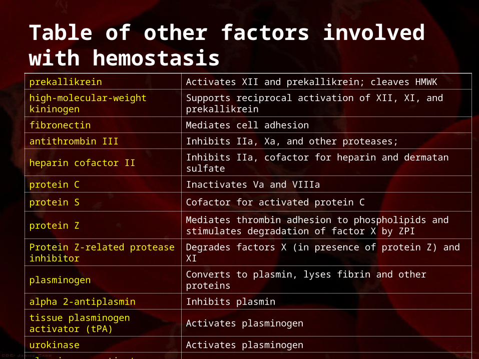
Table of other factors involved with hemostasisprekallikrein Activates XII and prekallikrein; cleaves HMWK
high-molecular-weight kininogen Supports reciprocal activation of XII, XI, and prekallikrein
fibronectin Mediates cell adhesion
antithrombin III Inhibits IIa, Xa, and other proteases;
heparin cofactor II Inhibits IIa, cofactor for heparin and dermatan sulfate
protein C Inactivates Va and VIIIa
protein S Cofactor for activated protein C
protein ZMediates thrombin adhesion to phospholipids and stimulates degradation of factor X by ZPI
Protein Z-related protease inhibitor Degrades factors X (in presence of protein Z) and XI
plasminogen Converts to plasmin, lyses fibrin and other proteins
alpha 2-antiplasmin Inhibits plasmin
tissue plasminogen activator (tPA) Activates plasminogen
urokinase Activates plasminogen
plasminogen activator inhibitor-1 Inactivates tPA & urokinase (endothelial PAI)
plasminogen activator inhibitor-2 Inactivates tPA & urokinase (placental PAI)
cancer procoagulant Pathological factor X activator linked to thrombosis in cancer

Summary
• Blood as a transport, regulative, hydraulic and protective medium
• Production of RBCs involves a recycling aspect (Fe conservation)
• Hemostasis involves– Vascular spasm– Platelet plug formation– Coagulation– Functionally a positive feedback system