Embed Size (px)
Citation preview
Cardiovascular System
Biol 105
Lecture 15
Reading: Ch 12
Copyright © 2009 Pearson Education, Inc.
Outline
I. Functions of cardiovascular system
II. Components of the cardiovascular system
III. The heart
IV. Regulation of the heartbeat
V. ECG/EKG
VI. Blood pressure
VII.Circulatory circuits
VIII.Cardiovascular diseases
IX. Lymphatic system

Copyright © 2009 Pearson Education, Inc.
Cardiovascular
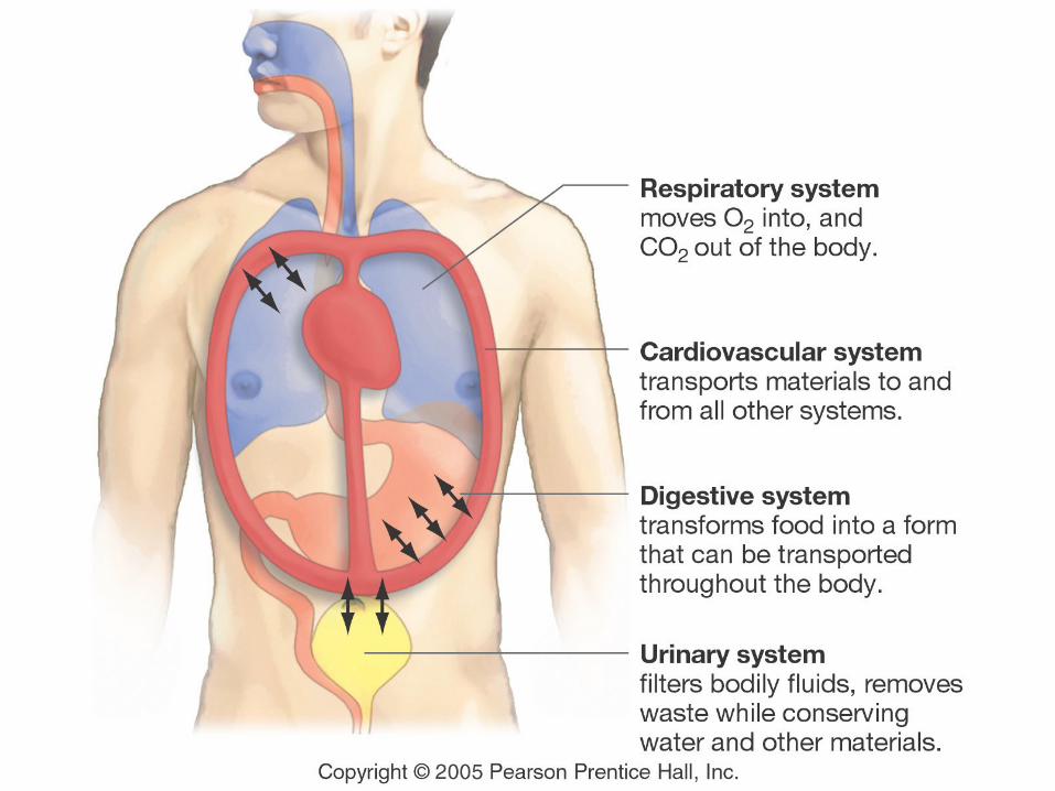
Function of the cardiovascular system is to
transport blood containing:
Nutrients
Waste
Hormones
Immune cells
Oxygen
Copyright © 2009 Pearson Education, Inc.
Cardiovascular system
Cardiovascular System consists of three
components:
1. Blood
2. The heart, which pumps blood.
3. The blood vessels, through which blood flows.
5-2
Copyright © 2009 Pearson Education, Inc.
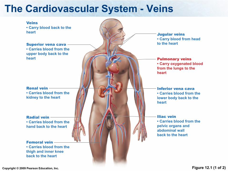
The Cardiovascular System - Veins
Figure 12.1 (1 of 2)
Veins
• Carry blood back to the
heartJugular veins
• Carry blood from headto the heart
Renal vein
• Carries blood from thekidney to the heart
Pulmonary veins
• Carry oxygenated blood
from the lungs to theheart
Inferior vena cava
• Carries blood from the
lower body back to theheart
Superior vena cava
• Carries blood from the
upper body back to the
heart
Iliac vein
• Carries blood from the
pelvic organs and
abdominal wallback to the heart
Radial vein
• Carries blood from the
hand back to the heart
Femoral vein
• Carries blood from the
thigh and inner kneeback to the heart
Copyright © 2009 Pearson Education, Inc.
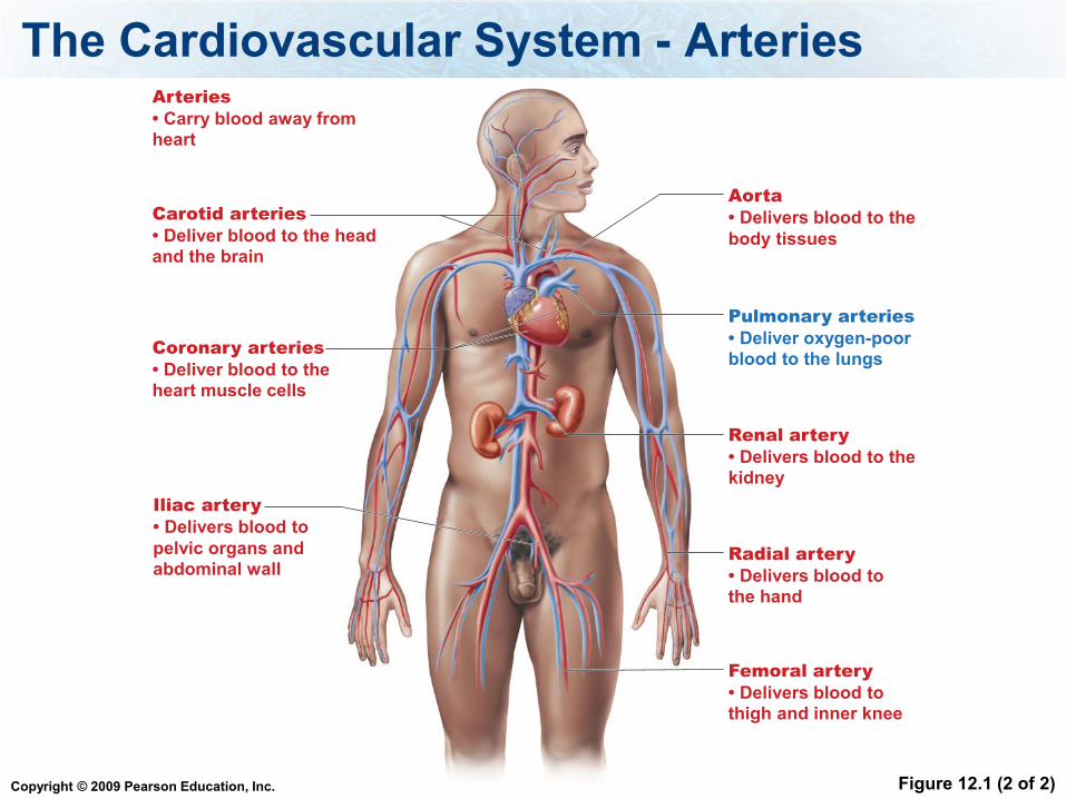
The Cardiovascular System - Arteries
Figure 12.1 (2 of 2)
Arteries
• Carry blood away fromheart
Carotid arteries
• Deliver blood to the headand the brain
Aorta
• Delivers blood to the
body tissues
Pulmonary arteries
• Deliver oxygen-poorblood to the lungs
Coronary arteries
• Deliver blood to theheart muscle cells
Renal artery
• Delivers blood to thekidney
Iliac artery
• Delivers blood to
pelvic organs andabdominal wall
Radial artery
• Delivers blood tothe hand
Femoral artery
• Delivers blood tothigh and inner knee
Copyright © 2009 Pearson Education, Inc.
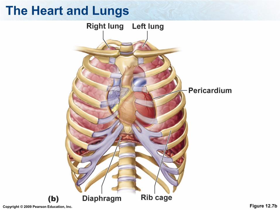
The Heart and Lungs
Figure 12.7b
Copyright © 2009 Pearson Education, Inc.
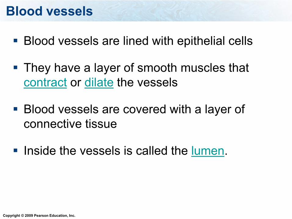
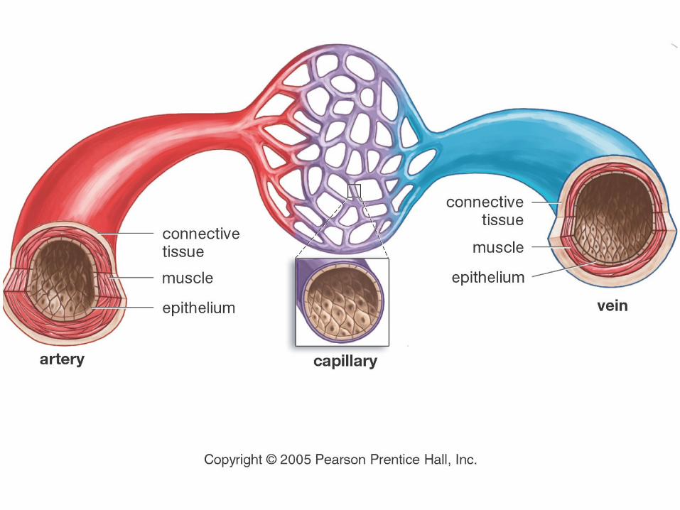
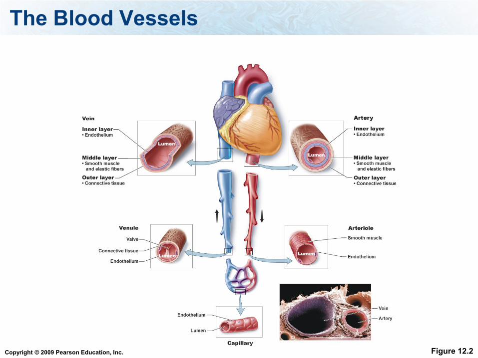
Blood vessels
Blood vessels are lined with epithelial cells
They have a layer of smooth muscles that
contract or dilate the vessels
Blood vessels are covered with a layer of
connective tissue
Inside the vessels is called the lumen.
Copyright © 2009 Pearson Education, Inc.
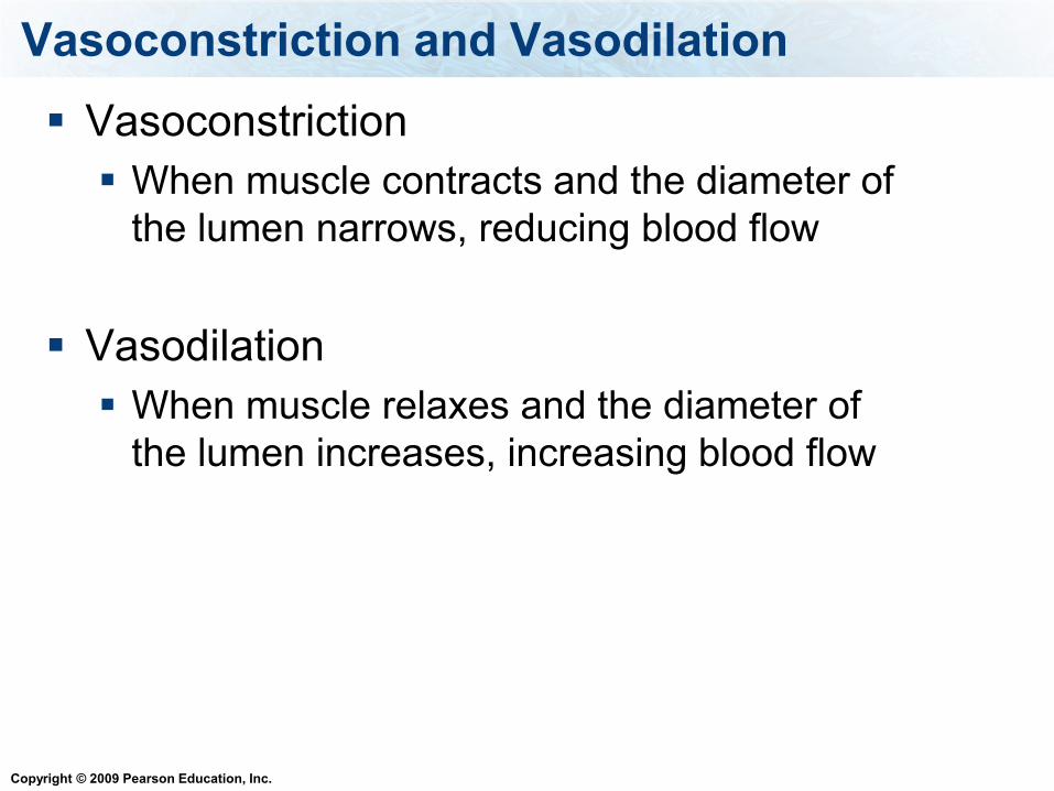
Vasoconstriction and Vasodilation
Vasoconstriction
When muscle contracts and the diameter of
the lumen narrows, reducing blood flow
Vasodilation
When muscle relaxes and the diameter of
the lumen increases, increasing blood flow
Copyright © 2009 Pearson Education, Inc.
The Blood Vessels
Arteries
Arterioles
Capillaries
Venules
Veins
Copyright © 2009 Pearson Education, Inc.
The Blood Vessels – Arteries and Veins
Arteries - Always carry blood away from the
heart and usually carry O2-rich blood.
Veins - Always returns blood to the heart
and usually carry O2-poor blood.
5-4
Copyright © 2009 Pearson Education, Inc.
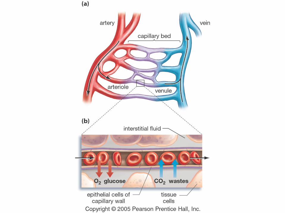
The Blood Vessels – Arterioles and Venules
Arteries break down into smaller vessels called
arterioles, bringing O2, water, and nutrients to the
tissues
Arterioles break down into small vessels called
capillaries
Blood leaves the capillaries and enters venules
Venules take CO2, water, and wastes away from
the tissues.
Venules join together to form veins.
5-4
Copyright © 2009 Pearson Education, Inc.
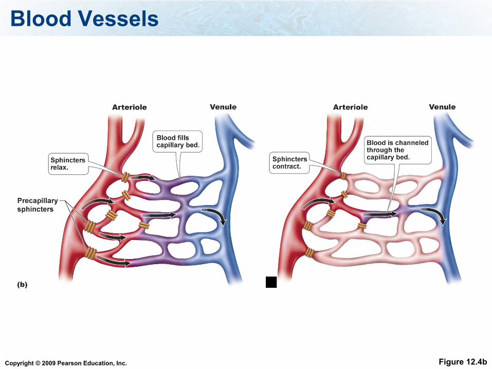
The Blood Vessels – Arterioles
There are sphincter muscles that contract
to reduce blood flow to the capillaries
or they dilate to increase blood flow to the
capillaries.
5-4
Copyright © 2009 Pearson Education, Inc.
Capillaries
Small vessels are called capillaries
It is here that components (O2, CO2,
nutrients, waste) can pass from the blood
vessels to other tissues
Capillaries do not have a smooth muscle
layer
Copyright © 2009 Pearson Education, Inc.
Can gas freely pass through the plasma membrane?
1. True
2. False
Copyright © 2009 Pearson Education, Inc.
Capillaries
The RBCs stay in the blood vessels but the
oxygen leaves the RBCs and the capillaries
and goes into the tissues.
The oxygen leaves the capillaries because
there is a gradient – there is more oxygen in
the capillaries than in the tissues.
Copyright © 2009 Pearson Education, Inc.
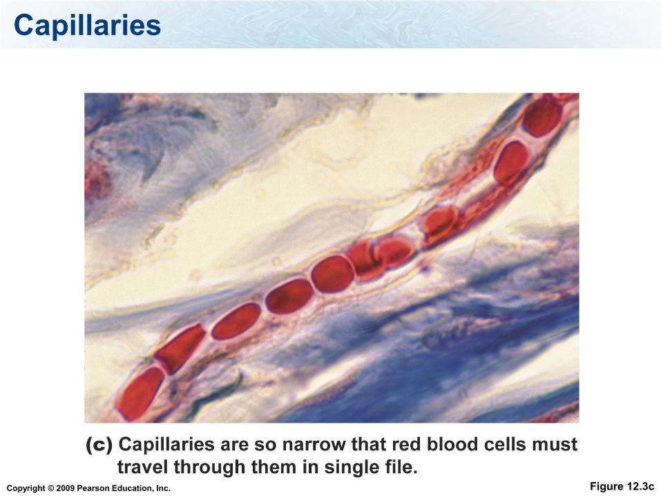
Blood flow in capillaries is slow.
This is important because it allows time
for the exchange of substances between
the blood and surrounding tissues.
5-18
Capillaries
Copyright © 2009 Pearson Education, Inc.
The Blood Vessels
Figure 12.2
Copyright © 2009 Pearson Education, Inc.
Blood Vessels
Figure 12.4b
Copyright © 2009 Pearson Education, Inc.
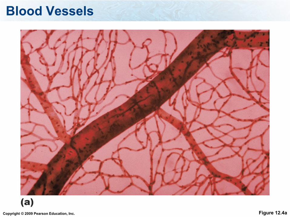
Blood Vessels
Figure 12.4a
Copyright © 2009 Pearson Education, Inc.
Capillaries
Figure 12.3c
Copyright © 2009 Pearson Education, Inc.
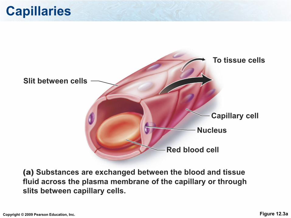
Capillaries
Figure 12.3a
Capillary cell
To tissue cells
Slit between cells
Nucleus
Red blood cell
(a) Substances are exchanged between the blood and tissue
fluid across the plasma membrane of the capillary or through
slits between capillary cells.
Copyright © 2009 Pearson Education, Inc.
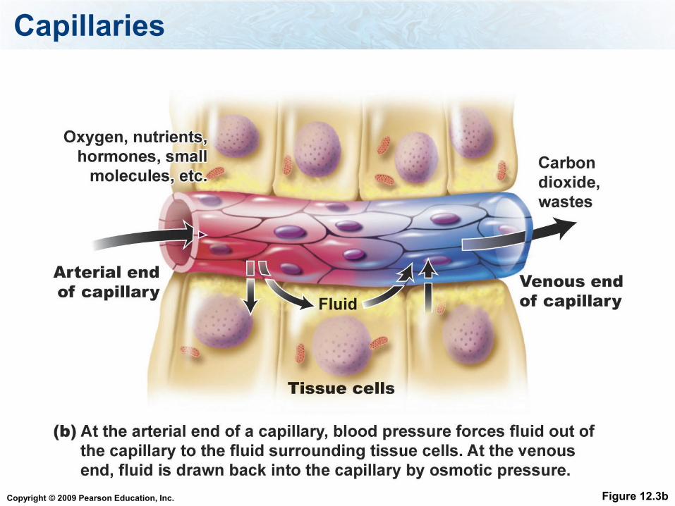
Capillaries
Figure 12.3b
Copyright © 2009 Pearson Education, Inc.
Do RBCs leave the capillaries?
1. Yes
2. No
Copyright © 2009 Pearson Education, Inc.
Pressures and Their Effect on Capillaries
At the arterial end of the capillaries blood
pressure forces fluid out of the capillary and into
the tissue
At the venous end, osmotic pressure draws fluid
back into the vessel from the tissue
Diffusion is the pressure that draws gasses
across the capillary
Copyright © 2009 Pearson Education, Inc.
The Blood Vessels
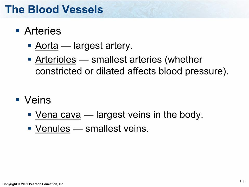
Arteries
Aorta — largest artery.
Arterioles — smallest arteries (whether
constricted or dilated affects blood pressure).
Veins
Vena cava — largest veins in the body.
Venules — smallest veins.
5-4
Copyright © 2009 Pearson Education, Inc.
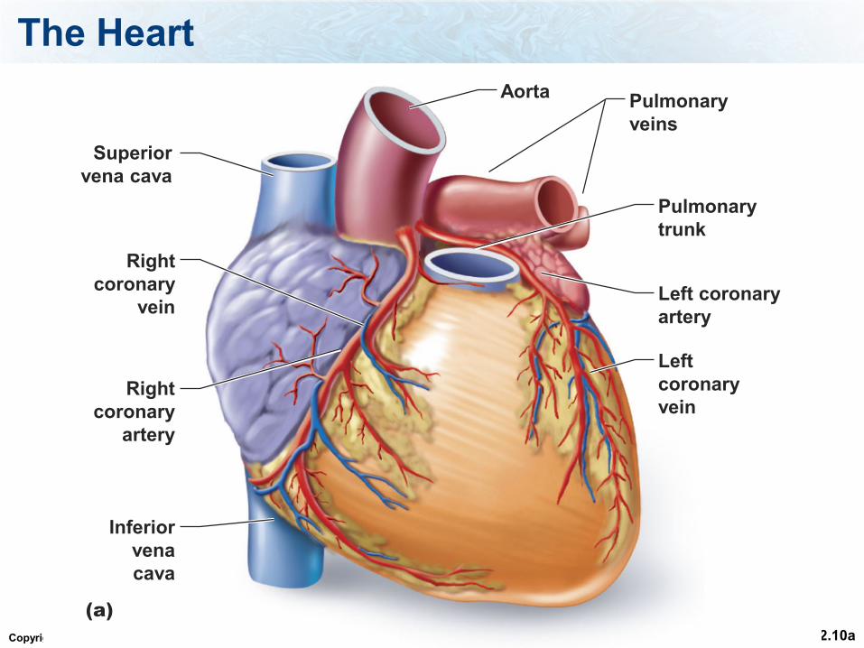
The Heart
Figure 12.10a
Inferior
vena
cava
Superior
vena cava
Pulmonary
trunk
Aorta
Right
coronary
vein
Right
coronary
artery
Left coronary
artery
Left
coronary
vein
(a)
Pulmonary
veins
Copyright © 2009 Pearson Education, Inc.
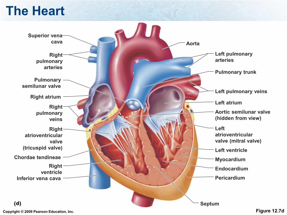
The Heart
Figure 12.7d
Superior vena
cava Aorta
Right
pulmonary
arteries
Pulmonary
semilunar valve
Right atrium
Right
pulmonary
veins
Right
atrioventricular
valve
(tricuspid valve)
Chordae tendineae
Right
ventricle
Inferior vena cava
Left pulmonary
arteries
Pulmonary trunk
Left pulmonary veins
Left atrium
Aortic semilunar valve
(hidden from view)
Left
atrioventricular
valve (mitral valve)
Left ventricle
Myocardium
Endocardium
Pericardium
Septum(d)
Copyright © 2009 Pearson Education, Inc.
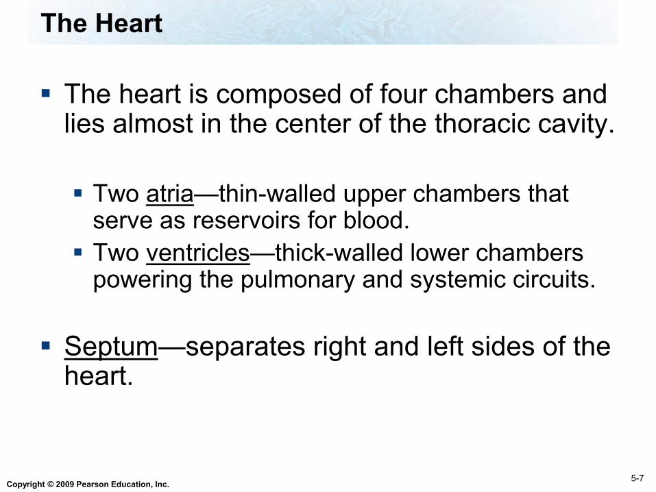
The Heart
The heart is composed of four chambers and lies almost in the center of the thoracic cavity.
Two atria—thin-walled upper chambers that serve as reservoirs for blood.
Two ventricles—thick-walled lower chambers powering the pulmonary and systemic circuits.
Septum—separates right and left sides of the heart.
5-7
Copyright © 2009 Pearson Education, Inc.
The Heart
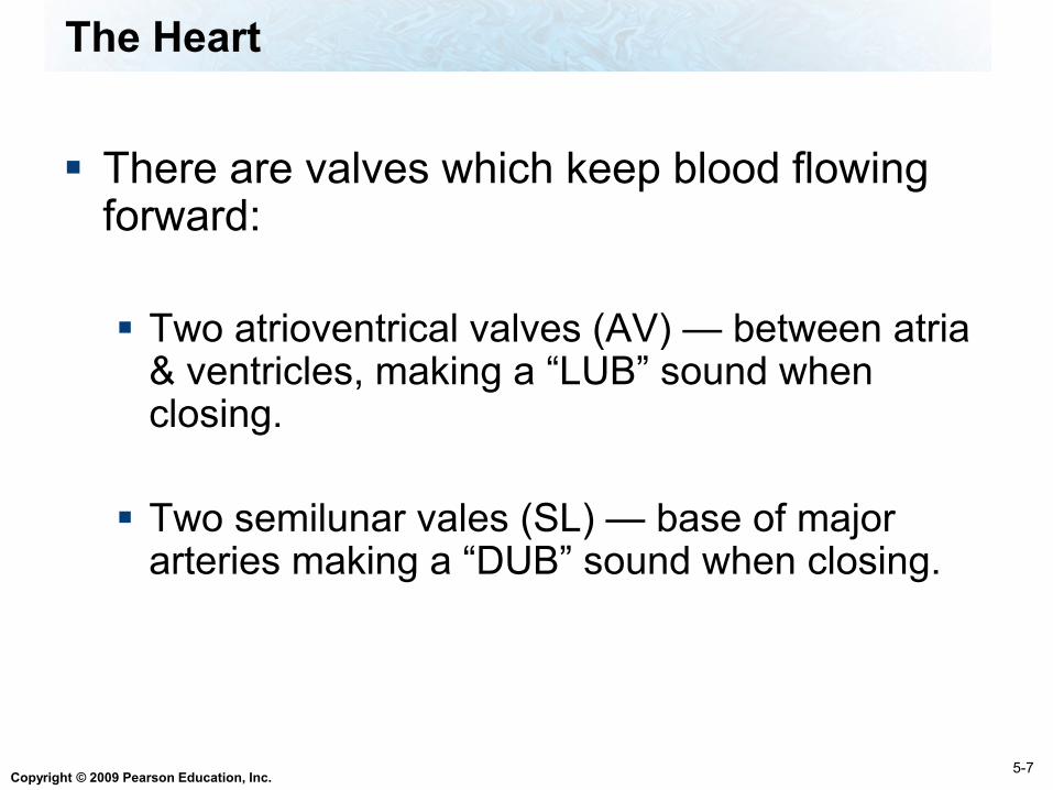
There are valves which keep blood flowing forward:
Two atrioventrical valves (AV) — between atria & ventricles, making a “LUB” sound when closing.
Two semilunar vales (SL) — base of major arteries making a “DUB” sound when closing.
5-7
Copyright © 2009 Pearson Education, Inc.
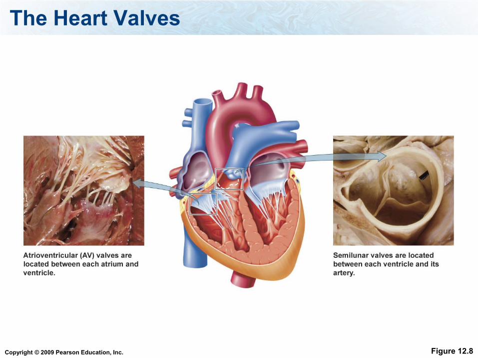
The Heart Valves
Figure 12.8
Copyright © 2009 Pearson Education, Inc.
The Heart
Pericardium — thick membranous sac surrounding the heart (secretes serous fluid).
Myocardium — consists of cardiac muscle tissue, which contracts to pump blood.
The interior of the heart is lined by endocardium
5-7
Copyright © 2009 Pearson Education, Inc.
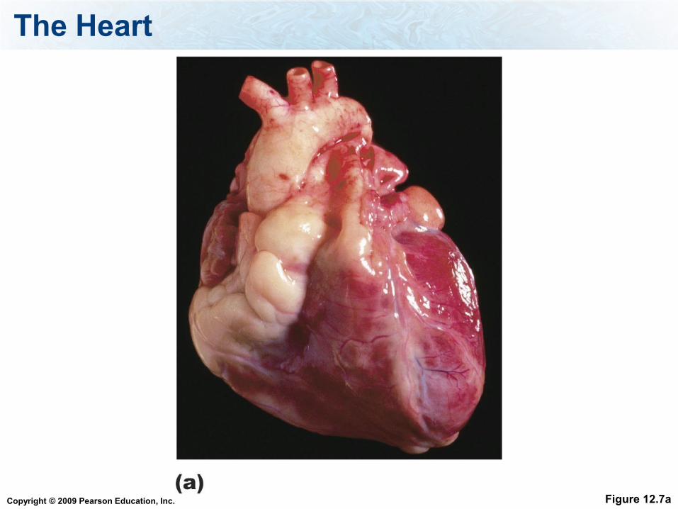
The Heart
Figure 12.7a
Copyright © 2009 Pearson Education, Inc.
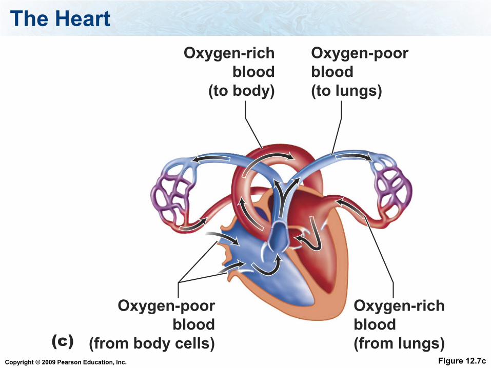
The Heart
Figure 12.7c
Oxygen-rich
blood
(to body)
Oxygen-poor
blood
(to lungs)
Oxygen-rich
blood
(from lungs)
Oxygen-poor
blood
(from body cells)(c)
Copyright © 2009 Pearson Education, Inc.
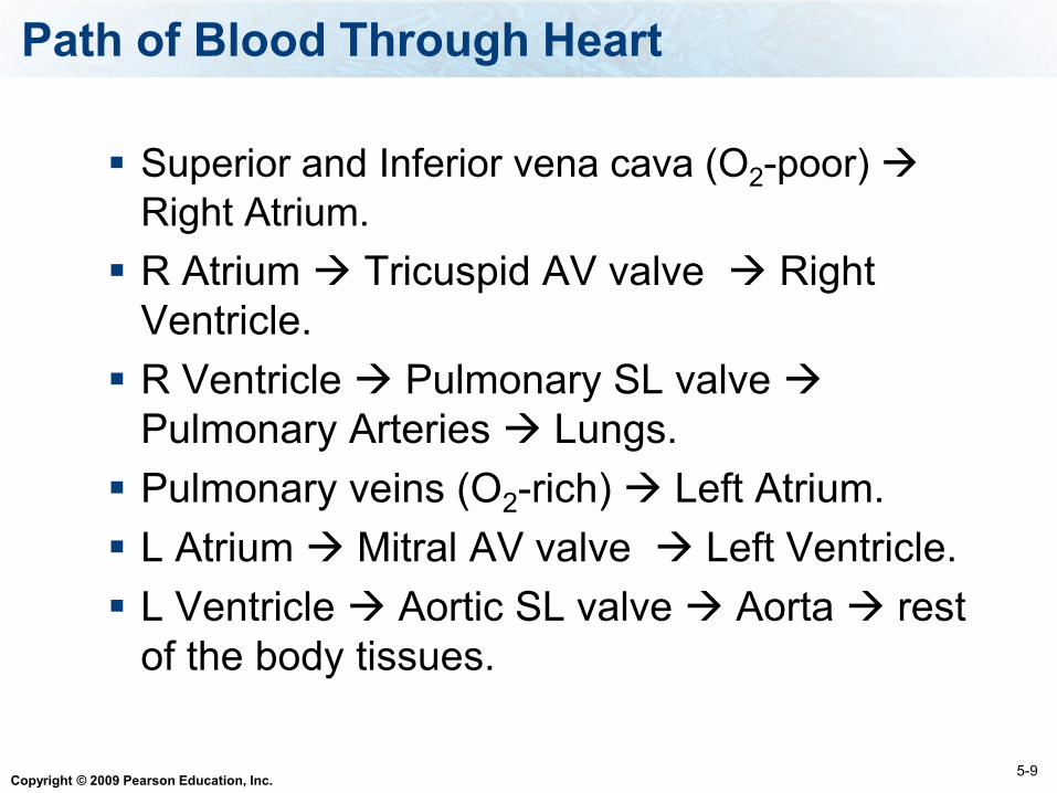
Path of Blood Through Heart
Superior and Inferior vena cava (O2-poor)
Right Atrium.
R Atrium Tricuspid AV valve Right
Ventricle.
R Ventricle Pulmonary SL valve
Pulmonary Arteries Lungs.
Pulmonary veins (O2-rich) Left Atrium.
L Atrium Mitral AV valve Left Ventricle.
L Ventricle Aortic SL valve Aorta rest
of the body tissues.
5-9
Copyright © 2009 Pearson Education, Inc.
Cardiac Cycle
Cardiac cycle - one complete heart beat
where both atria contract simultaneously (at
the same time) followed by both ventricles
contracting simultaneously.
a. Systole - when ventricles contract and pump
blood out of the heart.
b. Diastole - when ventricles relax and receive
blood from atria.
5-11
Copyright © 2009 Pearson Education, Inc.
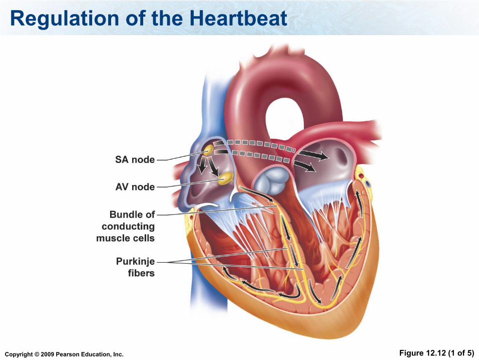
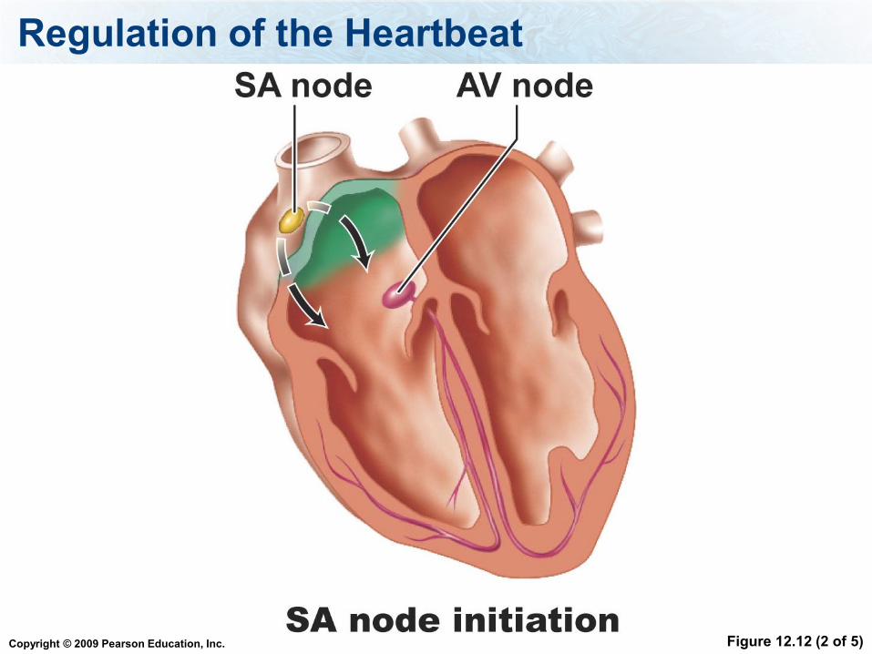
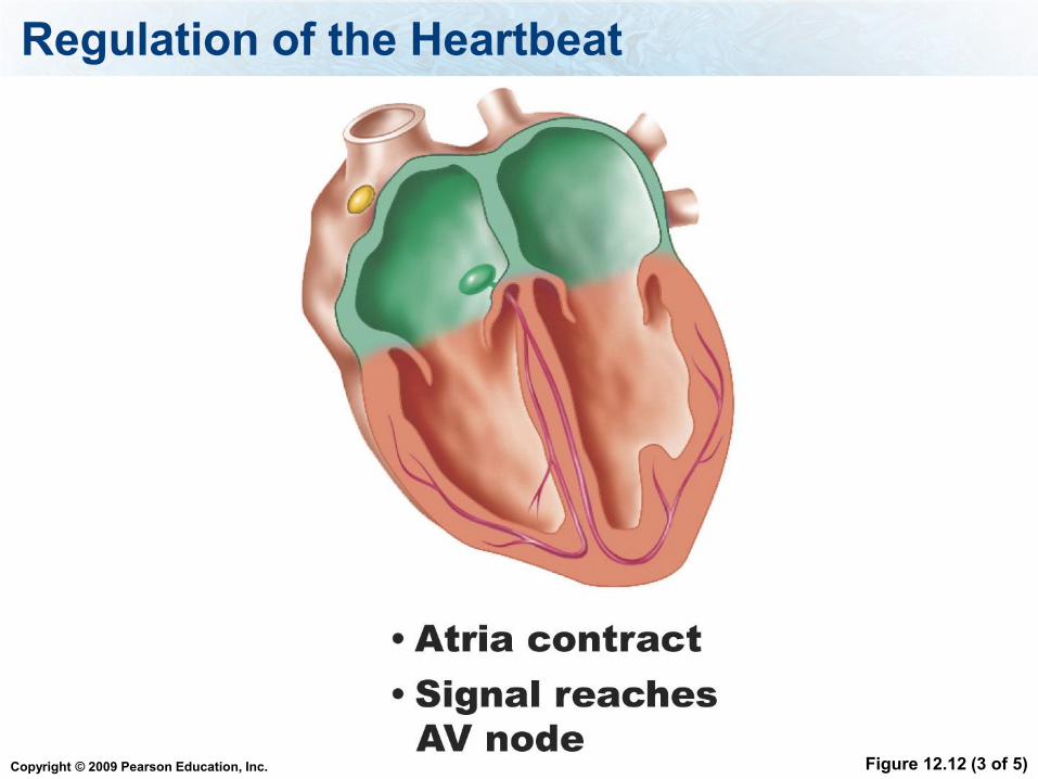
Intrinsic Control:
Sinoatrial node (SA) (pacemaker)—
initiates the heartbeat and causes the
atria to contract.
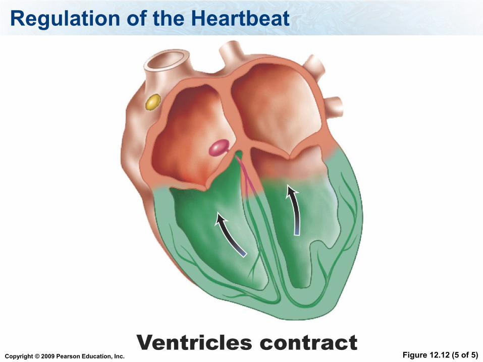
Atrioventricular node (AV) - causes the
ventricles to contract.
Heartbeat regulation - Intrinsic

Copyright © 2009 Pearson Education, Inc.
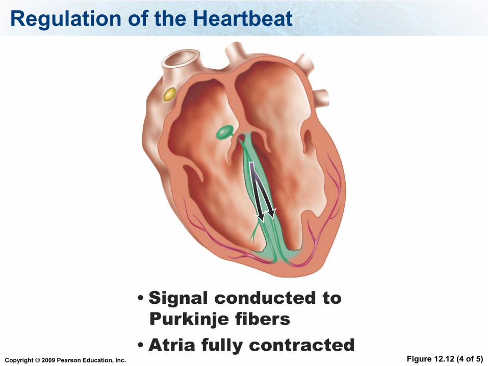
The AV node relays the message to the
ventricles using bundles of specialized
muscle cells = atrioventricular bundle
The bundle divides into smaller bundles
of specialized cardiac muscle cells
called Purkinje fibers
Heartbeat regulation - Intrinsic
Copyright © 2009 Pearson Education, Inc.
When the ventricles contract, which valves are closed?
1. AV valves
2. SL valves
Copyright © 2009 Pearson Education, Inc.
Extrinsic Control of Heartbeat - the
autonomic nervous system and
hormones can modify the rate of the
heartbeat.
Heartbeat regulation - Extrinsic
Copyright © 2009 Pearson Education, Inc.
Which part of the autonomic NS controls the heart most of the time?
1. Sympathetic
2. Parasympathetic
Copyright © 2009 Pearson Education, Inc.
Regulation of the Heartbeat
Figure 12.12 (1 of 5)
Copyright © 2009 Pearson Education, Inc.
Regulation of the Heartbeat
Figure 12.12 (2 of 5)
Copyright © 2009 Pearson Education, Inc.
Regulation of the Heartbeat
Figure 12.12 (3 of 5)
Copyright © 2009 Pearson Education, Inc.
Regulation of the Heartbeat
Figure 12.12 (4 of 5)
Copyright © 2009 Pearson Education, Inc.
Regulation of the Heartbeat
Figure 12.12 (5 of 5)
Copyright © 2009 Pearson Education, Inc.
Electrocardiogram (ECG) - a recording
of the electrical changes that occur in
the myocardium during a cardiac cycle.
5-13
Recording the Heartbeat
Copyright © 2009 Pearson Education, Inc.
ECG/EKG
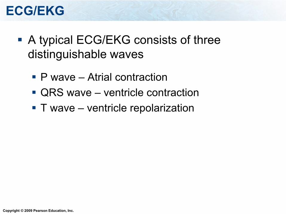
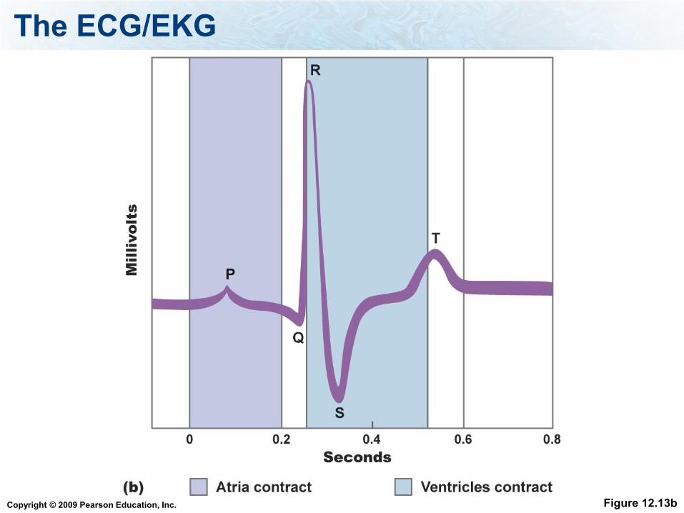
A typical ECG/EKG consists of three
distinguishable waves
P wave – Atrial contraction
QRS wave – ventricle contraction
T wave – ventricle repolarization
Copyright © 2009 Pearson Education, Inc.
The ECG/EKG
Figure 12.13b
Copyright © 2009 Pearson Education, Inc.
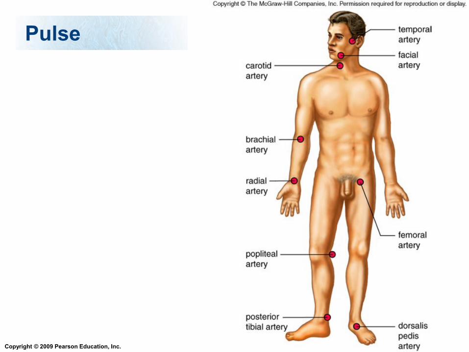
Pulse
As the heart pumps blood into the arteries,
they expand such that one is able to feel a
pulse
The pulse rate is the same as the heart
rate
Copyright © 2009 Pearson Education, Inc.
Pulse
5-15
Copyright © 2009 Pearson Education, Inc.
Blood pressure
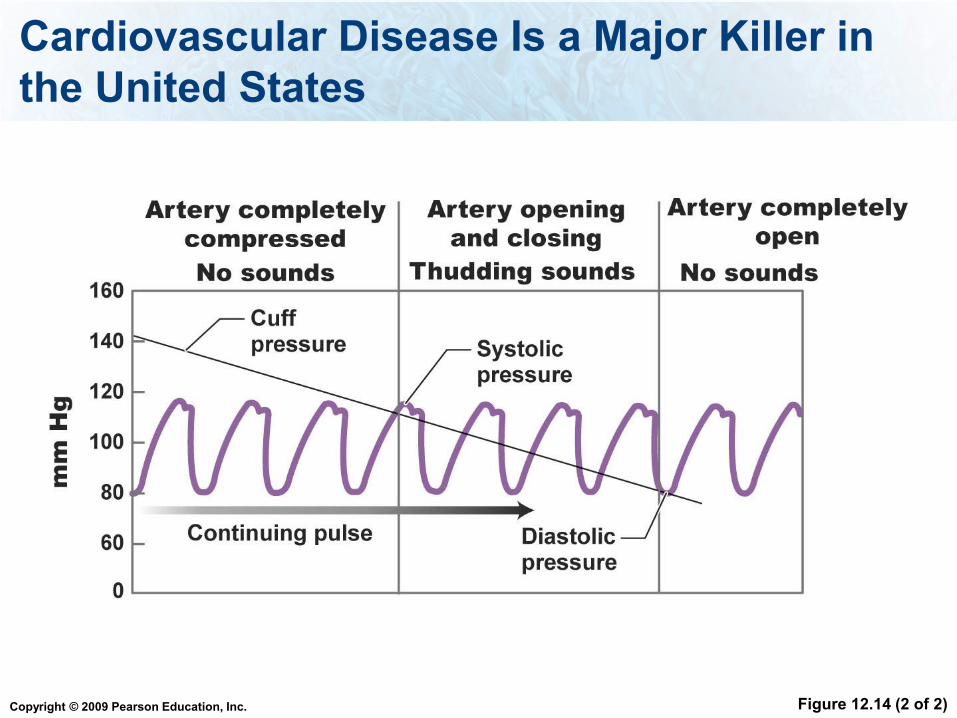
Blood pressure
Systolic - when the ventricles contract,
sending blood into the arteries
Diastolic - when the heart relaxes between
beats
Copyright © 2009 Pearson Education, Inc.
Which blood pressure would be the highest:
1. systolic
2. diastolic
Copyright © 2009 Pearson Education, Inc.
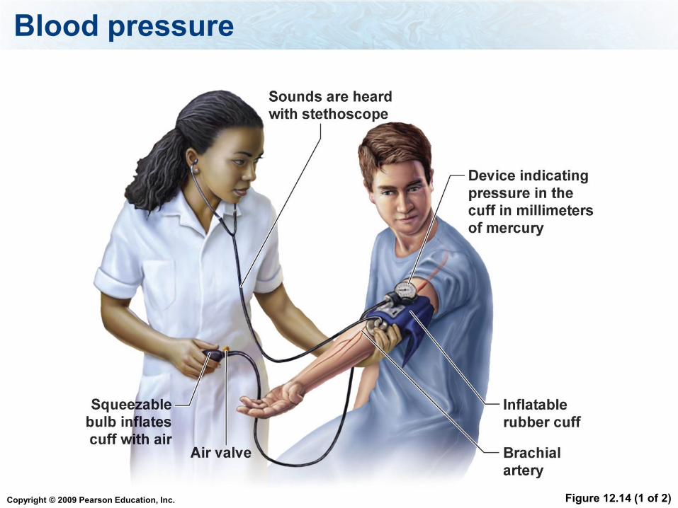
Blood pressure
Sphygmomanometer
Measures blood pressure
Can provide early identification of
hypertension, or high blood pressure, the
silent killer
Copyright © 2009 Pearson Education, Inc.
Blood pressure
Figure 12.14 (1 of 2)
Copyright © 2009 Pearson Education, Inc.
Cardiovascular Disease Is a Major Killer in
the United States
Figure 12.14 (2 of 2)
Copyright © 2009 Pearson Education, Inc.
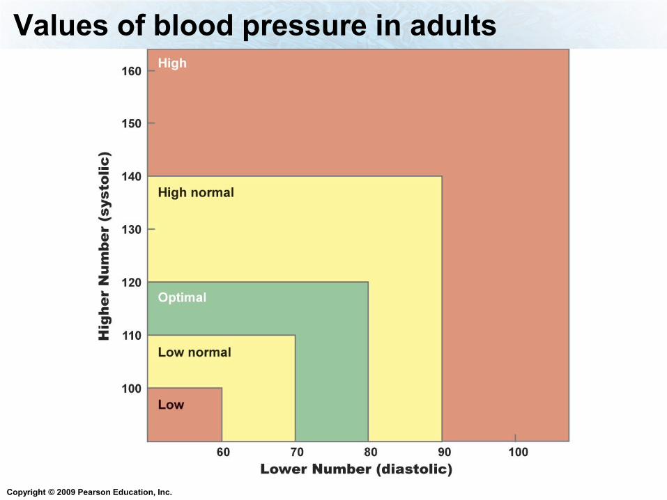
Values of blood pressure in adults

Copyright © 2009 Pearson Education, Inc.
Blood flow
Blood flow in the arteries is from the blood
pressure due to the heart pumping.
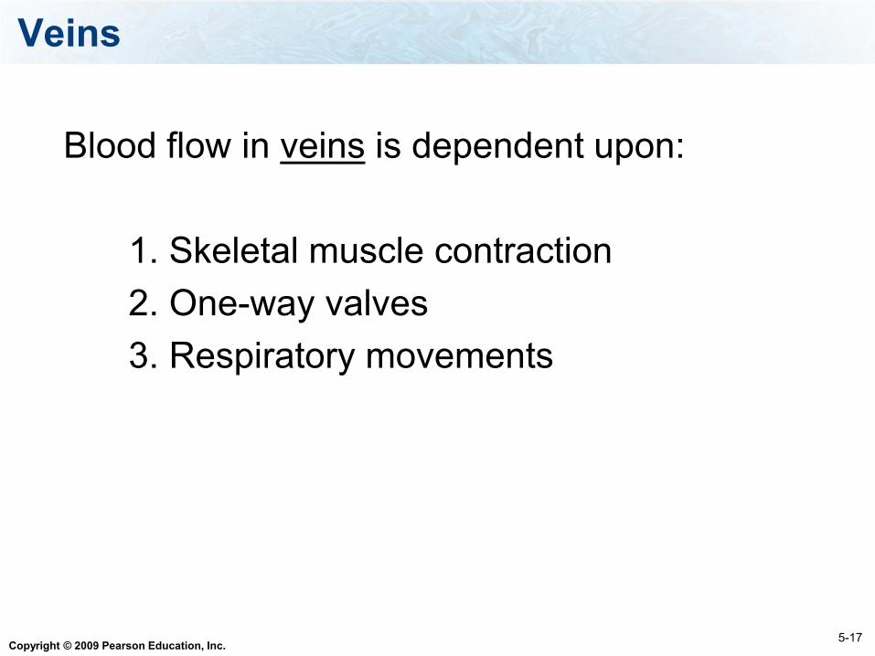
The blood pressure in veins is very low
Copyright © 2009 Pearson Education, Inc.
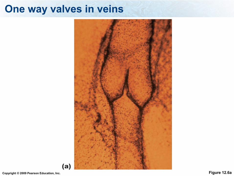
Blood flow in veins is dependent upon:
1. Skeletal muscle contraction
2. One-way valves
3. Respiratory movements
5-17
Veins
Copyright © 2009 Pearson Education, Inc.
One way valves in veins
Figure 12.6a
Copyright © 2009 Pearson Education, Inc.
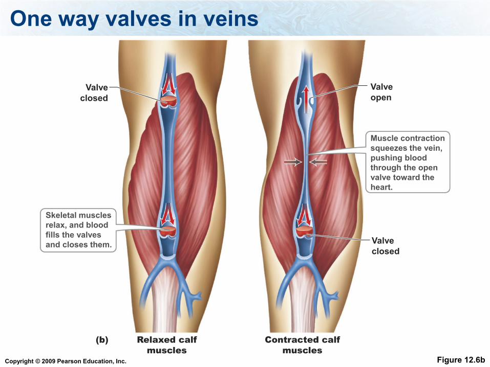
One way valves in veins
Figure 12.6b
Valve
closed
Valve
open
Relaxed calf
muscles
Skeletal muscles
relax, and blood
fills the valves
and closes them.
Muscle contraction
squeezes the vein,
pushing blood
through the open
valve toward the
heart.
Valve
closed
Contracted calf
muscles
(b)
Copyright © 2009 Pearson Education, Inc.
Cardiovascular system circuits
Pulmonary circuit - flow of blood from
the heart, to the lungs and back to the
heart, powered by the right ventricle.
Systemic circuit - flow of blood through
the rest of the body, powered by the
left ventricle.
5-2
Copyright © 2009 Pearson Education, Inc.
Pulmonary circuit
Pulmonary arteries—carry O2-poor
blood to the lungs.
Pulmonary veins—carry O2-rich
blood from lungs to the left atrium.
5-19
Copyright © 2009 Pearson Education, Inc.
Systemic circuit
Aorta - carries O2-rich blood to all
body tissues.
Vena cava - returns O2-poor blood to
the right atrium.
5-19
Copyright © 2009 Pearson Education, Inc.
Systemic circuits
Renal circuit - supplies blood to the
kidneys.
Hepatic portal circuit - supplies
blood to the digestive organs
especially the liver.
5-21
Copyright © 2009 Pearson Education, Inc.
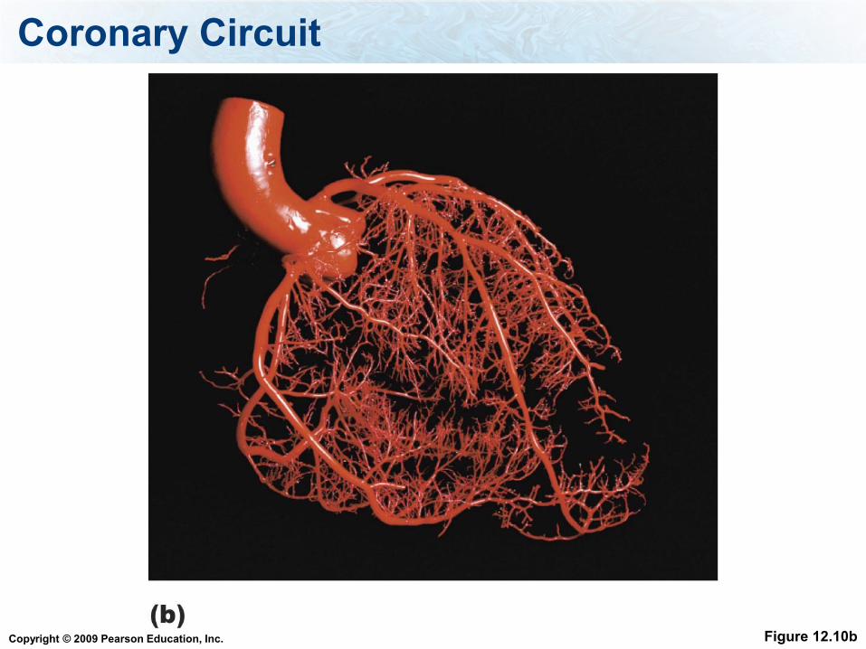
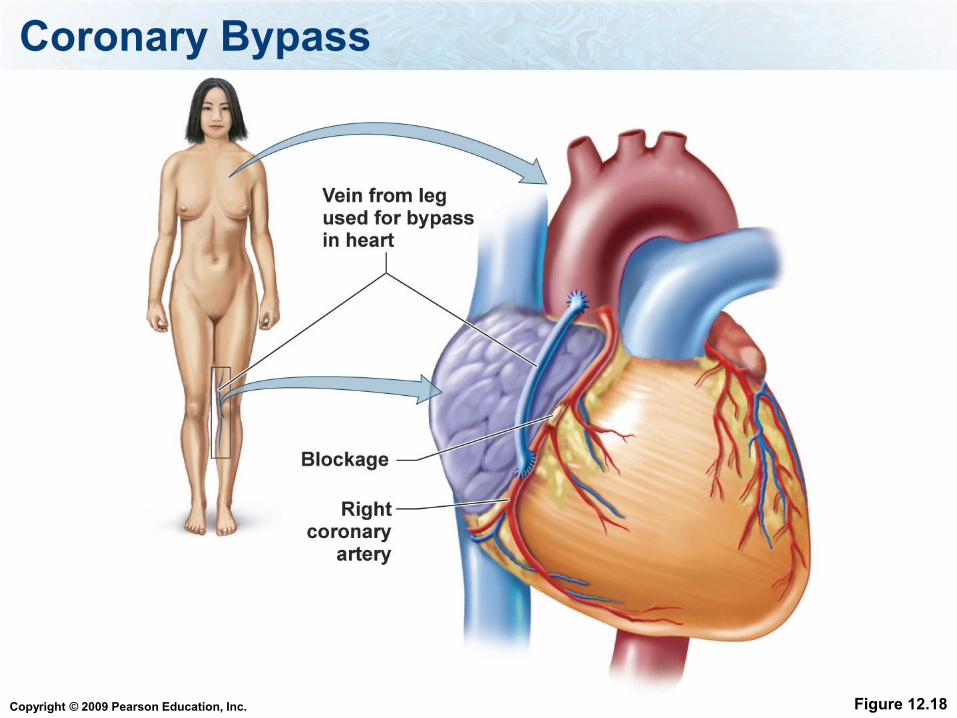
Systemic - Coronary circuit
Supplies blood to the heart muscle itself.
Coronary arteries branch off the aorta.
Coronary arteries can become clogged
and by-pass surgery may be necessary.
Coronary veins return blood to the heart
5-21
Copyright © 2009 Pearson Education, Inc.
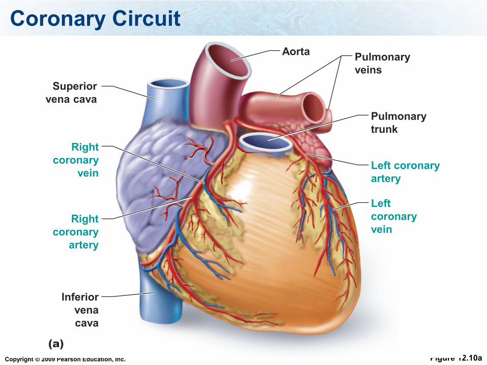
Coronary Circuit
Figure 12.10a
Inferior
vena
cava
Superior
vena cava
Pulmonary
trunk
Aorta
Right
coronary
vein
Right
coronary
artery
Left coronary
artery
Left
coronary
vein
(a)
Pulmonary
veins
Copyright © 2009 Pearson Education, Inc.
Coronary Circuit
Figure 12.10b
Copyright © 2009 Pearson Education, Inc.
This pressure draws fluid back into the capillaries
1. Blood Pressure
2. Osmotic Pressure
3. Diffusion
Copyright © 2009 Pearson Education, Inc.
Blood flow in veins is dependent upon: one way valves, respiratory
movements and:
1. Smooth muscle
2. Skeletal muscle
Copyright © 2009 Pearson Education, Inc.
When ventricles relax and receive blood from atria it is:
1. Systole
2. Diastole
Copyright © 2009 Pearson Education, Inc.
Disorders of the Cardiovascular System
1. High Blood Pressure
2. Atherosclerosis and coronary artery disease
3. Heart attack
4. Thromboembolism
5. Stroke
6. Aneurism
Copyright © 2009 Pearson Education, Inc.
Disorders – High Blood Pressure
High blood pressure is also called hypertension
Causes:
90% of high blood pressure has no known cause.
Can be caused by kidney not being able to balance
the sodium concentration. Increased fluid in blood
increases blood pressure.
Stress can lead to high blood pressure.
Copyright © 2009 Pearson Education, Inc.
Disorders – High Blood Pressure
Result: high blood pressure causes the heart to
work too hard, leads to heart failure, kidney
problems, blood vessel problems and death.
Prevention includes: lower salt intake, lose
weight, exercise, and stop smoking.
Copyright © 2009 Pearson Education, Inc.
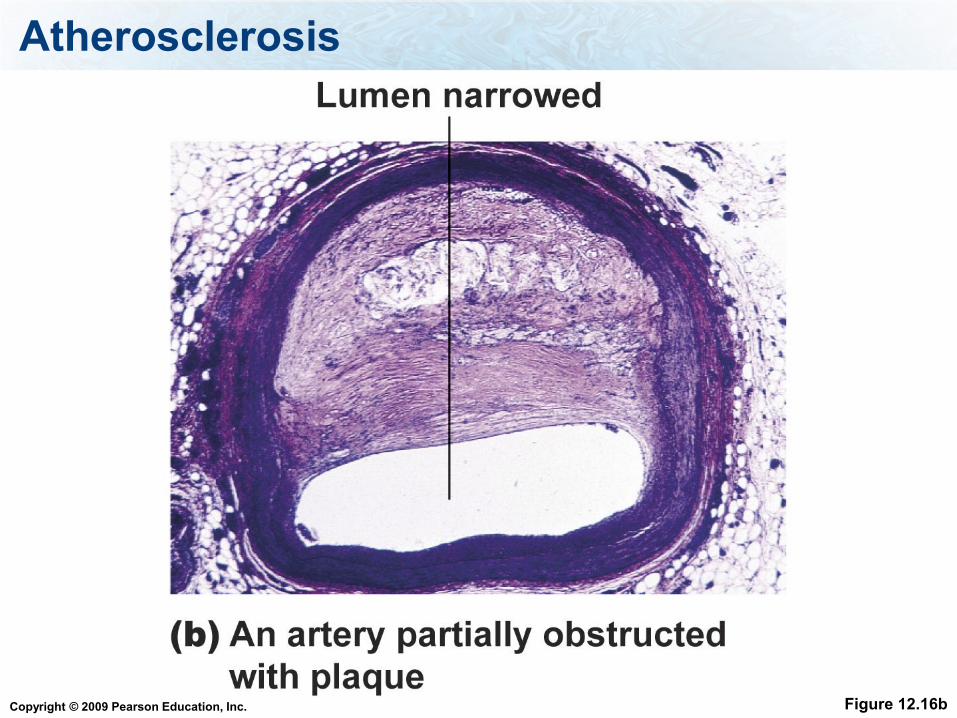
Disorders - Atherosclerosis
Atherosclerosis is a narrowing of the arteries
due to fatty deposits and thickening of the
wall
Can lead to heart attack or stroke
When this occurs in the arteries of the heart
muscle, it is called coronary artery disease
Copyright © 2009 Pearson Education, Inc.
Cholesterol
Remember that lipoproteins are proteins that
carry cholesterol in the blood.
Low density lipoproteins (LDL)
High density lipoproteins (HDL)
Copyright © 2009 Pearson Education, Inc.
This type of lipoprotein carries cholesterol away from the liver
1. LDL
2. HDL
Copyright © 2009 Pearson Education, Inc.
Coronary Artery Blockage
Some of the LDLs can become damaged
through oxidative stress. The damaged LDL
can get stuck in these coronary arteries.
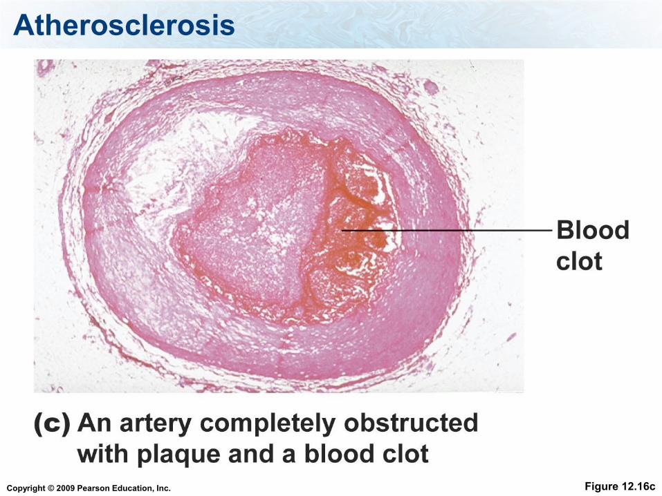
The immune system reacts to this by mounting
an inflammatory response = blood clot.
The oxidized material can build up and reduce
the flow of blood to the heart = coronary artery
blockage.
Copyright © 2009 Pearson Education, Inc.
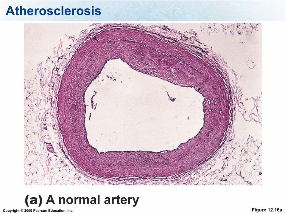
Atherosclerosis
Figure 12.16a
Copyright © 2009 Pearson Education, Inc.
Atherosclerosis
Figure 12.16b
Copyright © 2009 Pearson Education, Inc.
Atherosclerosis
Figure 12.16c

Copyright © 2009 Pearson Education, Inc.
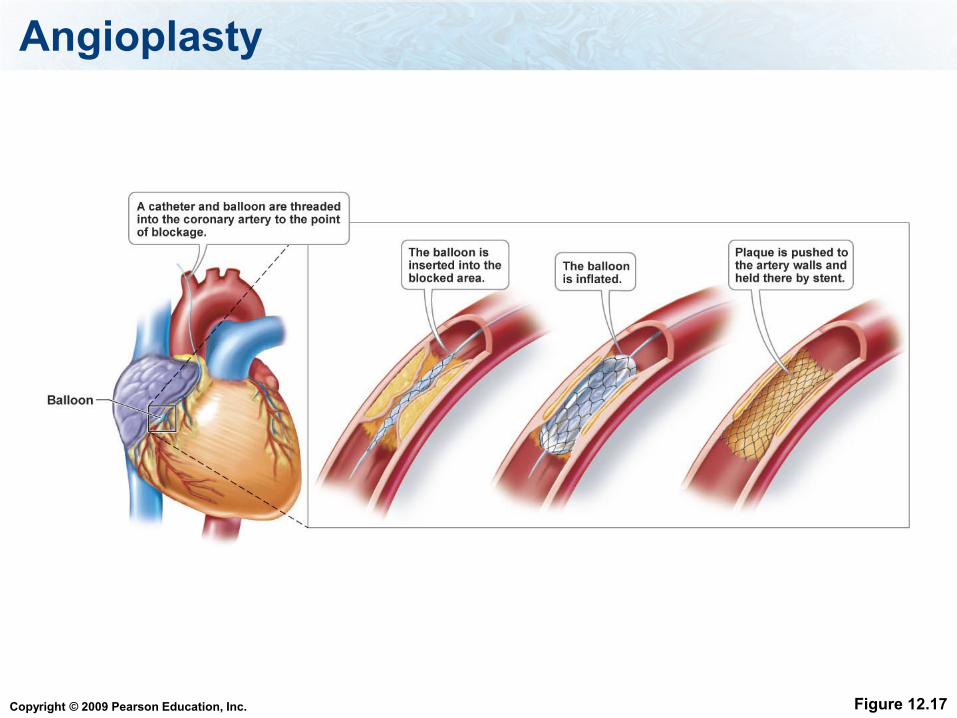
Treatments of Coronary Artery Blockage
Angiography can show coronary artery
blockage, which can then be treated with
medicines or surgical operations such as
angioplasty or coronary bypass surgery
See pages 235-236
Copyright © 2009 Pearson Education, Inc.
Angioplasty
Figure 12.17
Copyright © 2009 Pearson Education, Inc.
Coronary Bypass
Figure 12.18
Copyright © 2009 Pearson Education, Inc.
Disorders - Heart Attack - myocardial infarction
Heart muscle dies because of an insufficient
blood supply during a heart attack
(myocardial infarction) and is gradually
replaced by scar tissue
Can be caused by coronary artery blockage
Scar tissue cannot contract, so part of the
heart permanently loses its pumping ability
Copyright © 2009 Pearson Education, Inc.
Disorders - Thromboembolism
Thromboembolism is a clot that has
been carried in the bloodstream but is
now stationary.
Can result in a stroke
5-22
Copyright © 2009 Pearson Education, Inc.
Disorders - Stroke
Stroke - cranial arteriole bursts or is
blocked, reducing blood supply to an
area of the brain.
The result is that a portion of the brain
dies, and may result in paralysis or
death.
5-22
Copyright © 2009 Pearson Education, Inc.
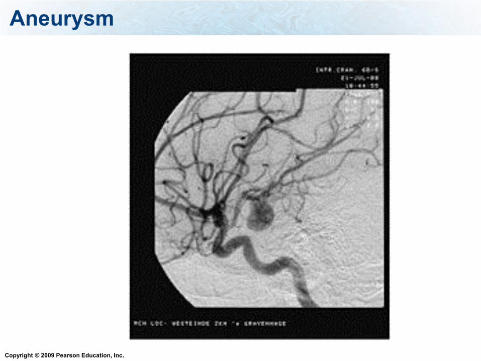
Disorders - Aneurysm
Aneurysm - weak spot in a blood vessel
where it balloons out and may rupture
May cause a stroke if in the brain or
death if in aorta
5-22
Copyright © 2009 Pearson Education, Inc.
Aneurysm
Copyright © 2009 Pearson Education, Inc.
The blood supply to the kidneys is the:
1. Hepatic portal circuit
2. Renal circuit
3. Cardiac circuit
Copyright © 2009 Pearson Education, Inc.
Lymphatic System
Lymphatic system - system that takes
excess tissue fluid to the subclavian veins.
Skeletal muscles and valves keep fluid
moving
5-26
Copyright © 2009 Pearson Education, Inc.
The Lymphatic System Functions
Functions
1. Return interstitial fluid from tissues to the
blood stream
2. Transport products of fat digestion lacteals
3. Defend the body against disease-causing
organisms and abnormal cells
Copyright © 2009 Pearson Education, Inc.
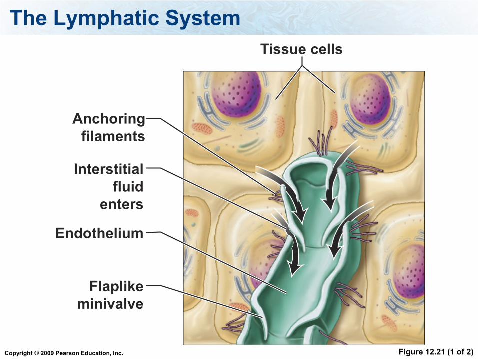
The Lymphatic System
Figure 12.21 (1 of 2)
Anchoring
filaments
Interstitial
fluid
enters
Endothelium
Flaplike
minivalve
Tissue cells
Copyright © 2009 Pearson Education, Inc.
Components of the Lymphatic System
Lymph
lymphatic vessels – including lacteals
lymphoid organs.
5-26
Copyright © 2009 Pearson Education, Inc.
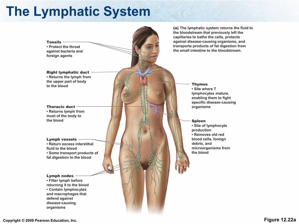
The Lymphatic System
Figure 12.22a
Tonsils
• Protect the throat
against bacteria and
foreign agents
Right lymphatic duct
• Returns the lymph from
the upper part of body
to the blood Thymus
• Site where T
lymphocytes mature,
enabling them to fight
specific disease-causing
organisms Thoracic duct
• Returns lymph from
most of the body to
the blood Spleen
• Site of lymphocyte
production
• Removes old red
blood cells, foreign
debris, and
microorganisms from
the blood
Lymph vessels
• Return excess interstitial
fluid to the blood
• Some transport products of
fat digestion to the blood
Lymph nodes
• Filter lymph before
returning it to the blood
• Contain lymphocytes
and macrophages that
defend against
disease-causing
organisms
(a) The lymphatic system returns the fluid to
the bloodstream that previously left the
capillaries to bathe the cells, protects
against disease-causing organisms, and
transports products of fat digestion from
the small intestine to the bloodstream.
Copyright © 2009 Pearson Education, Inc.
Lymphoid Organs:
1. Lymph nodes - cleanse lymph of debris and
pathogens and store lymphocytes and
macrophages to fight infection.
2. Spleen - cleanses the blood, remove old
blood cells.
3. Red bone marrow - produces both B cells
and T cells.
4. Thymus gland - where T cells mature.
5. Tonsils - function to recognize infectious
agents entering the body.

Copyright © 2009 Pearson Education, Inc.
Lymph Node
Figure 12.22b
Copyright © 2009 Pearson Education, Inc.
These vessels always carry blood away from the heart
1. Arteries
2. Veins
Copyright © 2009 Pearson Education, Inc.
What are the small blood vessels where the oxygen transfers into the
tissues and carbon dioxide is taken up
1. Arterioles
2. Venules
3. Capillaries
4. Lacteals
Copyright © 2009 Pearson Education, Inc.
What are the small lymphatic vessels where the fat is absorbed from the
digestive tract
1. Arterioles
2. Venules
3. Capillaries
4. Lacteals
Copyright © 2009 Pearson Education, Inc.
Important concepts
Read Chapter 13 for next lecture
What are the functions of the circulatory system?
What are the components of the circulatory
system?
What are the components of the blood vessel
and their functions, what would the cross section
of a vein, artery and capillary look like?
Copyright © 2009 Pearson Education, Inc.
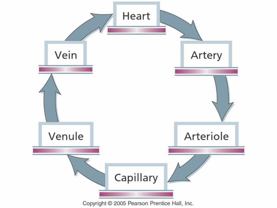
Important concepts
What is the path of the blood through the body, starting when it leaves the heart through the aorta, into arteries, then arterioles, then capillaries, then venules, to the veins, to the vena cava, back to the heart?
How do arterioles affect blood pressure?
What are the pressures that cause fluid to enter and leave the capillaries and what pressure causes gasses to enter and leave the capillaries. Be able to discuss in detail the transport of fluid, gasses, nutrients and waste across the capillaries?
Copyright © 2009 Pearson Education, Inc.
Important concepts
What is the function of capillaries?
What are the chambers of the heart, which
are the lower chambers and which are the
more muscular chambers?
What cavity is the heart located in?
What is the path of the blood through the
heart?
Be able to describe the cardiac cycle.
Copyright © 2009 Pearson Education, Inc.
Important concepts
What are the valves in the heart, where are
they located, when are they opened, when
are they closed?
How is the heartbeat is regulated, both
intrinsically and extrinsically?
What records the electrical changes that
occur in the myocardium during a
cardiac cycle?
Copyright © 2009 Pearson Education, Inc.
Important concepts
What are the three waves on the ECG and be
able to describe the events that happen
during each of the waves on the ECG?
What measures blood pressure?
What causes blood to flow in the arteries and
in the veins?
What are the pulmonary, systemic renal,
hepatic portal, and coronary circuits, what
tissues to they go to?
Copyright © 2009 Pearson Education, Inc.
Important concepts
What is the function of the aorta, vena cava,
pulmonary arteries, pulmonary veins,
coronary arteries and coronary veins?
What is the role of LDL and HDL in coronary
artery disease?
What are causes and effects of the
cardiovascular diseases discussed in lecture
How can you prevent high blood pressure.
Copyright © 2009 Pearson Education, Inc.
Important concepts
What are two treatments of coronary artery
blockage
How is coronary artery blockage detected?
What are the function of the lymphatic system?
Copyright © 2009 Pearson Education, Inc.
Important concepts
What are the components of the lymphatic
system and their functions
What are lacteals are what is their function?
What causes fluids to travel through lymphatic
vessels?
Copyright © 2009 Pearson Education, Inc.
Definitions
Lumen, vasoconstriction, vasodilation,
osmotic pressure, blood pressure, low
density lipoproteins (LDL), high density
lipoproteins (HDL), septum, capillaries,
arteries, veins, arteriole, venule, vena cava,
aorta, sinoatrial node (SA), atrioventricular
node (AV), pericardium, myocardium,
endocardium,
Copyright © 2009 Pearson Education, Inc.
Definitions
Cardiac cycle, systole, diastole,
atrioventricular bundle, purkinje fibers,
extrinsic control, intrinsic control,
electrocardiogram, pulse, systolic pressure,
diastolic pressure, sphygmomanometer,
coronary arteries, renal circuit, hepatic portal
circuit, coronary circuit, hypertension,
interstitial fluid, lacteal