Embed Size (px)
Citation preview
28WWW.CEN-ONLINE.ORG JUNE 29, 2009
SOME QUESTIONS are best answered with a picture: Where are particular chemicals located in a sample? How are they arranged relative to one another? What path do they follow through the sample? The developers of CARS (co-herent anti-Stokes Raman scattering) microscopy and SRS (stimulated Raman scattering) microscopy hope their tech-niques can answer these and other ques-tions in a broad range of applications, from medical diagnostics to biofuels engineering.
CARS and SRS paint pictures in similar ways. Two pulsed laser beams—tuned so that the difference in their frequen-cies matches the frequency of a particular Raman band that’s characteristic of a chemical species or a compound class—are aimed at a sample, and the scattered light is detected. Images are constructed by mapping the distribution of that Ra-man band, and thus the chemical species
or compound class, across the sample one spot at a time.
Background signals and spectral dis-tortions have restricted CARS to high-concentration species with well-separated Raman bands, such as the CH 2 stretches in lipids. In contrast, SRS, because it is back-ground-free, gives access to the weaker and densely packed Raman bands throughout the spectrum—including the “fingerprint” region—and a wider variety of samples ( Science 2008, 322, 1857).
Each form of microscopy is suited to particular analytical needs. “If it’s impor-tant for your application to make a quanti-tative statement, then SRS is the method of choice,” says Andreas Volkmer, a physicist at the University of Stuttgart, in Germany, who is developing SRS microscopy ( New J. Phys. 2009, 11, 033026). “If you want to do very fast imaging of lipids, you use CARS.”
For example, collaborating with re-searchers at the German Cancer Research
Center, Volkmer is developing SRS meth-ods to quantitatively differentiate cancer-ous and even precancerous cells from healthy cells in skin cancer. “If you just want to qualitatively identify cancerous versus healthy tissue, CARS will give you the answer as well.”
With various applications in the works for cancer and other diseases, CARS and SRS are set to shine in biomedical imaging. Significant credit for the methods’ rising popularity goes to Harvard University chemistry professor X. Sunney Xie, who has championed these techniques since rescuing CARS microscopy from obscu-rity a decade ago ( Phys. Rev. Lett. 1999, 82, 4142).
THESE TECHNIQUES offer the possibil-ity of cellular or even subcellular imaging with three-dimensional spatial resolution surpassing that of magnetic resonance im-aging (MRI), currently the most powerful tool in the medical imager’s toolbox, notes Geoffrey S. Young, a neuroimaging special-ist at Harvard Medical School and Brigham & Women’s Hospital and Xie’s longtime collaborator.
Working with Eric Seibel, an engineer at the University of Washington, Seattle, Young and Xie are developing an endo-scope for brain imaging during stereotactic biopsies. In this type of biopsy, surgeons drill a hole in the skull, insert a needle, and, guided by some form of imaging, deter-mine which tissue to remove. To add CARS or SRS, they would feed a tiny optical fiber through the biopsy needle. The real-time images that CARS or SARS generate would distinguish diseased from normal tissue before any samples are removed for con-ventional pathology workups.
The promise of these methods goes even further. “If you could acquire images without extracting tissue, you may make the procedure safer,” Young says. The
SCIENCE & TECHNOLOGY
CARS AND SRS PAINT VIVID PICTURES
Images based on RAMAN SCATTERING give clear answers to many questions CELIA HENRY ARNAUD , C&EN WASHINGTON
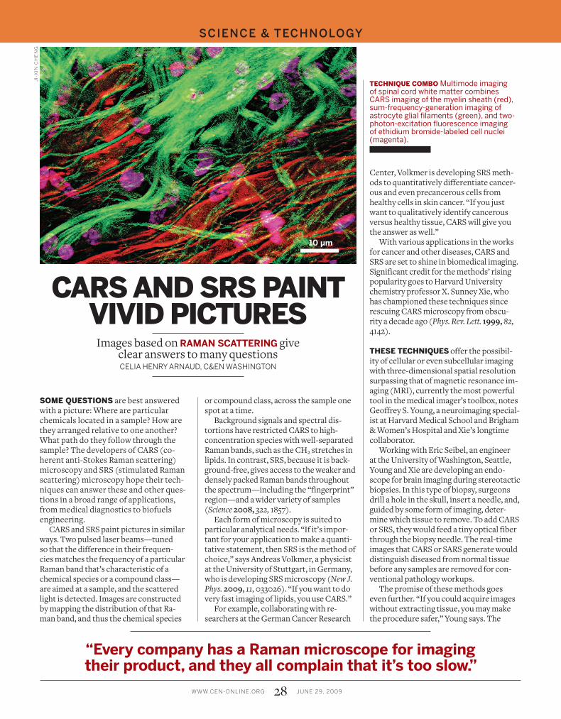
TECHNIQUE COMBO Multimode imaging of spinal cord white matter combines CARS imaging of the myelin sheath (red), sum-frequency-generation imaging of astrocyte glial filaments (green), and two-photon-excitation fluorescence imaging of ethidium bromide-labeled cell nuclei (magenta).
JI-
XIN
CH
EN
G
“Every company has a Raman microscope for imaging their product, and they all complain that it’s too slow.”
29WWW.CEN-ONLINE.ORG JUNE 29, 2009
hope is that Raman will provide contrast equivalent to that achieved with staining in traditional pathology tests and eliminate the need for removing tissue, at least in some cases.
Xie and Young are currently using CARS and SRS imaging to study animal models of various diseases, including brain cancer, metastatic cancer, and multiple sclerosis. In this work, they ask pathologists to in-terpret Raman images of tissue samples from the animal disease models. As they learn from the pathologists’ mistakes, Xie and Young will tweak the data collection to make better diagnostic images.
Young expects that the first application in people will be in patients with suspected brain cancer for which noninvasive methods, such as MRI, are inconclusive. But Young
cautions that these techniques will not deliver chemists’ dreams of seeing a unique molecular sig-nature for every disease that takes all guesswork out of diagnosis, be-cause no disease has a truly unique molecular signal. Instead, the goal of CARS and SRS imaging, he says, should be to produce images that pathologists can use for diagnosis.
“We want to make the lipids one color, the water another color, the protein a third color, and overlay them,” Young says. “If you can do that, you’ve got an image that an expert interpreter can use to make a gold-standard diagnosis.”
CARS is also being used to study the central nervous system. Collaborating with Stephen D. Miller, an immunologist at Northwestern University Medical School, Ji-Xin Cheng, a biomedical engineering professor at Purdue University, uses CARS to study the lipid-rich myelin sheaths that encase axons in the brain and spinal cord.
CARS is an excellent tool to visualize ax-ons with high resolution, Cheng says. With CARS, Cheng can see myelin on individual
axons, “something that used to require electron microscopy,” he notes.
Degradation of the myelin sheath is a hallmark of the neurological disease mul-tiple sclerosis. Cheng’s current project in-volves watching individual axons in a mouse model of multiple sclerosis over the course of a month to follow disease progression.
Meanwhile, Miller is trying to promote repair of myelin. CARS imaging “offers us the opportunity to see with very high reso-lution what myelin repair really looks like,” he says.
Cheng also collaborates with Michael S. Sturek, a physiologist at the Indiana Uni-versity School of Medicine, to study lesions in atherosclerosis, the hardening of arterial walls due to fatty deposits. They combine CARS imaging of foam cells (white blood
cells that have absorbed oxidized low-density lipoproteins) with simul-taneous sum-frequency-generation imaging of the collagen matrix in which they reside. Such collagen-embedded foam cells are the main constituents of plaques, which consist of lipid cores with fibrous caps that connect to the artery wall. “If we could ac-tually see in a human that the fibrous cap was very thin and there was a big lipid core, we could imme-diately identify it as an un-stable plaque,” Sturek says. “Those unstable plaques cause the majority of heart attacks.”
Pharmaceutical imag-ing may turn out to be the biggest application of these
techniques, particularly SRS, according to both Xie and Cheng.
“Every company has a Raman micro-scope for imaging their product, and they all complain that it’s too slow,” Cheng says. CARS and SRS can reduce imaging time from hours to one second, he says. “I hope that one day every pharmaceutical com-pany will have such a system,” Cheng says.
Because SRS has sufficient spectral reso-lution to pick out bands in the fingerprint region of the spectrum, it can distinguish between a drug molecule and the surround-ing matrix. SRS can also reveal details about the diffusion of drugs through skin.
“When you’re developing a drug delivery
patch, SRS will be a very valuable technique to understand the pharmacokinetics of the drug molecule’s diffusion across barriers,” says Jason Tsai, a physician who works at Pfizer and collaborates with Xie. They have used the technique to show retinoic acid diffusing through skin.
More examples of the clinical applica-tions of SRS are needed before it will be broadly adopted in pharma, Tsai says. “You need people who understand the technol-ogy and understand where a tool could be useful in a clinical setting” to bridge the gap between lab and clinic, he says.
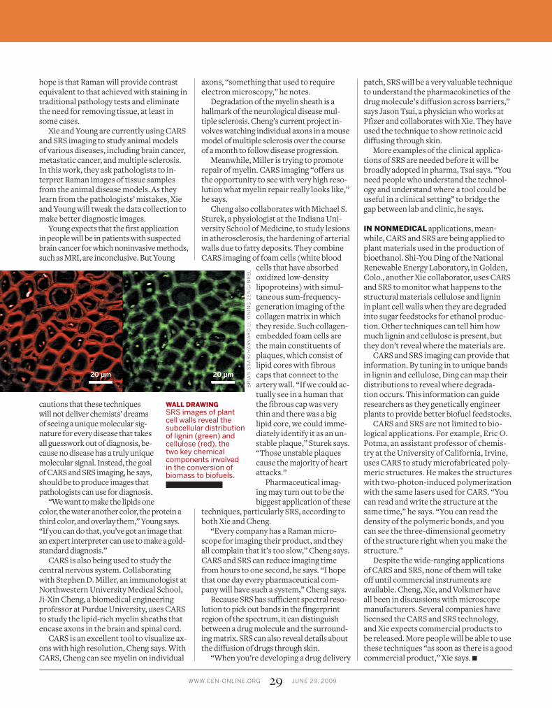
IN NONMEDICAL applications, mean-while, CARS and SRS are being applied to plant materials used in the production of bioethanol. Shi-You Ding of the National Renewable Energy Laboratory, in Golden, Colo., another Xie collaborator, uses CARS and SRS to monitor what happens to the structural materials cellulose and lignin in plant cell walls when they are degraded into sugar feedstocks for ethanol produc-tion. Other techniques can tell him how much lignin and cellulose is present, but they don’t reveal where the materials are.
CARS and SRS imaging can provide that information. By tuning in to unique bands in lignin and cellulose, Ding can map their distributions to reveal where degrada-tion occurs. This information can guide researchers as they genetically engineer plants to provide better biofuel feedstocks.
CARS and SRS are not limited to bio-logical applications. For example, Eric O. Potma, an assistant professor of chemis-try at the University of California, Irvine, uses CARS to study microfabricated poly-meric structures. He makes the structures with two-photon-induced polymerization with the same lasers used for CARS. “You can read and write the structure at the same time,” he says. “You can read the density of the polymeric bonds, and you can see the three-dimensional geometry of the structure right when you make the structure.”
Despite the wide-ranging applications of CARS and SRS, none of them will take off until commercial instruments are available. Cheng, Xie, and Volkmer have all been in discussions with microscope manufacturers. Several companies have licensed the CARS and SRS technology, and Xie expects commercial products to be released. More people will be able to use these techniques “as soon as there is a good commercial product,” Xie says. ■
BR
IAN
SA
AR
/H
AR
VA
RD
U;
YIN
ING
ZE
NG
/N
RE
L
WALL DRAWING SRS images of plant cell walls reveal the subcellular distribution of lignin (green) and cellulose (red), the two key chemical components involved in the conversion of biomass to biofuels.