Embed Size (px)
Citation preview

Case ReportBabinski-Nageotte Syndrome Diagnosed in Postpartum Period
Serdar Oruç,1 Hayri DemirbaG,1 Abdullah Güzel,1
Mehtap Beker Acay,2 and Mehmet Yaman1
1Department of Neurology, Afyon Kocatepe University School of Medicine, 03200 Afyonkarahisar, Turkey2Department of Radiology, Afyon Kocatepe University School of Medicine, 03200 Afyonkarahisar, Turkey
Correspondence should be addressed to Serdar Oruc; [email protected]
Received 19 October 2015; Revised 13 January 2016; Accepted 26 January 2016
Academic Editor: Chin-Chang Huang
Copyright © 2016 Serdar Oruc et al. This is an open access article distributed under the Creative Commons Attribution License,which permits unrestricted use, distribution, and reproduction in any medium, provided the original work is properly cited.
Babinski-Nageotte Syndrome (BNS) is one of the brainstem syndromes characterized by muscle weakness in the opposite half ofthe body with classicWallenberg findings. According to our literature survey, only a few cases have been reported and none of themwas in the postpartum period. We report a case of a typical BNS in a postpartum woman with an ischemic lesion in the medullaoblongata shown on magnetic resonance imaging.
1. Introduction
Firstly diagnosed in 1902, Babinski-Nageotte Syndrome(BNS) is one of the brainstem syndromes characterized bymedulla oblongata ischemia [1]. In BNS, which has beendescribed in very few cases in the literature, lateral medullarysyndrome symptoms as well as contralateral motor deficithave been observed. Here, we have reported a case diagnosedwith BNS which was developed on the 10th day of thepostpartum period.
2. Case Report
A 30-year-old woman was evaluated with complaints ofdysarthria, dysphagia, dizziness, nausea, vomiting, andweak-ness of left arm and leg. According to her medical history,therewas sudden development of dizziness, nausea, and vom-iting symptoms one hour before her admission to the hospitaland later complaints of dysphagia, dysarthria, and weaknessof left arm and leg were added to her presenting complaints.Therewas no background or family history of the patient withthe exception that she had given birth by Caesarean section,10 days before, and was diagnosed with preeclampsia in the33rdweek of her pregnancy. In her first evaluation, the patientwas in stuporous state and she had dysarthric speech. Duringexamination of her eye movements, vertical and horizontalnystagmus were observed. There was flattening of left sided
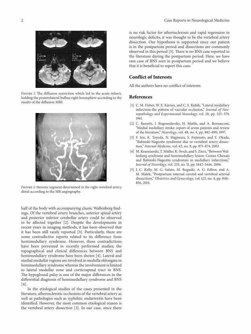
nasolabial sulcus with abnormal gag reflex observed duringcranial nerve examination. In themotor system examination,the left upper and lower extremity muscle power were 3/5level and her Babinski reflex was found to be an exten-sor response on the left side. During the sensory systemexamination, pain and thermal senses of the patient weredecreased on the left side of the body and cerebellar tests wereabnormal on the right side. Evaluation of the cranial MRIscreening of the patient with themisdoubt of cerebrovasculardisease showed results that were consistent with diffusionrestrictionwhich was thought to be acute infarct extending toinferior cerebellar peduncle with involvement of right sidedposterolateral medulla oblongata (Figure 1). In magneticresonance angiography (MRA), stenosis was seen in the distalsegment of right vertebral artery (Figure 2).
In addition to lateral medullary syndrome symptoms,there was also contralateral muscle weakness observed in thepatient. Therefore, we reached a conclusion that the patienthad BNS and medical treatment was initiated accordingly.The patient was hospitalized for 15 days in our clinic. At theend of the fifteenth day her neurological symptoms improvedexcept ataxia and dysarthria.
3. Discussion
Babinski-Nageotte Syndrome (BNS) is one of the brainstemsyndromes characterized by muscle weakness in the opposite
Hindawi Publishing CorporationCase Reports in Neurological MedicineVolume 2016, Article ID 5206430, 2 pageshttp://dx.doi.org/10.1155/2016/5206430
2 Case Reports in Neurological Medicine
Figure 1: The diffusion restriction which led to the acute infarct,holding the posterolateral bulbus right hemisphere according to theresults of the diffusion MRI.
Figure 2: Stenotic segment determined in the right vertebral arterydistal according to the MR angiography.
half of the body with accompanying classic Wallenberg find-ings. Of the vertebral artery branches, anterior spinal arteryand posterior inferior cerebellar artery could be observedto be affected together [2]. Despite the developments inrecent years in imaging methods, it has been observed thatit has been still rarely reported [3]. Particularly, there aresome contradictive reports related to its difference fromhemimedullary syndrome. However, these contradictionshave been prevented in recently performed studies; thetopographical and clinical differences between BNS andhemimedullary syndrome have been shown [4]. Lateral andmedialmedullar regions are involved inmedulla oblongata inhemimedullary syndromewhereas the involvement is limitedto lateral medullar zone and corticospinal tract in BNS.The hypoglossal palsy is one of the major differences in thedifferential diagnosis of hemimedullary syndrome and BNS[4].
In the etiological studies of the cases presented in theliterature, atherosclerotic occlusions of the vertebral artery aswell as pathologies such as syphilitic endarteritis have beenidentified. However, the most common etiological reason isthe vertebral artery dissection [3]. In our case, since there
is no risk factor for atherosclerosis and rapid regression inneurologic deficits, it was thought to be the vertebral arterydissection. Our hypothesis is supported since our patientis in the postpartum period and dissections are commonlyobserved in this period [5]. There is no BNS case reported inthe literature during the postpartum period. Here, we haverare case of BNS seen in postpartum period and we believethat it is beneficial to report this case.
Conflict of Interests
All the authors have no conflict of interests.
References
[1] C. M. Fisher, W. E. Karnes, and C. S. Kubik, “Lateral medullaryinfarction-the pattern of vascular occlusion,” Journal of Neu-ropathology and Experimental Neurology, vol. 20, pp. 323–379,1961.
[2] C. Bassetti, J. Bogousslavsky, H. Mattle, and A. Bernasconi,“Medial medullary stroke: report of seven patients and reviewof the literature,” Neurology, vol. 48, no. 4, pp. 882–890, 1997.
[3] F. Irie, K. Toyoda, N. Hagiwara, S. Fujimoto, and Y. Okada,“Babinski-Nageotte syndrome due to vertebral artery dissec-tion,” Internal Medicine, vol. 42, no. 9, pp. 871–874, 2003.
[4] M. Krasnianski, T.Muller, K. Stock, and S. Zierz, “BetweenWal-lenberg syndrome and hemimedullary lesion: Cestan-Chenaisand Babinski-Nageotte syndromes in medullary infarctions,”Journal of Neurology, vol. 253, no. 11, pp. 1442–1446, 2006.
[5] J. C. Kelly, M. G. Safain, M. Roguski, A. G. Edlow, and A.M. Malek, “Postpartum internal carotid and vertebral arterialdissections,” Obstetrics and Gynecology, vol. 123, no. 4, pp. 848–856, 2014.

Submit your manuscripts athttp://www.hindawi.com
Stem CellsInternational
Hindawi Publishing Corporationhttp://www.hindawi.com Volume 2014
Hindawi Publishing Corporationhttp://www.hindawi.com Volume 2014
MEDIATORSINFLAMMATION
of
Hindawi Publishing Corporationhttp://www.hindawi.com Volume 2014
Behavioural Neurology
EndocrinologyInternational Journal of
Hindawi Publishing Corporationhttp://www.hindawi.com Volume 2014
Hindawi Publishing Corporationhttp://www.hindawi.com Volume 2014
Disease Markers
Hindawi Publishing Corporationhttp://www.hindawi.com Volume 2014
BioMed Research International
OncologyJournal of
Hindawi Publishing Corporationhttp://www.hindawi.com Volume 2014
Hindawi Publishing Corporationhttp://www.hindawi.com Volume 2014
Oxidative Medicine and Cellular Longevity
Hindawi Publishing Corporationhttp://www.hindawi.com Volume 2014
PPAR Research
The Scientific World JournalHindawi Publishing Corporation http://www.hindawi.com Volume 2014
Immunology ResearchHindawi Publishing Corporationhttp://www.hindawi.com Volume 2014
Journal of
ObesityJournal of
Hindawi Publishing Corporationhttp://www.hindawi.com Volume 2014
Hindawi Publishing Corporationhttp://www.hindawi.com Volume 2014
Computational and Mathematical Methods in Medicine
OphthalmologyJournal of
Hindawi Publishing Corporationhttp://www.hindawi.com Volume 2014
Diabetes ResearchJournal of
Hindawi Publishing Corporationhttp://www.hindawi.com Volume 2014
Hindawi Publishing Corporationhttp://www.hindawi.com Volume 2014
Research and TreatmentAIDS
Hindawi Publishing Corporationhttp://www.hindawi.com Volume 2014
Gastroenterology Research and Practice
Hindawi Publishing Corporationhttp://www.hindawi.com Volume 2014
Parkinson’s Disease
Evidence-Based Complementary and Alternative Medicine
Volume 2014Hindawi Publishing Corporationhttp://www.hindawi.com