Embed Size (px)
Citation preview

Int J Clin Exp Pathol 2014;7(11):8190-8197www.ijcep.com /ISSN:1936-2625/IJCEP0002608
Case ReportDiffuse large B-cell lymphoma solely involving bilateral adrenal glands and stomach: report of an extremely rare case with review of the literature
Mutsumi Wakabayashi1, Yasunobu Sekiguchi1, Asami Shimada1,5, Kunimoto Ichikawa1,5, Keiji Sugimoto1, Shigeki Tomita2, Hiroshi Izumi2, Noriko Nakamura3, Tomohiro Sawada3, Yasunori Ohta4, Norio Komatsu5, Masaaki Noguchi1
Departments of 1Hematology, 2Pathology, 3Clinical Laboratory, Juntendo University Urayasu Hospital, Japan; 4Department of Pathology, Research Hospital, The Institute of Medical Science, The University of Tokyo, Japan; 5Department of Hematology, Juntendo University Hospital, Japan
Received September 18, 2014; Accepted November 1, 2014; Epub October 15, 2014; Published November 1, 2014
Abstract: A 60-year-old man complained of nausea, vomiting, decreased appetite, and a feeling of abdominal full-ness in August 2013. Based on biopsy findings from an upper gastrointestinal endoscopy examination, a diag-nosis of non-Hodgkin’s lymphoma (NHL), diffuse large B-cell lymphoma (DLBCL), non-GC type, was made. F18-fluorodeoxyglucose-positron emission tomography/computed tomography (FDG-PET/CT) revealed abnormal accu-mulations solely in the gastric wall (SUVmax = 14.5), the left adrenal gland (SUVmax = 14.3), and the right adrenal gland (SUVmax = 8.5). The clinical stage (Ann Arbor) was IVA, the serum LDH level was within the reference range, and the International Prognostic Index (IPI) was low-intermediate. The serum soluble IL-2 receptor level was within the reference range, and there was no evidence of HIV, EB virus, or autoimmune disease. After the completion of 4 cycles of R-CHOP (rituximab, cyclophosphamide, doxorubicin, vincristine, and prednisone) and 2 parallel cycles of prophylactic intrathecal (I.T.), an upper gastrointestinal endoscopy and a FDG-PET/CT examination showed com-plete remission (CR). The patient received 8 cycles of ritsuximab therapy, 6 cycles of CHOP, and 3 cycles of I.T. The patient has maintained a CR for about 14 months. A literature search revealed that malignant lymphoma with involvement confined to the adrenal gland and gastrointestinal tract is exceedingly rare, and only 3 cases of malig-nant lymphoma have been reported, with involvement of the stomach in 2 cases and the duodenum in 1 case. All of the cases were diagnosed as DLBCL. The case described herein represents the third case with involvement of the stomach.
Keywords: Diffuse large B-cell lymphoma (DLBCL), adrenal gland, stomach
Introduction
Tumors of the adrenal gland may be either pri-mary or secondary neoplasms. Primary adrenal tumors mainly occur as adenomas or adrenal carcinomas [1], while primary adrenal lympho-mas are rare [2]. Among cases of secondary adrenal tumors, 90% were carcinomas and 49% were bilateral tumors [3]. Lymphomatous invasion of the adrenal gland was discovered by computed tomography (CT) examination in 5% of cases [4] and by a morbid anatomy in 25%-35% of cases [5, 6]. Secondary adrenal lymphoma is considered to be not uncommon. Almost all secondary adrenal lymphomas have
been documented to arise in the retroperitone-al lymph node or the ipsilateral kidney [7], and lymphoma with the involvement of both adrenal glands and the stomach alone, as seen in the present case, is thought to be uncommon. No more than 4 case reports, including the present case, have been described in which tumor inva-sion of the adrenal gland and the digestive tract was evident [8-10]. All these cases consisted of diffuse large B-cell lymphoma (DLBCL). A litera-ture review has suggested that the prognosis may be favorable insofar as the malignant involvement is confined to the gastrointestinal tract and adrenal glands. While reports docu-menting a poor prognosis of patients with pri-

DLBCL involving bilateral adrenal glands and stomach
8191 Int J Clin Exp Pathol 2014;7(11):8190-8197
mary adrenal lymphoma are relatively common [11-13], the prognosis and clinical features of secondary adrenal lymphoma remain unclear. Further exploration and assessments of accu-mulated cases are needed.
Case report
The patient was a 60-year-old man whose chief complaints were nausea, vomiting, decreased appetite, and a sense of abdominal fullness; he had been diagnosed as having hypertension at the age of 58 years. His family history was non-contributory. Regarding the present illness, the patient had sought medical advice at a local clinic because of his chief complaints, which had developed in early August 2013. A suspi-
cion of Borrmann type III gastric carcinoma was entertained based on the results of an upper gastrointestinal endoscopy (U-GIS); therefore, he was referred to the Department of Surgery of this hospital in late August. A re-examination using U-GIS also roused a suspicion of gastric cancer (Figure 1A, 1B), but hematoxylin-eosin (HE)-stained biopsy specimens showed the sheet-like growth of large atypical cells with prominent nucleoli (Figure 2A, 2B) and immu-nostaining revealed these atypical cells to be positive for cluster of differentiation (CD) 20, B-cell lymphoma (BCL) 2, and multiple myelo-ma oncogene (MUM) 1 (Figure 2C-E). The cells were negative for BCL-6, CD5, and CD10 (Figure 2F-H). As for the MIB-1 labeling index, 90% of the cells were found to be positive (Figure 2I).
Figure 1. Upper gastrointestinal endoscopy findings. A. Conventional. B. Indigocarmine chromoendoscopy: a dis-crete ulcer with marginal elevation was noted on the lesser curvature at the angulus before the treatment. C. Con-ventional. D. Indigocarmine chromoendoscopy: after the treatment, the lesion had almost completely disappeared, with only a scar left on the lesser curvature at the angulus.

DLBCL involving bilateral adrenal glands and stomach
8192 Int J Clin Exp Pathol 2014;7(11):8190-8197
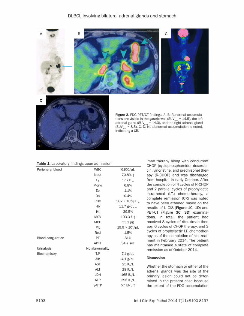
The patient was therefore diagnosed as having non-Hodgkin’s lymphoma, DLBCL, non-GC type, and the patient was examined in the Department of Hematology in early September. F18-fluorodeoxyglucose-positron emission to- mography/computed tomography (FDG-PET/CT) revealed abnormal FDG accumulations in the gastric wall (SUVmax = 14.5), the left adrenal gland (SUVmax = 14.3), and the right adrenal gland (SUVmax = 8.5) (Figure 3A, 3B). Adrenal biopsies from the abnormal uptake sites were considered but could not be performed because of a lack of consent from the patient and his family members. No abnormalities were noted in a bone marrow examination or in a cerebrospinal fluid examination. The clinical stage was IVA according to the Ann Arbor stag-ing system, and the International Prognostic Index (IPI) was low-intermediate (clinical stage and extranodal lesions). The patient was admit-ted to this hospital in mid-September.
The patient’s status upon admission was as fol-lows: height, 164 cm; body weight, 63.0 kg; temperature, 36.4°C; blood pressure, 132/78 mmHg; pulse rate, 74 beats/min (regular); clear consciousness; palpebral conjunctiva, slightly anemic; bulbar conjunctiva, no sign of jaundice; oral cavity, no abnormalities; both lungs, clear; heart sounds, clear; and liver and spleen, impalpable. No neurological abnormalities were observed. No superficial lymph nodes were palpable.
The patient’s laboratory findings upon admis-sion were as follows (see Table 1): negative for anti-human immunodeficiency virus (HIV) anti-bodies, and lactate dehydrogenase (LDH) and soluble interleukin (IL) -2 receptor levels and adrenal function tests were within the respec-tive reference ranges.
The patient’s clinical course is shown in Figure 4. The patient received an initial cycle of ritsux-
Figure 2. Pathologic findings of gastric biopsy specimens. A. (HE, × 40): Sheet-like growth of large atypical cells with prominent nucleoli. B. (HE, × 600): Sheet-like growth of large atypical cells with prominent nucleoli. C. (CD20, × 600): Positive. D. (bcl-2, × 600): Weakly positive. E. (MUM-1, × 600): Positive. F. (bcl-6, × 600): Negative. G. (CD5, × 600): Negative. H. (CD10, × 600): Negative. I. (MIB-1 index, × 600): 90% positive.
DLBCL involving bilateral adrenal glands and stomach
8193 Int J Clin Exp Pathol 2014;7(11):8190-8197
imab therapy along with concurrent CHOP (cyclophosphamide, doxorubi-cin, vincristine, and prednisone) ther-apy (R-CHOP) and was discharged from hospital in early October. After the completion of 4 cycles of R-CHOP and 2 parallel cycles of prophylactic intrathecal (I.T.) chemotherapy, a complete remission (CR) was noted to have been attained based on the results of U-GIS (Figure 1C, 1D) and PET-CT (Figure 3C, 3D) examina-tions. In total, the patient had received 8 cycles of ritsuximab ther-apy, 6 cycles of CHOP therapy, and 3 cycles of prophylactic I.T. chemother-apy as of the completion of his treat-ment in February 2014. The patient has maintained a state of complete remission as of October 2014.
Discussion
Whether the stomach or either of the adrenal glands was the site of the primary lesion could not be deter-mined in the present case because the extent of the FDG accumulation
Table 1. Laboratory findings upon admissionPeripheral blood WBC 6100/μL
Neut 70.8% ↑ Ly 17.7% ↓
Mono 6.8%Eo 1.1%Ba 0.4%
RBC 382 × 104/μL ↓Hb 11.7 g/dL ↓Ht 39.5%
MCV 103.3 fl ↑MCH 33.1 pgPlt 19.9 × 10⁴/μL
Reti 1.5%Blood coagulation PT 81%
APTT 34.7 secUrinalysis No abnormalityBiochemistry T.P 7.1 g/dL
Alb 4.1 g/dLAST 25 IU/LALT 28 IU/LLDH 165 IU/LALP 296 IU/L
γ-GTP 57 IU/L ↑
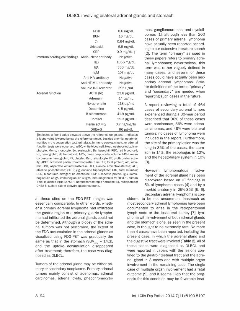
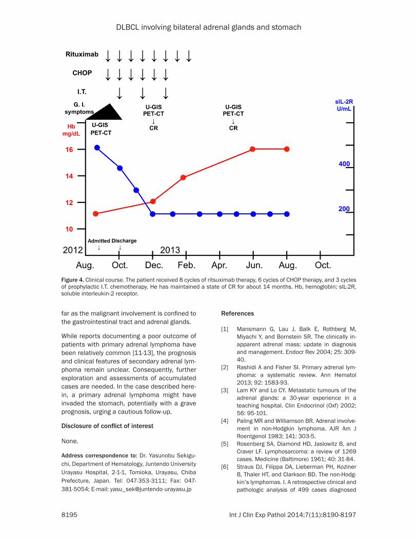
Figure 3. FDG-PET/CT findings. A, B. Abnormal accumula-tions are visible in the gastric wall (SUVmax = 14.5), the left adrenal gland (SUVmax = 14.3), and the right adrenal gland (SUVmax = 8.5). C, D. No abnormal accumulation is noted, indicating a CR.
DLBCL involving bilateral adrenal glands and stomach
8194 Int J Clin Exp Pathol 2014;7(11):8190-8197
at these sites on the FDG-PET images was essentially comparable. In other words, wheth-er a primary adrenal lymphoma had infiltrated the gastric region or a primary gastric lympho-ma had infiltrated the adrenal glands could not be determined. Although a biopsy of the adre-nal tumors was not performed, the extent of the FDG accumulation in the adrenal glands as visualized using FDG-PET was practically the same as that in the stomach (SUVmax = 14.3), and the uptake accumulation disappeared after treatment; therefore, the case was diag-nosed as DLBCL.
Tumors of the adrenal gland may be either pri-mary or secondary neoplasms. Primary adrenal tumors mainly consist of adenomas, adrenal carcinomas, adrenal cysts, pheochromocyto-
sidered to be not uncommon. Inasmuch as most secondary adrenal lymphomas have been documented to arise in the retroperitoneal lymph node or the ipsilateral kidney [7], lym-phoma with involvement of both adrenal glands and the stomach alone, as seen in the present case, is thought to be extremely rare. No more than 4 cases have been reported, including the present case, in which the adrenal gland and the digestive tract were involved (Table 2). All of these cases were diagnosed as DLBCL and were reported in Japan, with the lesions con-fined to the gastrointestinal tract and the adre-nal gland in 3 cases and with multiple organ involvement in the remaining case. The single case of multiple organ involvement had a fatal outcome [9], and it seems likely that the prog-nosis for this condition may be favorable inso-
T-Bill 0.6 mg/dLBUN 10 mg/dLCr 0.64 mg/dL
Uric acid 6.9 mg/dLCRP 0.9 mg/dL ↑
Immuno-serological findings Antinuclear antibody NegativeIgG 1056 mg/dLIgA 333 mg/dLIgM 107 mg/dL
Anti-HIV antibody NegativeAnti-HTLV-1 antibody NegativeSoluble IL-2 receptor 395 U/mL
Adrenal function ACTH (RI) 23.8 pg/mLAdrenalin 14 pg/mL
Noradrenalin 218 pg/mLDopamine ≤ 5 pg/mL
B aldosterone 41.9 pg/mLCortisol 15.3 μg/mL
Renin activity 0.7 ng/mL/hrDHEA-S 96 μg/dL
↑indicates a found value elevated above the reference range, and ↓indicates a found value lowered below the reference range. Besides anemia, no abnor-malities in the coagulation test, urinalysis, immuno-serologic tests, or adrenal function tests were observed. WBC, white blood cell; Neut, neutrocyte; Ly, lym-phocyte; Mono, monocyte; Eo, eosinophil; Ba, basophil; RBC, red blood cell; Hb, hemoglobin; Ht, hematocrit; MCV, mean corpuscular volume; MCH, mean corpuscular hemoglobin; Plt, platelet; Reti, reticulocyte; PT, prothrombin activ-ity; APTT, activated partial thromboplastin time; T.P, total protein; Alb, albu-min; AST, aspartate aminotransferase; ALT, alanine aminotransferase; ALP, alkaline phosphatase; γ-GTP, γ-guanosine triphosphate; T-Bil, total bilirubin; BUN, blood urea nitrogen; Cr, creatinine; CRP, C-reactive protein; IgG, immu-noglobulin G; IgA, immunoglobulin A; IgM, immunoglobulin M; HTVL-1, human T-cell leukemia virus-1; ACTH, adrenocorticotropic hormone; RI, radioisotope; DHEA-S, sulfate salt of dehydroepiandrosterone.
mas, ganglioneuromas, and myeloli-pomas [1], although less than 200 cases of primary adrenal lymphoma have actually been reported accord-ing to our extensive literature search [2]. The term “primary” as used in these papers refers to primary adre-nal lymphomas; nevertheless, this term was rather vaguely defined in many cases, and several of these cases could have actually been sec-ondary adrenal lymphomas. Stric- ter definitions of the terms “primary” and “secondary” are needed when reporting such cases in the future.
A report reviewing a total of 464 cases of secondary adrenal tumors experienced during a 30-year period described that 90% of these cases were carcinomas, 56% were adeno-carcinomas, and 49% were bilateral tumors; no cases of lymphoma were included in the report. Furthermore, the site of the primary lesion was the lung in 35% of the cases, the stom-ach in 14%, the esophagus in 12%, and the hepatobiliary system in 10% [3].
However, lymphomatous involve-ment of the adrenal gland has been discovered based on CT findings in 5% of lymphoma cases [4] and by a morbid anatomy in 25%-35% [5, 6]. Secondary adrenal lymphoma is con-
DLBCL involving bilateral adrenal glands and stomach
8195 Int J Clin Exp Pathol 2014;7(11):8190-8197
Figure 4. Clinical course. The patient received 8 cycles of ritsuximab therapy, 6 cycles of CHOP therapy, and 3 cycles of prophylactic I.T. chemotherapy. He has maintained a state of CR for about 14 months. Hb, hemoglobin; sIL-2R, soluble interleukin-2 receptor.
far as the malignant involvement is confined to the gastrointestinal tract and adrenal glands.
While reports documenting a poor outcome of patients with primary adrenal lymphoma have been relatively common [11-13], the prognosis and clinical features of secondary adrenal lym-phoma remain unclear. Consequently, further exploration and assessments of accumulated cases are needed. In the case described here-in, a primary adrenal lymphoma might have invaded the stomach, potentially with a grave prognosis, urging a cautious follow-up.
Disclosure of conflict of interest
None.
Address correspondence to: Dr. Yasunobu Sekigu- chi, Department of Hematology, Juntendo University Urayasu Hospital, 2-1-1, Tomioka, Urayasu, Chiba Prefecture, Japan. Tel: 047-353-3111; Fax: 047-381-5054; E-mail: [email protected]
References
[1] Mansmann G, Lau J, Balk E, Rothberg M, Miyachi Y, and Bornstein SR. The clinically in-apparent adrenal mass: update in diagnosis and management. Endocr Rev 2004; 25: 309-40.
[2] Rashidi A and Fisher SI. Primary adrenal lym-phoma: a systematic review. Ann Hematol 2013; 92: 1583-93.
[3] Lam KY and Lo CY. Metastatic tumours of the adrenal glands: a 30-year experience in a teaching hospital. Clin Endocrinol (Oxf) 2002; 56: 95-101.
[4] Paling MR and Williamson BR. Adrenal involve-ment in non-Hodgkin lymphoma. AJR Am J Roentgenol 1983; 141: 303-5.
[5] Rosenberg SA, Diamond HD, Jaslowitz B, and Craver LF. Lymphosarcoma: a review of 1269 cases. Medicine (Baltimore) 1961; 40: 31-84.
[6] Straus DJ, Filippa DA, Lieberman PH, Koziner B, Thaler HT, and Clarkson BD. The non-Hodg-kin’s lymphomas. I. A retrospective clinical and pathologic analysis of 499 cases diagnosed
DLBCL involving bilateral adrenal glands and stomach
8196 Int J Clin Exp Pathol 2014;7(11):8190-8197
Table 2. Reported cases of lymphoma with involvement confined to the gastrointestinal tract and adrenal glands
Case Age & Sex Symptoms Histological
featuresAdrenal lesions G.I. lesions Other lesions Treatment Therapeutic
response Outcome Reference
1 53/F Upper abdominal pain, weight loss
DLBCL Left Elevation of posterior wall adjacent to antral pyloric ring
None 4 cycles of CHOP therapy, gastrectomy, left adrenal-ectomy
CR Alive [8]
2 93/F Decreased appetite, disturbance of consciousness
DLBCL Details unknown
Intragastric multiple masses
Spleen, jejunum, liver, pancreas,
lumbar vertebrae
Parenteral fluid infusion, diuretics, elcitonin
PD Died [9]
3 73/M General malaise, high fever, skin pigmentation
DLBCL Bilateral Hemorrhage in duodenal mucosa
None Hydrocortisone, 1 cycle of R-THPCOP therapy
PR Alive [10]
4 60/M Decreased appetite, vomiting, feeling of abdominal fullness
DLBCL Bilateral Ulcer on lesser curvature at angulus
None 8 cycles of R, 6 cycles of CHOP therapy, 3 cycles of I.T. therapy
CR Alive Present case
F, female; M, man; DLBCL, diffuse large B cell lymphoma; R-THPCOP, rituximab, pirarubicin, cyclophosphamide, vincristine, and prednisone; PD, progression disease; PR, partial remission.

DLBCL involving bilateral adrenal glands and stomach
8197 Int J Clin Exp Pathol 2014;7(11):8190-8197
between 1958 and 1969. Cancer 1983; 51: 101-9.
[7] Glazer HS, Lee JK, Balfe DM, Mauro MA, Griffith R, and Sagel SS. Non-Hodgkin lymphoma: computed tomographic demonstration of un-usual extranodal involvement. Radiology 1983; 149: 211-7.
[8] Matsuda T, Okihama Y, Egami K, Wada M, Yoshioka M, Maeda S, and Onda M. Complete cure of malignant lymphoma of the stomach with a huge adrenal lesion achieved by preop-erative chemotherapy and surgery: report of a case. Surg Today 2001; 31: 62-7.
[9] Ota H, Azuma K, Horiuchi T, Kazama H, Araki A, Hosoi T, Sawabe M, Amizuka N, and Orimo H. [An elderly case of non-Hodgkin’s lymphoma (NHL) with hypercalcemia]. Nippon Ronen Igakkai Zasshi 2003; 40: 167-71.
[10] Nishiuchi T, Imachi H, Fujiwara M, Murao K, Onishi H, Kiguchi T, Takimoto H, Kushida Y, Haba R, and Ishida T. A case of non-Hodgkin’s lymphoma primary arising in both adrenal glands associated with adrenal failure. Endocrine 2009; 35: 34-7.
[11] Takahashi H, Tomita N, Yokoyama M, Tsunoda S, Yano T, Murayama K, Hashimoto C, Tamura K, Sato K, and Ishigatsubo Y. Prognostic im-pact of extranodal involvement in diffuse large B-cell lymphoma in the rituximab era. Cancer 2012; 118: 4166-72.
[12] Ezer A, Parlakgümüş A, Kocer NE, Colakoğlu T, Nursal GN, and Yildirim S. Primary adrenal non-Hodgkin’s lymphoma: report of two cases. Turk J Gastroenterol 2011; 22: 643-7.
[13] Yang Y, Li Q, and Pan Y. Bilateral primary adre-nal lymphoma. Br J Haematol 2010; 150: 250.