Embed Size (px)
Citation preview
Case ReportFasciitis Necroticans after Elective Hernia Inguinal Surgery
T. A. Sigterman,1 Kim J. Gorissen,2 and Dennis E. J. G. J. Dolmans3
1 Atrium Medical Centre, Department of General Surgery, Henri Dunantstraat 5, 6419 PC Heerlen, The Netherlands2Oxford University Hospitals, Department of Surgery, Old Road, Headington, Oxford OX3 7LE, UK3Diakonessenhuis, Department of Surgery, Bosboomstraat 3, 3582 KE Utrecht, The Netherlands
Correspondence should be addressed to T. A. Sigterman; [email protected]
Received 12 September 2013; Accepted 26 November 2013; Published 5 January 2014
Academic Editors: G. Sandblom, Y.-B. Tang, and F. Turegano
Copyright © 2014 T. A. Sigterman et al.This is an open access article distributed under the Creative Commons Attribution License,which permits unrestricted use, distribution, and reproduction in any medium, provided the original work is properly cited.
Necrotising fasciitis is a rare but disastrous complication after elective surgery. We present two patients (both male, 58 and 18 yearsold) who developed necrotising fasciitis following elective inguinal hernia repair according to Lichtenstein. The importance ofboth recognition and time interval between symptom occurrence and surgical intervention is illustrated, emphasising the need forimmediate action when necrotising fasciitis is suspected. A high index of suspicion of necrotising fasciitis should be maintainedwhen a wound infection is accompanied by disproportional pain, lethargy, or sepsis. Epidermolysis and subcutaneous emphysemaare often very late symptoms. Recognition and immediate intervention decrease mortality and morbidity.
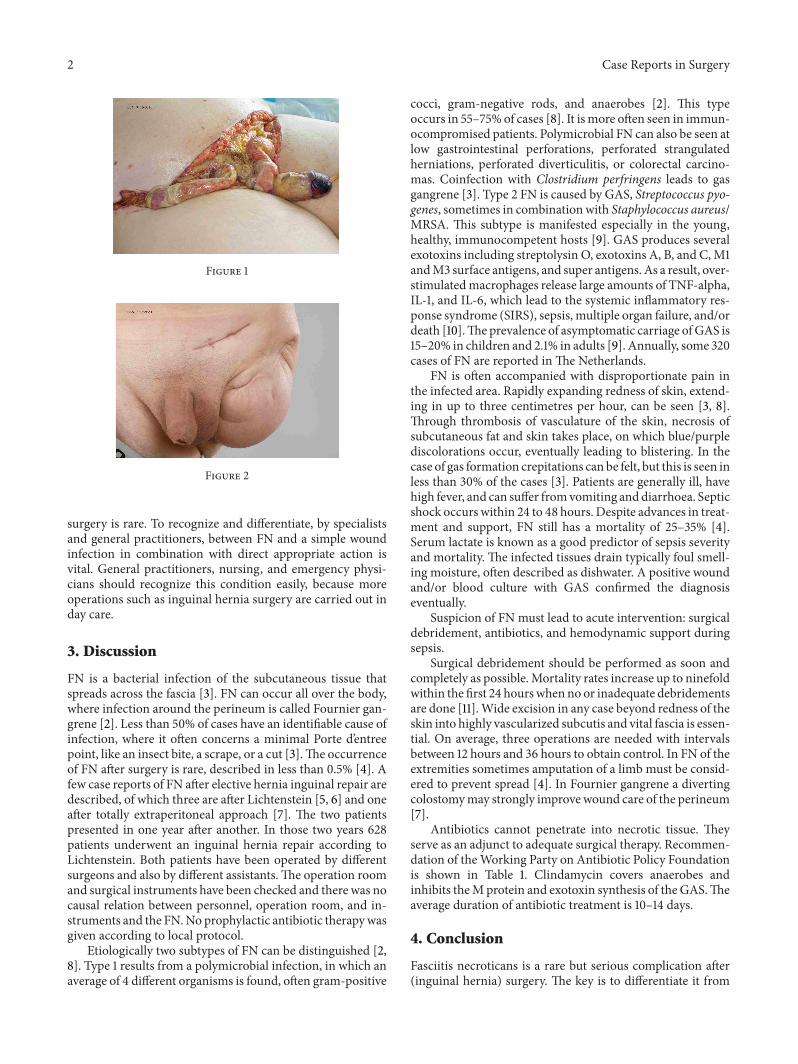
1. Case ReportA 58-year-old man undergoes an elective correction of aninguinal hernia according to Lichtenstein [1].Hismedical his-tory includes insulin dependent diabetes and hypertension.The procedure was straightforward and the patient was dis-charged the sameday. Twodays after the operation the patientvisited our emergency room with severe pain and swelling atthe operation site. On physical examination we saw a moder-ately ill man, with a temperature of 38.7 degrees Celsius and apulse rate of 101 beats per minute. Blood examination showedleukocyte numbers of 9.9 × 10⁹ and a CRP 179mg/L.The sur-gical site showed a hematoma without redness or pus. Thepatient was admitted and reassessed after eight hours. More-over an ill man was seen with blistering, livid discoloration ofthe scrotum.With the suspicion of a fasciitis necroticans (FN)the patient was brought to the operation theatre and antibi-otics were started. Perioperatively a fulminant Fournier gan-grene [2] was seen, for which an extensive necrosectomy wasperformedwith the formation of a colostomy (Figure 1). Peri-operative cultures showed group A beta-hemolytic streptococ-cus (GAS).The patient was admitted to the intensive care unit(ICU). After a total of nine reinterventions and twomonths inhospital, he was discharged to a rehabilitation centre.
Secondly, an 18-year-old male underwent elective ingui-nal hernia surgery according to Lichtenstein. Medical history
showed a through-the-hip amputation in its first weeks of lifebecause of iatrogenic dissection of the femoral artery.The sur-gical procedure was straightforward, whereafter the patientwas discharged the same day.That evening he felt unwell; aftera few hours he visited the emergency room with groin painand fever. The patient was severely ill with a temperature of39.1 degrees Celsius and a pulse rate of 92 beats per minute.Blood tests showed leukocyte numbers of 23 × 109 and a CRPof 365mg/L. There was redness of the wound, which waspainful on palpation, without blisters or crepitation. On ex-ploration of the wound in the operation theatre, a largeamount of foul smelling fluid was drained. Blood and woundcultures showed a GAS. After three days in the ICU thepatient was transferred to the surgical ward. The patient wasalso treated for one week intravenously and two weeksorally with amoxicillin/clavulanic acid and clindamycin. Thepatient recovered well without any sequelae and is dischargedin good clinical condition (Figure 2).
2. Introduction
We describe the case histories of two patients followingelective inguinal hernia correction complicated by fasciitisnecroticans, to focus attention on rapid recognition of thispotentially lethal complication. The occurrence of FN after
Hindawi Publishing CorporationCase Reports in SurgeryVolume 2014, Article ID 981262, 3 pageshttp://dx.doi.org/10.1155/2014/981262
2 Case Reports in Surgery
Figure 1
Figure 2
surgery is rare. To recognize and differentiate, by specialistsand general practitioners, between FN and a simple woundinfection in combination with direct appropriate action isvital. General practitioners, nursing, and emergency physi-cians should recognize this condition easily, because moreoperations such as inguinal hernia surgery are carried out inday care.
3. Discussion
FN is a bacterial infection of the subcutaneous tissue thatspreads across the fascia [3]. FN can occur all over the body,where infection around the perineum is called Fournier gan-grene [2]. Less than 50% of cases have an identifiable cause ofinfection, where it often concerns a minimal Porte d’entreepoint, like an insect bite, a scrape, or a cut [3].The occurrenceof FN after surgery is rare, described in less than 0.5% [4]. Afew case reports of FN after elective hernia inguinal repair aredescribed, of which three are after Lichtenstein [5, 6] and oneafter totally extraperitoneal approach [7]. The two patientspresented in one year after another. In those two years 628patients underwent an inguinal hernia repair according toLichtenstein. Both patients have been operated by differentsurgeons and also by different assistants.The operation roomand surgical instruments have been checked and there was nocausal relation between personnel, operation room, and in-struments and the FN.No prophylactic antibiotic therapywasgiven according to local protocol.
Etiologically two subtypes of FN can be distinguished [2,8]. Type 1 results from a polymicrobial infection, in which anaverage of 4 different organisms is found, often gram-positive
cocci, gram-negative rods, and anaerobes [2]. This typeoccurs in 55–75% of cases [8]. It ismore often seen in immun-ocompromised patients. Polymicrobial FN can also be seen atlow gastrointestinal perforations, perforated strangulatedherniations, perforated diverticulitis, or colorectal carcino-mas. Coinfection with Clostridium perfringens leads to gasgangrene [3]. Type 2 FN is caused by GAS, Streptococcus pyo-genes, sometimes in combinationwith Staphylococcus aureus/MRSA. This subtype is manifested especially in the young,healthy, immunocompetent hosts [9]. GAS produces severalexotoxins including streptolysin O, exotoxins A, B, and C,M1andM3 surface antigens, and super antigens. As a result, over-stimulatedmacrophages release large amounts of TNF-alpha,IL-1, and IL-6, which lead to the systemic inflammatory res-ponse syndrome (SIRS), sepsis, multiple organ failure, and/ordeath [10].Theprevalence of asymptomatic carriage ofGAS is15–20% in children and 2.1% in adults [9]. Annually, some 320cases of FN are reported inThe Netherlands.
FN is often accompanied with disproportionate pain inthe infected area. Rapidly expanding redness of skin, extend-ing in up to three centimetres per hour, can be seen [3, 8].Through thrombosis of vasculature of the skin, necrosis ofsubcutaneous fat and skin takes place, on which blue/purplediscolorations occur, eventually leading to blistering. In thecase of gas formation crepitations can be felt, but this is seen inless than 30% of the cases [3]. Patients are generally ill, havehigh fever, and can suffer fromvomiting anddiarrhoea. Septicshock occurs within 24 to 48 hours. Despite advances in treat-ment and support, FN still has a mortality of 25–35% [4].Serum lactate is known as a good predictor of sepsis severityand mortality. The infected tissues drain typically foul smell-ing moisture, often described as dishwater. A positive woundand/or blood culture with GAS confirmed the diagnosiseventually.
Suspicion of FN must lead to acute intervention: surgicaldebridement, antibiotics, and hemodynamic support duringsepsis.
Surgical debridement should be performed as soon andcompletely as possible.Mortality rates increase up to ninefoldwithin the first 24 hourswhen no or inadequate debridementsare done [11].Wide excision in any case beyond redness of theskin into highly vascularized subcutis and vital fascia is essen-tial. On average, three operations are needed with intervalsbetween 12 hours and 36 hours to obtain control. In FN of theextremities sometimes amputation of a limb must be consid-ered to prevent spread [4]. In Fournier gangrene a divertingcolostomymay strongly improvewound care of the perineum[7].
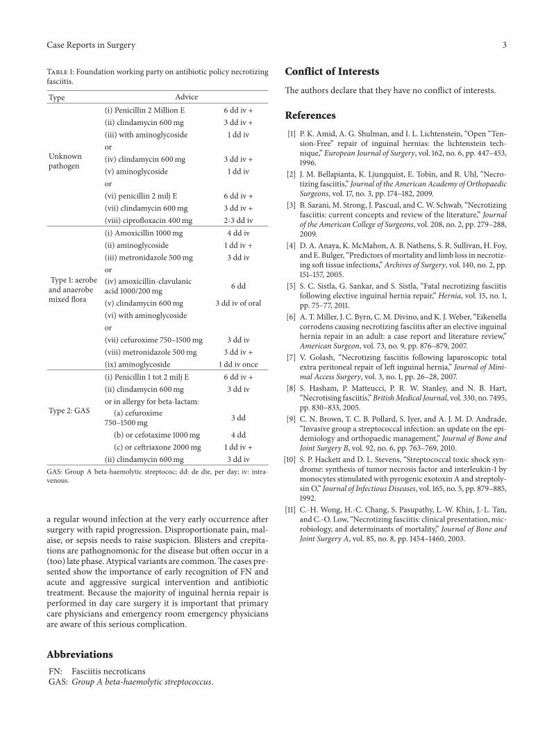
Antibiotics cannot penetrate into necrotic tissue. Theyserve as an adjunct to adequate surgical therapy. Recommen-dation of theWorking Party on Antibiotic Policy Foundationis shown in Table 1. Clindamycin covers anaerobes andinhibits theMprotein and exotoxin synthesis of the GAS.Theaverage duration of antibiotic treatment is 10–14 days.
4. Conclusion
Fasciitis necroticans is a rare but serious complication after(inguinal hernia) surgery. The key is to differentiate it from
Case Reports in Surgery 3
Table 1: Foundation working party on antibiotic policy necrotizingfasciitis.
Type Advice
Unknownpathogen
(i) Penicillin 2 Million E 6 dd iv +(ii) clindamycin 600mg 3 dd iv +(iii) with aminoglycoside 1 dd ivor(iv) clindamycin 600mg 3 dd iv +(v) aminoglycoside 1 dd ivor(vi) penicillin 2 milj E 6 dd iv +(vii) clindamycin 600mg 3 dd iv +(viii) ciprofloxacin 400mg 2-3 dd iv
Type 1: aerobeand anaerobemixed flora
(i) Amoxicillin 1000mg 4 dd iv(ii) aminoglycoside 1 dd iv +(iii) metronidazole 500mg 3 dd ivor(iv) amoxicillin-clavulanicacid 1000/200mg 6 dd
(v) clindamycin 600mg 3 dd iv of oral(vi) with aminoglycosideor(vii) cefuroxime 750–1500mg 3 dd iv(viii) metronidazole 500mg 3 dd iv +(ix) aminoglycoside 1 dd iv once
Type 2: GAS
(i) Penicillin 1 tot 2 milj E 6 dd iv +(ii) clindamycin 600mg 3 dd ivor in allergy for beta-lactam:
(a) cefuroxime750–1500mg 3 dd
(b) or cefotaxime 1000mg 4 dd(c) or ceftriaxone 2000mg 1 dd iv +
(ii) clindamycin 600mg 3 dd ivGAS: Group A beta-haemolytic streptococ; dd: de die, per day; iv: intra-venous.
a regular wound infection at the very early occurrence aftersurgery with rapid progression. Disproportionate pain, mal-aise, or sepsis needs to raise suspicion. Blisters and crepita-tions are pathognomonic for the disease but often occur in a(too) late phase. Atypical variants are common.The cases pre-sented show the importance of early recognition of FN andacute and aggressive surgical intervention and antibiotictreatment. Because the majority of inguinal hernia repair isperformed in day care surgery it is important that primarycare physicians and emergency room emergency physiciansare aware of this serious complication.
Abbreviations
FN: Fasciitis necroticansGAS: Group A beta-haemolytic streptococcus.
Conflict of Interests
The authors declare that they have no conflict of interests.
References
[1] P. K. Amid, A. G. Shulman, and I. L. Lichtenstein, “Open “Ten-sion-Free” repair of inguinal hernias: the lichtenstein tech-nique,” European Journal of Surgery, vol. 162, no. 6, pp. 447–453,1996.
[2] J. M. Bellapianta, K. Ljungquist, E. Tobin, and R. Uhl, “Necro-tizing fasciitis,” Journal of the AmericanAcademy of OrthopaedicSurgeons, vol. 17, no. 3, pp. 174–182, 2009.
[3] B. Sarani, M. Strong, J. Pascual, and C.W. Schwab, “Necrotizingfasciitis: current concepts and review of the literature,” Journalof the American College of Surgeons, vol. 208, no. 2, pp. 279–288,2009.
[4] D. A. Anaya, K.McMahon, A. B. Nathens, S. R. Sullivan, H. Foy,and E. Bulger, “Predictors ofmortality and limb loss in necrotiz-ing soft tissue infections,”Archives of Surgery, vol. 140, no. 2, pp.151–157, 2005.
[5] S. C. Sistla, G. Sankar, and S. Sistla, “Fatal necrotizing fasciitisfollowing elective inguinal hernia repair,” Hernia, vol. 15, no. 1,pp. 75–77, 2011.
[6] A. T.Miller, J. C. Byrn, C.M. Divino, and K. J.Weber, “Eikenellacorrodens causing necrotizing fasciitis after an elective inguinalhernia repair in an adult: a case report and literature review,”American Surgeon, vol. 73, no. 9, pp. 876–879, 2007.
[7] V. Golash, “Necrotizing fasciitis following laparoscopic totalextra peritoneal repair of left inguinal hernia,” Journal of Mini-mal Access Surgery, vol. 3, no. 1, pp. 26–28, 2007.
[8] S. Hasham, P. Matteucci, P. R. W. Stanley, and N. B. Hart,“Necrotising fasciitis,”BritishMedical Journal, vol. 330, no. 7495,pp. 830–833, 2005.
[9] C. N. Brown, T. C. B. Pollard, S. Iyer, and A. J. M. D. Andrade,“Invasive group a streptococcal infection: an update on the epi-demiology and orthopaedic management,” Journal of Bone andJoint Surgery B, vol. 92, no. 6, pp. 763–769, 2010.
[10] S. P. Hackett and D. L. Stevens, “Streptococcal toxic shock syn-drome: synthesis of tumor necrosis factor and interleukin-1 bymonocytes stimulatedwith pyrogenic exotoxinA and streptoly-sin O,” Journal of Infectious Diseases, vol. 165, no. 5, pp. 879–885,1992.
[11] C.-H. Wong, H.-C. Chang, S. Pasupathy, L.-W. Khin, J.-L. Tan,andC.-O. Low, “Necrotizing fasciitis: clinical presentation,mic-robiology, and determinants of mortality,” Journal of Bone andJoint Surgery A, vol. 85, no. 8, pp. 1454–1460, 2003.

Submit your manuscripts athttp://www.hindawi.com
Stem CellsInternational
Hindawi Publishing Corporationhttp://www.hindawi.com Volume 2014
Hindawi Publishing Corporationhttp://www.hindawi.com Volume 2014
MEDIATORSINFLAMMATION
of
Hindawi Publishing Corporationhttp://www.hindawi.com Volume 2014
Behavioural Neurology
EndocrinologyInternational Journal of
Hindawi Publishing Corporationhttp://www.hindawi.com Volume 2014
Hindawi Publishing Corporationhttp://www.hindawi.com Volume 2014
Disease Markers
Hindawi Publishing Corporationhttp://www.hindawi.com Volume 2014
BioMed Research International
OncologyJournal of
Hindawi Publishing Corporationhttp://www.hindawi.com Volume 2014
Hindawi Publishing Corporationhttp://www.hindawi.com Volume 2014
Oxidative Medicine and Cellular Longevity
Hindawi Publishing Corporationhttp://www.hindawi.com Volume 2014
PPAR Research
The Scientific World JournalHindawi Publishing Corporation http://www.hindawi.com Volume 2014
Immunology ResearchHindawi Publishing Corporationhttp://www.hindawi.com Volume 2014
Journal of
ObesityJournal of
Hindawi Publishing Corporationhttp://www.hindawi.com Volume 2014
Hindawi Publishing Corporationhttp://www.hindawi.com Volume 2014
Computational and Mathematical Methods in Medicine
OphthalmologyJournal of
Hindawi Publishing Corporationhttp://www.hindawi.com Volume 2014
Diabetes ResearchJournal of
Hindawi Publishing Corporationhttp://www.hindawi.com Volume 2014
Hindawi Publishing Corporationhttp://www.hindawi.com Volume 2014
Research and TreatmentAIDS
Hindawi Publishing Corporationhttp://www.hindawi.com Volume 2014
Gastroenterology Research and Practice
Hindawi Publishing Corporationhttp://www.hindawi.com Volume 2014
Parkinson’s Disease
Evidence-Based Complementary and Alternative Medicine
Volume 2014Hindawi Publishing Corporationhttp://www.hindawi.com