Embed Size (px)
Citation preview
Case ReportLittoral Cell Angioma in a Patient with Crohn’s Disease
Joel Johansson,1 Bergthor Björnsson,2,3 Simone Ignatova,4
Per Sandström,2,3 and Mattias Ekstedt1
1Department of Gastroenterology and Department of Clinical and Experimental Medicine, Linkoping University,58185 Linkoping, Sweden2Department of Surgery, County Council of Ostergotland, 58185 Linkoping, Sweden3Division of Surgery, Department of Clinical and Experimental Medicine, Faculty of Health Sciences, Linkoping University,581 85 Linkoping, Sweden4Department of Pathology, Linkoping University Hospital, 581 85 Linkoping, Sweden
Correspondence should be addressed to Joel Johansson; [email protected]
Received 24 November 2014; Revised 19 January 2015; Accepted 20 January 2015
Academic Editor: Haruhiko Sugimura
Copyright © 2015 Joel Johansson et al. This is an open access article distributed under the Creative Commons Attribution License,which permits unrestricted use, distribution, and reproduction in any medium, provided the original work is properly cited.
Littoral cell angioma is a rare vascular tumor of the spleen. The pathogenesis is unknown but the lesion is associated with severalmalignancies and immunological disorders. The diagnosis requires histopathological examination. The malignant potential of thislesion is unknown, which is why splenectomy is recommend for all cases. Symptomatic cases generally suffer from hypersplenismand pyrexia. A previously healthy 20-year-old female was diagnosed with colonic Crohn’s disease; as part of the work-up amagnetic resonance enterography was performedwhich showedmultiple signal changes of the spleen.The patient reported chronicabdominal pain in the left upper quadrant, malaise, and fever. The unknown splenic lesions prompted a laparoscopic splenectomy;pathology revealed a littoral cell angioma.Theabdominal pain andmalaise remitted but the fever persisted one year despite adequatetreatment of the patient’s Crohn’s disease. Littoral cell angioma is associated with immune-dysregulation including Crohn’s diseasewith several reported cases. Signs and symptoms of hypersplenism and splenic lesions on imaging should raise suspicion of littoralcell angioma in patients with Crohn’s disease. Magnetic resonance enterography to assess disease severity in Crohn’s disease mayprovide an opportunity to study the prevalence and natural history of this rare splenic tumor.
1. Introduction
Littoral cell angioma, LCA, is a primary vascular tumor ofthe spleen, first described by Falk et al. in 1991 [1]. It is a rareneoplasm which is thought to arise from the splenic cordsshowing both endothelial and histiocytic phenotype [1]. Ithas been associated with bothmalignancies [2] and immune-dysregulation [3]. Around 150 cases have been reportedand although radiological features have been described forUS, MRI, and CT, findings are unspecific and diagnosisusually requires histopathology [4] on image guided biopsyspecimen [5] or splenectomy specimen. The LCA is fre-quently an incidental finding on imaging but some cases aresymptomatic. Symptomatic cases typically develop signs andsymptoms of hypersplenism including anemia, splenomegaly,and thrombocytopenia as well as constitutional symptomsincluding fever [3]. Treatment is splenectomy as it cures
symptoms and malignant lesions cannot be excluded withimaging [6]. We report a case of LCA in a patient with newonset of Crohn’s disease, naive to biologics, and with littleexposure to immunosuppressive agents before the incidentalfinding of LCA on magnetic resonance enterography, MRE.
2. Case Presentation
A 20-year-old, previously healthy female with a two-monthhistory of abdominal pain, bloody diarrhoea, and tenesmuswas referred to the Gastroenterology Clinic at the LinkopingUniversityHospital by her primary care physician. She under-went colonoscopy that revealed a friable erythematous areawith contact bleeding that spanned from the splenic flexureand 15 cm down the descending colon; biopsies were takenthroughout the procedure.These findings were interpreted asCrohn’s disease because of the distribution of inflammation
Hindawi Publishing CorporationCase Reports in Gastrointestinal MedicineVolume 2015, Article ID 474969, 4 pageshttp://dx.doi.org/10.1155/2015/474969
2 Case Reports in Gastrointestinal Medicine
(a)
Arterial phase Venous phase
3min 10min
(b)
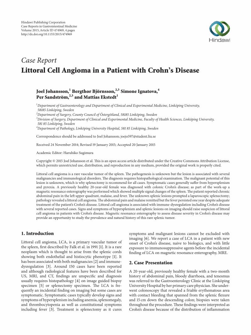
Figure 1: (a) Multiple lesions throughout the spleen up to 15mm in size. They have a high signal on T2 and SPIR. (b) Signal intensity waslow on T1 sequences. After gadolinium contrast, no attenuation was seen in the arterial phase, heterogeneous contrast enhancement in theportal venous phase, and after 3 minutes the enhancement is stronger in the periphery than central in the lesions. After 10 minutes thesignal is stronger in the lesions than the surrounding splenic tissue.The signal pattern is in keeping with hemangiomas. On histopathologicalexamination the diagnosis of LCA was confirmed.
although histology-pathology showed unspecific inflamma-tion more closely related to ulcerative colitis. The patient wasstarted on prednisolone which was tapered as planned over 8weeks. Follow-up with fecal calprotectin showed no responseand the patient was started on balsalazide.
When the patient came for follow-up at 9 monthsshe suffered from fever and bloody diarrhoea once daily.This was regarded as colonic Crohn’s disease and budes-onide was added to balsalazide, An MRE was made onemonth after the follow-up and it revealed no small bowelengagement; however it showed multiple signal changesin the spleen, benign cysts where suspected. A contrastenhanced ultrasound was made that showed indentations inthe superior portion of the spleen as well as multiple minorcystic lesions and a central 1 cm lesion with late contrastloading, possibly a hemangioma. After two months the
effect of budesonide wore off and the patient again sufferedfrombloody diarrhoea. Sigmoidoscopy showed proctitis withdiminished vascularity and contact bleeding. Budesonidewas switched to mesalamine which had no effect; thereforeazathioprine was tried but a colonoscopy one month latershowed inflammation from the sigmoid colon till the splenicflexure. The MRE images were once again examined and itwas decided that anMRIwas necessary to establish the natureof the splenic lesions. The MRI showed multiple lesionswith a contrast pattern of hemangiomas (Figures 1(a) and1(b)). Two months after the MRI she presented acutely tothe gastroenterology outpatient clinic with suddenworseningof abdominal pain that had been present for one month.There were no associated GI or micturition symptoms, bodytemperature of 38, 3∘C with abdominal tenderness in theupper left quadrant, no guarding, rigidity, or mass.
Case Reports in Gastrointestinal Medicine 3
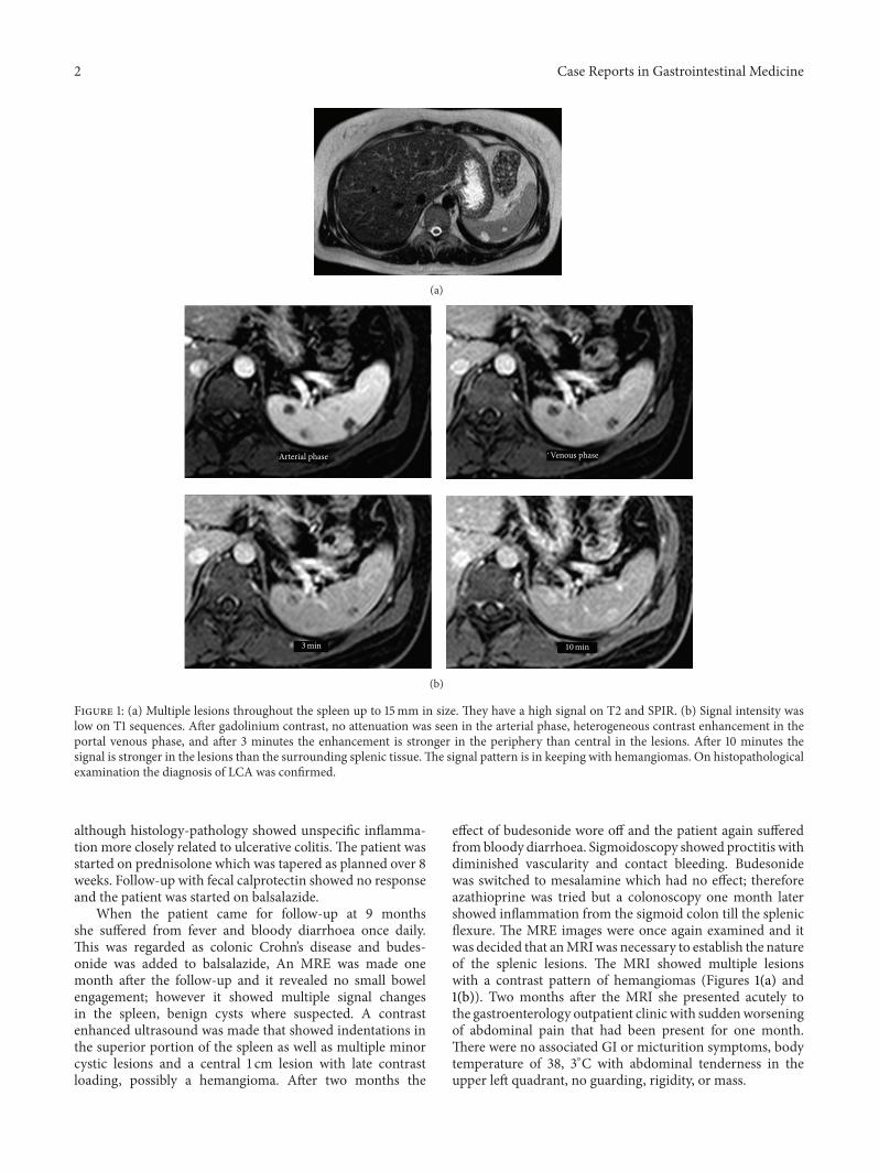
Figure 2: Photomicrograph ×20 magnification. Littoral cell angi-oma of the spleen composed of small anastomosing and irregularchannels reminiscent of splenic sinuses and covered of neoplasticplump “littoral” cells with low mitotic activity.
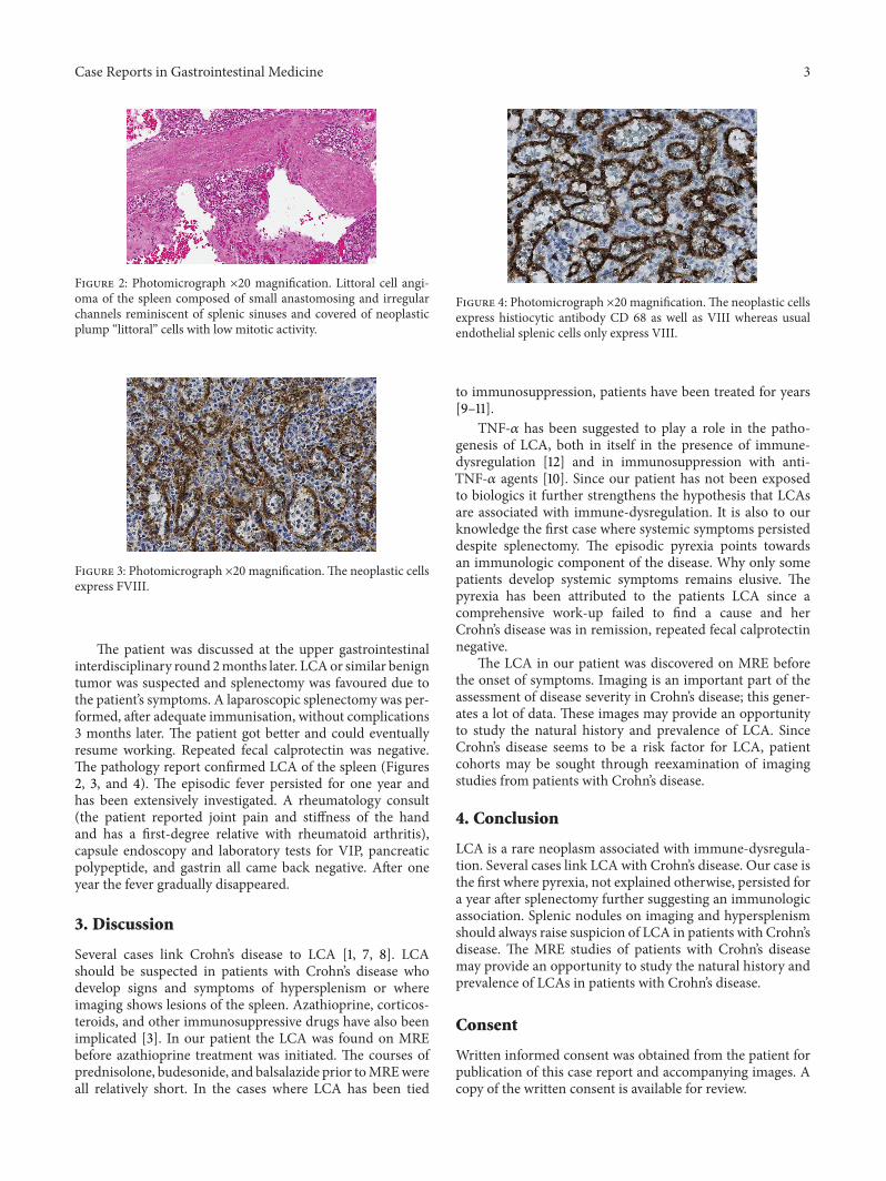
Figure 3: Photomicrograph ×20 magnification.The neoplastic cellsexpress FVIII.
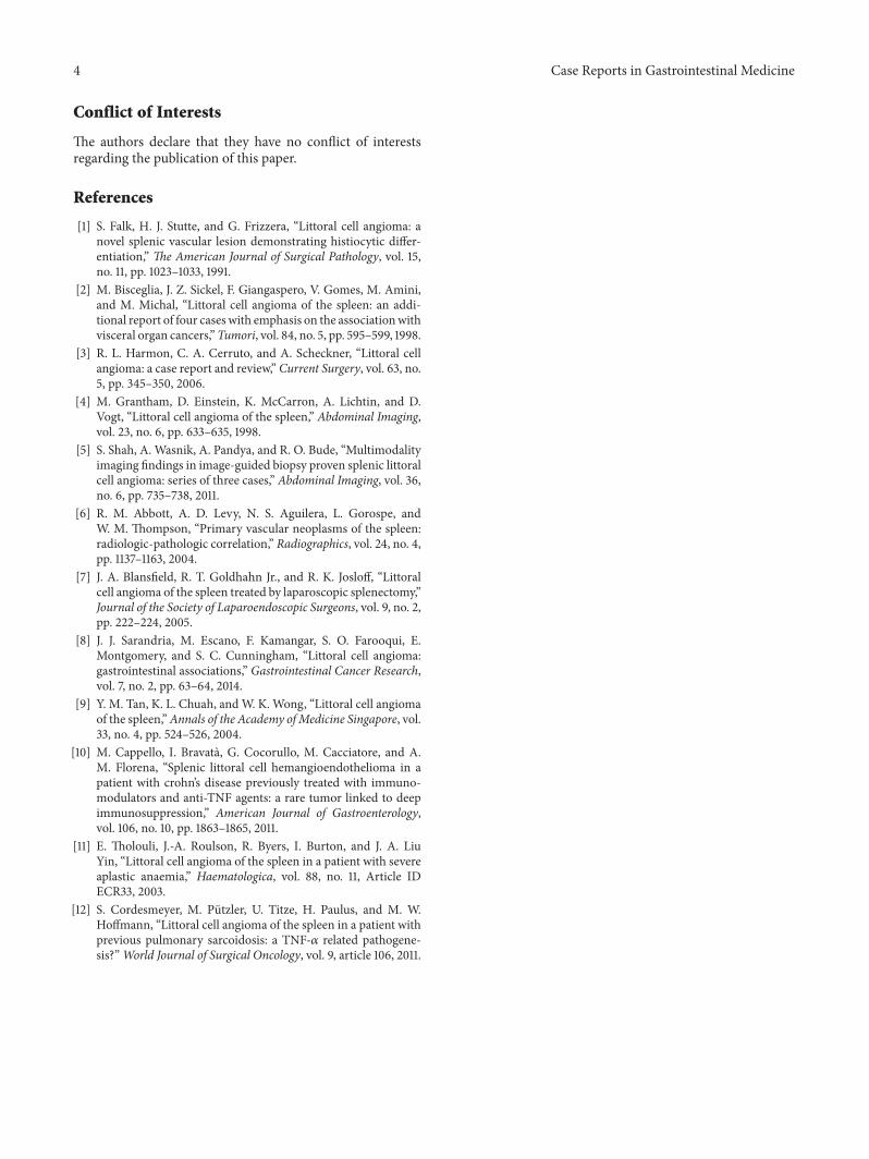
The patient was discussed at the upper gastrointestinalinterdisciplinary round 2months later. LCAor similar benigntumor was suspected and splenectomy was favoured due tothe patient’s symptoms. A laparoscopic splenectomy was per-formed, after adequate immunisation, without complications3 months later. The patient got better and could eventuallyresume working. Repeated fecal calprotectin was negative.The pathology report confirmed LCA of the spleen (Figures2, 3, and 4). The episodic fever persisted for one year andhas been extensively investigated. A rheumatology consult(the patient reported joint pain and stiffness of the handand has a first-degree relative with rheumatoid arthritis),capsule endoscopy and laboratory tests for VIP, pancreaticpolypeptide, and gastrin all came back negative. After oneyear the fever gradually disappeared.
3. Discussion
Several cases link Crohn’s disease to LCA [1, 7, 8]. LCAshould be suspected in patients with Crohn’s disease whodevelop signs and symptoms of hypersplenism or whereimaging shows lesions of the spleen. Azathioprine, corticos-teroids, and other immunosuppressive drugs have also beenimplicated [3]. In our patient the LCA was found on MREbefore azathioprine treatment was initiated. The courses ofprednisolone, budesonide, and balsalazide prior toMREwereall relatively short. In the cases where LCA has been tied
Figure 4: Photomicrograph ×20 magnification.The neoplastic cellsexpress histiocytic antibody CD 68 as well as VIII whereas usualendothelial splenic cells only express VIII.
to immunosuppression, patients have been treated for years[9–11].
TNF-𝛼 has been suggested to play a role in the patho-genesis of LCA, both in itself in the presence of immune-dysregulation [12] and in immunosuppression with anti-TNF-𝛼 agents [10]. Since our patient has not been exposedto biologics it further strengthens the hypothesis that LCAsare associated with immune-dysregulation. It is also to ourknowledge the first case where systemic symptoms persisteddespite splenectomy. The episodic pyrexia points towardsan immunologic component of the disease. Why only somepatients develop systemic symptoms remains elusive. Thepyrexia has been attributed to the patients LCA since acomprehensive work-up failed to find a cause and herCrohn’s disease was in remission, repeated fecal calprotectinnegative.
The LCA in our patient was discovered on MRE beforethe onset of symptoms. Imaging is an important part of theassessment of disease severity in Crohn’s disease; this gener-ates a lot of data. These images may provide an opportunityto study the natural history and prevalence of LCA. SinceCrohn’s disease seems to be a risk factor for LCA, patientcohorts may be sought through reexamination of imagingstudies from patients with Crohn’s disease.
4. Conclusion
LCA is a rare neoplasm associated with immune-dysregula-tion. Several cases link LCA with Crohn’s disease. Our case isthe first where pyrexia, not explained otherwise, persisted fora year after splenectomy further suggesting an immunologicassociation. Splenic nodules on imaging and hypersplenismshould always raise suspicion of LCA in patients with Crohn’sdisease. The MRE studies of patients with Crohn’s diseasemay provide an opportunity to study the natural history andprevalence of LCAs in patients with Crohn’s disease.
Consent
Written informed consent was obtained from the patient forpublication of this case report and accompanying images. Acopy of the written consent is available for review.
4 Case Reports in Gastrointestinal Medicine
Conflict of Interests
The authors declare that they have no conflict of interestsregarding the publication of this paper.
References
[1] S. Falk, H. J. Stutte, and G. Frizzera, “Littoral cell angioma: anovel splenic vascular lesion demonstrating histiocytic differ-entiation,” The American Journal of Surgical Pathology, vol. 15,no. 11, pp. 1023–1033, 1991.
[2] M. Bisceglia, J. Z. Sickel, F. Giangaspero, V. Gomes, M. Amini,and M. Michal, “Littoral cell angioma of the spleen: an addi-tional report of four cases with emphasis on the associationwithvisceral organ cancers,”Tumori, vol. 84, no. 5, pp. 595–599, 1998.
[3] R. L. Harmon, C. A. Cerruto, and A. Scheckner, “Littoral cellangioma: a case report and review,” Current Surgery, vol. 63, no.5, pp. 345–350, 2006.
[4] M. Grantham, D. Einstein, K. McCarron, A. Lichtin, and D.Vogt, “Littoral cell angioma of the spleen,” Abdominal Imaging,vol. 23, no. 6, pp. 633–635, 1998.
[5] S. Shah, A. Wasnik, A. Pandya, and R. O. Bude, “Multimodalityimaging findings in image-guided biopsy proven splenic littoralcell angioma: series of three cases,” Abdominal Imaging, vol. 36,no. 6, pp. 735–738, 2011.
[6] R. M. Abbott, A. D. Levy, N. S. Aguilera, L. Gorospe, andW. M. Thompson, “Primary vascular neoplasms of the spleen:radiologic-pathologic correlation,” Radiographics, vol. 24, no. 4,pp. 1137–1163, 2004.
[7] J. A. Blansfield, R. T. Goldhahn Jr., and R. K. Josloff, “Littoralcell angioma of the spleen treated by laparoscopic splenectomy,”Journal of the Society of Laparoendoscopic Surgeons, vol. 9, no. 2,pp. 222–224, 2005.
[8] J. J. Sarandria, M. Escano, F. Kamangar, S. O. Farooqui, E.Montgomery, and S. C. Cunningham, “Littoral cell angioma:gastrointestinal associations,” Gastrointestinal Cancer Research,vol. 7, no. 2, pp. 63–64, 2014.
[9] Y. M. Tan, K. L. Chuah, andW. K. Wong, “Littoral cell angiomaof the spleen,”Annals of the Academy ofMedicine Singapore, vol.33, no. 4, pp. 524–526, 2004.
[10] M. Cappello, I. Bravata, G. Cocorullo, M. Cacciatore, and A.M. Florena, “Splenic littoral cell hemangioendothelioma in apatient with crohn’s disease previously treated with immuno-modulators and anti-TNF agents: a rare tumor linked to deepimmunosuppression,” American Journal of Gastroenterology,vol. 106, no. 10, pp. 1863–1865, 2011.
[11] E. Tholouli, J.-A. Roulson, R. Byers, I. Burton, and J. A. LiuYin, “Littoral cell angioma of the spleen in a patient with severeaplastic anaemia,” Haematologica, vol. 88, no. 11, Article IDECR33, 2003.
[12] S. Cordesmeyer, M. Putzler, U. Titze, H. Paulus, and M. W.Hoffmann, “Littoral cell angioma of the spleen in a patient withprevious pulmonary sarcoidosis: a TNF-𝛼 related pathogene-sis?”World Journal of Surgical Oncology, vol. 9, article 106, 2011.

Submit your manuscripts athttp://www.hindawi.com
Stem CellsInternational
Hindawi Publishing Corporationhttp://www.hindawi.com Volume 2014
Hindawi Publishing Corporationhttp://www.hindawi.com Volume 2014
MEDIATORSINFLAMMATION
of
Hindawi Publishing Corporationhttp://www.hindawi.com Volume 2014
Behavioural Neurology
EndocrinologyInternational Journal of
Hindawi Publishing Corporationhttp://www.hindawi.com Volume 2014
Hindawi Publishing Corporationhttp://www.hindawi.com Volume 2014
Disease Markers
Hindawi Publishing Corporationhttp://www.hindawi.com Volume 2014
BioMed Research International
OncologyJournal of
Hindawi Publishing Corporationhttp://www.hindawi.com Volume 2014
Hindawi Publishing Corporationhttp://www.hindawi.com Volume 2014
Oxidative Medicine and Cellular Longevity
Hindawi Publishing Corporationhttp://www.hindawi.com Volume 2014
PPAR Research
The Scientific World JournalHindawi Publishing Corporation http://www.hindawi.com Volume 2014
Immunology ResearchHindawi Publishing Corporationhttp://www.hindawi.com Volume 2014
Journal of
ObesityJournal of
Hindawi Publishing Corporationhttp://www.hindawi.com Volume 2014
Hindawi Publishing Corporationhttp://www.hindawi.com Volume 2014
Computational and Mathematical Methods in Medicine
OphthalmologyJournal of
Hindawi Publishing Corporationhttp://www.hindawi.com Volume 2014
Diabetes ResearchJournal of
Hindawi Publishing Corporationhttp://www.hindawi.com Volume 2014
Hindawi Publishing Corporationhttp://www.hindawi.com Volume 2014
Research and TreatmentAIDS
Hindawi Publishing Corporationhttp://www.hindawi.com Volume 2014
Gastroenterology Research and Practice
Hindawi Publishing Corporationhttp://www.hindawi.com Volume 2014
Parkinson’s Disease
Evidence-Based Complementary and Alternative Medicine
Volume 2014Hindawi Publishing Corporationhttp://www.hindawi.com


![Cerebellar Venous Angioma - AJNRAny type of angioma can bleed. In the cerebellum, the angioma that bleeds most often is the AVM. Venous an giomas, however, are second most common [8]](https://img.pdfslide.net/doc/110x75/5e545329291e4f4c762be047/cerebellar-venous-angioma-any-type-of-angioma-can-bleed-in-the-cerebellum-the.jpg)