Embed Size (px)
Citation preview
Case Report of Posttraumatic Middle CerebralArtery Territory InfarctAjaya Kumar Ayyappan Unnithan1
1Mar Gregorios Memorial Muthoot Medical Centre, Kozhencherry,Kerala, India
Indian J Neurosurg 2017;6:50–54.
Address for correspondence Ajaya Kumar Ayyappan Unnithan, MBBS,MS, DNB, MCh, Muthoot Healthcare, Kozhencherry, Pathanamthitta,Kerala 689641, India (e-mail: [email protected]).
Introduction
Posttraumatic cerebral infarction indicates poor clinicaloutcome in head injury. Middle cerebral artery (MCA)territory infarction is rare and is caused by stretching andattenuation of MCA and increased intracranial pressure (ICP).This is a case report of a young patient who had posttraumaticMCA territory infarction.
Case Report
A 27-year-old man was brought to emergency servicesfollowing road traffic accident. The patient was riding pillionon a bike, and the driver died in casualty due to head injuryand pelvic fracture. He was in unconscious state with theGlasgow Coma Scale (GCS) of E1M3V1. The pupils weresluggishly reacting to light bilaterally. He was intubated and
ventilated. Computed tomography (CT) of the head showeddiffuse injury-subarachnoid hemorrhage, multiple smallcontusions, right MCA hyperdensity, right temporofrontalhypodensity, ventricular effacement, and small midbraincontusion.
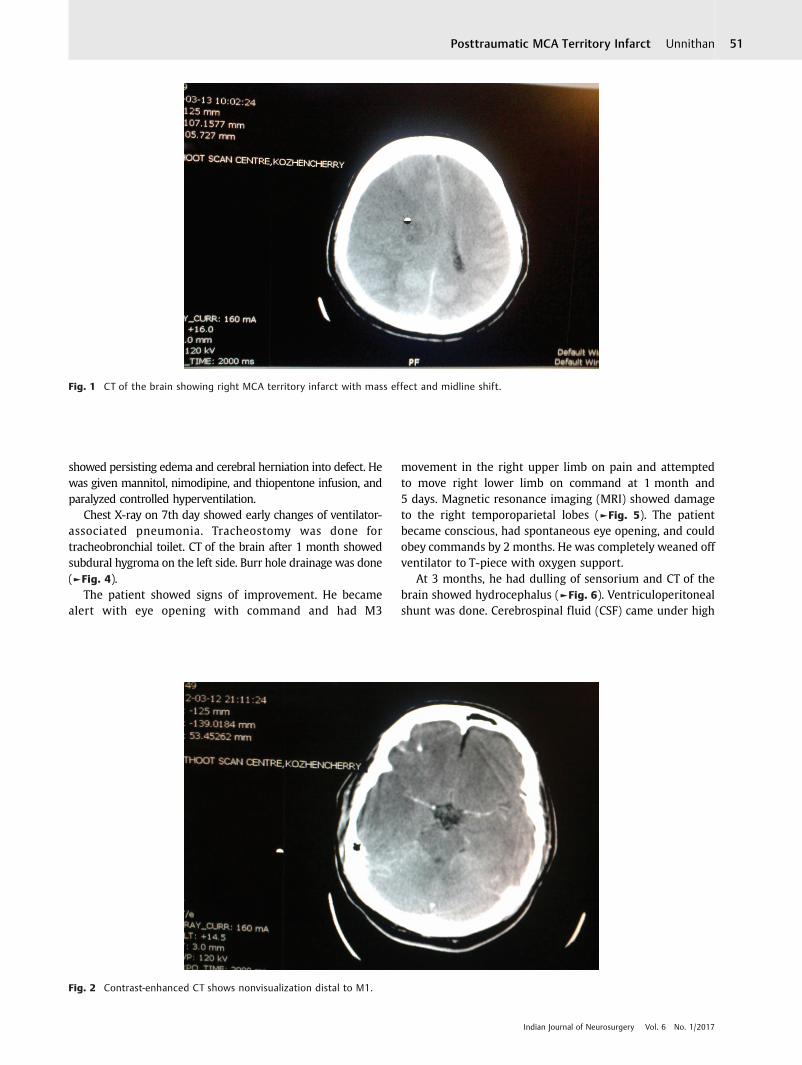
Antiedema and cerebral protectant therapy andhyperventilation were started and, nimodipine andciticoline were given. The next day he worsened with GCSE1M1 and dilated pupil on the right side, and repeat CT ofthe brain showed right (MCA) territory infarct with masseffect and midline shift (►Fig. 1). Contrast-enhanced CTshowed nonvisualization distal to M1 (►Fig. 2).
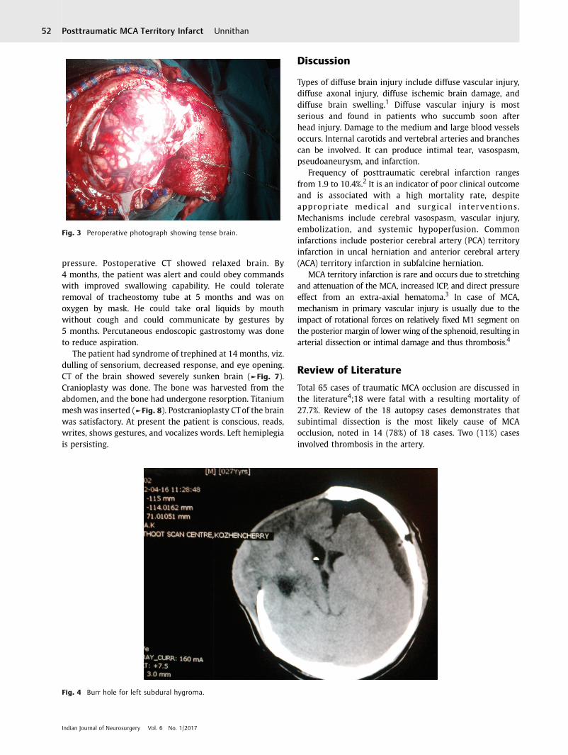
Emergency right frontotemporoparietal decompressivecraniectomy and infarct decompression were done. Duraplastywas done with G patch. The brain was very tense (►Fig. 3). Thepatient had GCS of E2M2 postoperatively and CT of the brain
Keywords
► diffuse brain injury► posttraumatic
cerebral infarction► middle cerebral
artery territoryinfarction
► decompressivecraniectomy
► subdural hygroma► burr hole► hydrocephalus► VP shunt► syndrome of
trephined► cranioplasty
Abstract A 27-year-old man had severe diffuse brain injury. The patient developed malignantright MCA territory infarction on second day. Emergency decompressive craniectomywas done. He was ventilated. He developed subdural hygroma on the opposite sidethat was drained. He improved slowly. He had hydrocephalus. VP shunt was done. Hebecame conscious but dependent. PEG was done for feeding. Cranioplasty was donefor syndrome of trephined. The patent improved to a state of good cognition withresidual motor aphasia and left hemiplegia.Posttraumatic cerebral infarction is an indicator of poor prognosis. Vasospasm, intimaldissection, and thrombosis are the mechanisms. MCA territorial infarction is rare.Usual mechanism is impact of rotational forces on relatively fixed M1 segment on theposterior margin of lower wing of sphenoid, resulting in arterial dissection or intimaldamage.
receivedJuly 28, 2015acceptedApril 4, 2016published onlineSeptember 1, 2016
DOI http://dx.doi.org/10.1055/s-0036-1584594.ISSN 2277-954X.
© 2017 Thieme Medical and ScientificPublishers Private Ltd.
Case ReportTHIEME
50
showed persisting edema and cerebral herniation into defect. Hewas given mannitol, nimodipine, and thiopentone infusion, andparalyzed controlled hyperventilation.
Chest X-ray on 7th day showed early changes of ventilator-associated pneumonia. Tracheostomy was done fortracheobronchial toilet. CT of the brain after 1 month showedsubdural hygroma on the left side. Burr hole drainage was done(►Fig. 4).
The patient showed signs of improvement. He becamealert with eye opening with command and had M3
movement in the right upper limb on pain and attemptedto move right lower limb on command at 1 month and5 days. Magnetic resonance imaging (MRI) showed damageto the right temporoparietal lobes (►Fig. 5). The patientbecame conscious, had spontaneous eye opening, and couldobey commands by 2 months. He was completely weaned offventilator to T-piece with oxygen support.
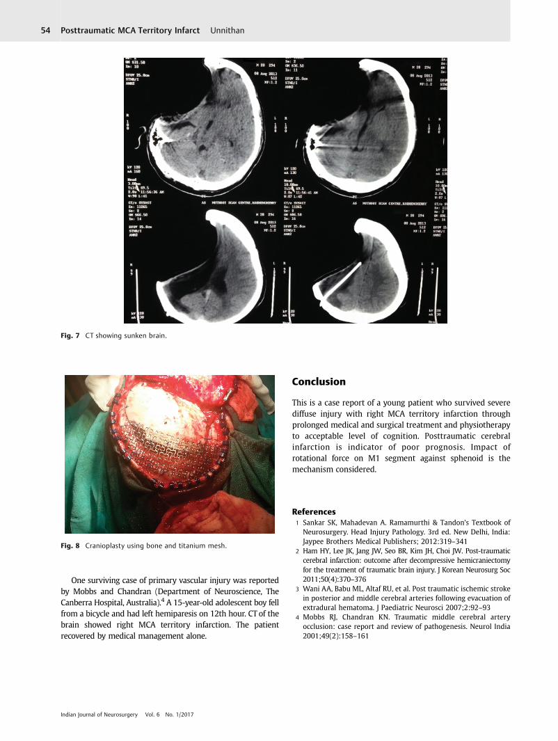
At 3 months, he had dulling of sensorium and CT of thebrain showed hydrocephalus (►Fig. 6). Ventriculoperitonealshunt was done. Cerebrospinal fluid (CSF) came under high
Fig. 1 CT of the brain showing right MCA territory infarct with mass effect and midline shift.
Fig. 2 Contrast-enhanced CT shows nonvisualization distal to M1.
Indian Journal of Neurosurgery Vol. 6 No. 1/2017
Posttraumatic MCA Territory Infarct Unnithan 51
pressure. Postoperative CT showed relaxed brain. By4 months, the patient was alert and could obey commandswith improved swallowing capability. He could tolerateremoval of tracheostomy tube at 5 months and was onoxygen by mask. He could take oral liquids by mouthwithout cough and could communicate by gestures by5 months. Percutaneous endoscopic gastrostomy was doneto reduce aspiration.
The patient had syndrome of trephined at 14 months, viz.dulling of sensorium, decreased response, and eye opening.CT of the brain showed severely sunken brain (►Fig. 7).Cranioplasty was done. The bone was harvested from theabdomen, and the bone had undergone resorption. Titaniummesh was inserted (►Fig. 8). Postcranioplasty CT of the brainwas satisfactory. At present the patient is conscious, reads,writes, shows gestures, and vocalizes words. Left hemiplegiais persisting.
Discussion
Types of diffuse brain injury include diffuse vascular injury,diffuse axonal injury, diffuse ischemic brain damage, anddiffuse brain swelling.1 Diffuse vascular injury is mostserious and found in patients who succumb soon afterhead injury. Damage to the medium and large blood vesselsoccurs. Internal carotids and vertebral arteries and branchescan be involved. It can produce intimal tear, vasospasm,pseudoaneurysm, and infarction.
Frequency of posttraumatic cerebral infarction rangesfrom 1.9 to 10.4%.2 It is an indicator of poor clinical outcomeand is associated with a high mortality rate, despiteappropriate medical and surgical interventions.Mechanisms include cerebral vasospasm, vascular injury,embolization, and systemic hypoperfusion. Commoninfarctions include posterior cerebral artery (PCA) territoryinfarction in uncal herniation and anterior cerebral artery(ACA) territory infarction in subfalcine herniation.
MCA territory infarction is rare and occurs due to stretchingand attenuation of the MCA, increased ICP, and direct pressureeffect from an extra-axial hematoma.3 In case of MCA,mechanism in primary vascular injury is usually due to theimpact of rotational forces on relatively fixed M1 segment onthe posterior margin of lower wing of the sphenoid, resulting inarterial dissection or intimal damage and thus thrombosis.4
Review of Literature
Total 65 cases of traumatic MCA occlusion are discussed inthe literature4;18 were fatal with a resulting mortality of27.7%. Review of the 18 autopsy cases demonstrates thatsubintimal dissection is the most likely cause of MCAocclusion, noted in 14 (78%) of 18 cases. Two (11%) casesinvolved thrombosis in the artery.
Fig. 3 Peroperative photograph showing tense brain.
Fig. 4 Burr hole for left subdural hygroma.
Indian Journal of Neurosurgery Vol. 6 No. 1/2017
Posttraumatic MCA Territory Infarct Unnithan52
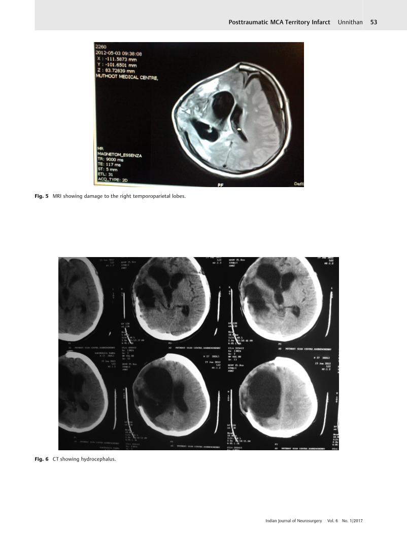
Fig. 5 MRI showing damage to the right temporoparietal lobes.
Fig. 6 CT showing hydrocephalus.
Indian Journal of Neurosurgery Vol. 6 No. 1/2017
Posttraumatic MCA Territory Infarct Unnithan 53
One surviving case of primary vascular injury was reportedby Mobbs and Chandran (Department of Neuroscience, TheCanberra Hospital, Australia).4 A 15-year-old adolescent boy fellfrom a bicycle and had left hemiparesis on 12th hour. CT of thebrain showed right MCA territory infarction. The patientrecovered by medical management alone.
Conclusion
This is a case report of a young patient who survived severediffuse injury with right MCA territory infarction throughprolonged medical and surgical treatment and physiotherapyto acceptable level of cognition. Posttraumatic cerebralinfarction is indicator of poor prognosis. Impact ofrotational force on M1 segment against sphenoid is themechanism considered.
References1 Sankar SK, Mahadevan A. Ramamurthi & Tandon’s Textbook of
Neurosurgery. Head Injury Pathology. 3rd ed. New Delhi, India:Jaypee Brothers Medical Publishers; 2012:319–341
2 Ham HY, Lee JK, Jang JW, Seo BR, Kim JH, Choi JW. Post-traumaticcerebral infarction: outcome after decompressive hemicraniectomyfor the treatment of traumatic brain injury. J Korean Neurosurg Soc2011;50(4):370–376
3 Wani AA, Babu ML, Altaf RU, et al. Post traumatic ischemic strokein posterior and middle cerebral arteries following evacuation ofextradural hematoma. J Paediatric Neurosci 2007;2:92–93
4 Mobbs RJ, Chandran KN. Traumatic middle cerebral arteryocclusion: case report and review of pathogenesis. Neurol India2001;49(2):158–161
Fig. 7 CT showing sunken brain.
Fig. 8 Cranioplasty using bone and titanium mesh.
Indian Journal of Neurosurgery Vol. 6 No. 1/2017
Posttraumatic MCA Territory Infarct Unnithan54