Embed Size (px)
Citation preview
Kang et al. BMC Research Notes 2013, 6:478http://www.biomedcentral.com/1756-0500/6/478
CASE REPORT Open Access
Cardiogenic shock accompanied by dynamic leftventricular outflow tract obstruction andmyocardial bridging after transient completeatrioventricular block mimicking ST-elevationmyocardial infarction: a case reportSeonghui Kang1, Sanghee An1, Hyung Min Yu1, Jiwan Kim1, Sung Hea Kim1,2*, Hyun-Joong Kim1,2
and Sang Man Chung1,2
Abstract
Background: Dynamic left ventricular outflow tract obstruction with or without mitral regurgitation is typicallyobserved in hypertrophic cardiomyopathy, but is also occasionally seen without left ventricular hypertrophy. In thisreport, we present a case of cardiogenic shock that mimics ST-elevation myocardial infarction, due to dynamic leftventricular outflow tract obstruction with transient mitral regurgitation and myocardial bridging after transientcomplete atrioventricular block.
Case presentation: A 65-year-old man with hypertension presented himself at the emergency department withsyncope after chest pain. His initial electrocardiography showed inferior ST elevation with profound precordial STdepression and transient complete atrioventricular block. Due to sustained hypotension, an intra-aortic balloonpump was applied. His coronary angiography revealed almost normal right coronary artery and left circumflex arteryand only a severe myocardial bridge in the mid-segment of his left anterior descending artery. Instead, severe mitralregurgitation was found without regional wall motion abnormality both in the left ventriculography and theportable echocardiography. However the severe mitral regurgitation completely disappeared in follow upechocardiography the day after. The pressure gradient across the left ventricular outflow tract was measured at8.95 mmHg during the resting state, and was increased to 38.95 mmHg during the Valsalva state.
Conclusions: The patient presented with a case of cardiogenic shock that mimicked ST-elevation myocardialinfarction due to dynamic left ventricular outflow tract obstruction combined with myocardial bridging in themid-left anterior descending artery.
Keywords: STEMI, Dynamic LVOT obstruction, Transient MR, Myocardial bridge, Complete AV block
* Correspondence: [email protected] of Internal Medicine, Konkuk University School of Medicine,Seoul, Korea2Department of cardiology, Konkuk University School of Medicine, Seoul,Korea
© 2013 Kang et al.; licensee BioMed Central Ltd. This is an open access article distributed under the terms of the CreativeCommons Attribution License (http://creativecommons.org/licenses/by/2.0), which permits unrestricted use, distribution, andreproduction in any medium, provided the original work is properly cited.
Kang et al. BMC Research Notes 2013, 6:478 Page 2 of 6http://www.biomedcentral.com/1756-0500/6/478
BackgroundDynamic left ventricular outflow tract (LVOT) obstructionwith or without mitral regurgitation is typically observedin hypertrophic cardiomyopathy, but is also seen inother diseases, including acute coronary syndrome [1],stress-induced cardiomyopathy [2] and left ventricularhypertrophy (LVH) [3]. Recent studies reported that it mayoccur even in the absence of LVH and cause symptomssuch as chest pain, syncope or both [4,5].Myocardial bridging is defined as a segment of a major
epicardial coronary artery, the ‘tunneled artery,’ that goesintramullary through the myocardium beneath the musclebridge [6]. The majority of such abnormalities were foundin the left anterior descending coronary artery, whereasthose that were found in the left circumflex (LCx) or theright coronary artery (RCA) were rare [7]. The estimatedfrequency of their discovery varies from 0.5% to 16% whenassessed via coronary angiography [8,9]; however in someautopsy series, it is as high as 85.7% [10]. Most clinicalfindings have shown that the myocardial bridge does notlead to major problems; however some cases have beenreported of unstable perfusion of the coronary arteriesthat caused serious cardiac disorders such as myocardialischemia, myocardial infarction, arrhythmia and evensudden cardiac death. In fact, several clinical studies haveshown that myocardial bridging occasionally causes acutemyocardial infarction [11-14]. In this report, a case ispresented of cardiogenic shock that mimicked ST-elevationmyocardial infarction (STEMI) due to dynamic LVOTobstruction combined with myocardial bridging in themid-left anterior descending artery (LAD) after transientcomplete atrioventricular block (AV block).
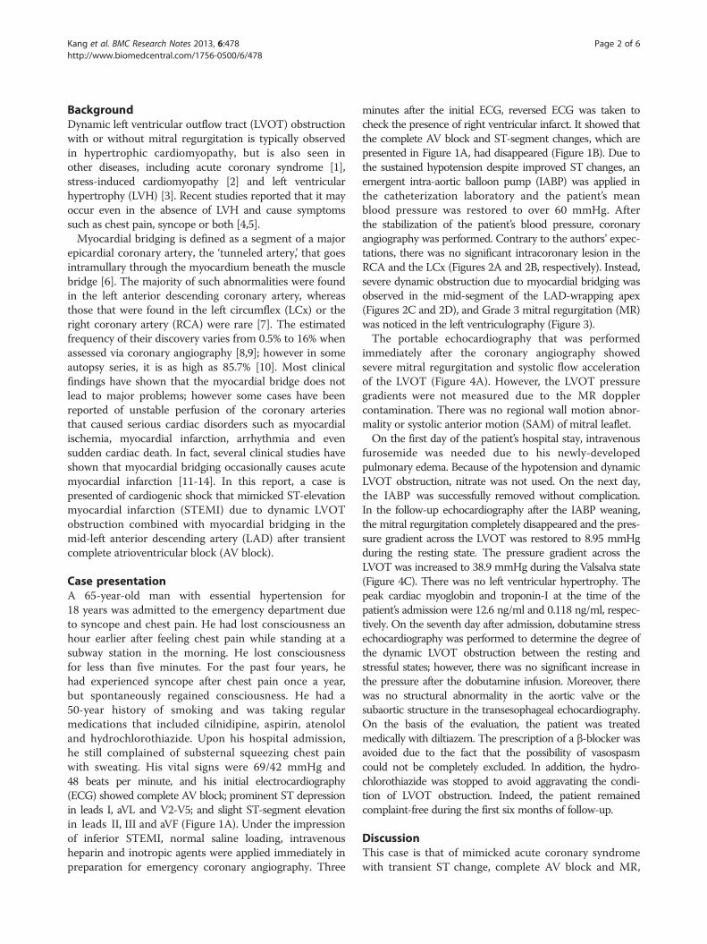
Case presentationA 65-year-old man with essential hypertension for18 years was admitted to the emergency department dueto syncope and chest pain. He had lost consciousness anhour earlier after feeling chest pain while standing at asubway station in the morning. He lost consciousnessfor less than five minutes. For the past four years, hehad experienced syncope after chest pain once a year,but spontaneously regained consciousness. He had a50-year history of smoking and was taking regularmedications that included cilnidipine, aspirin, atenololand hydrochlorothiazide. Upon his hospital admission,he still complained of substernal squeezing chest painwith sweating. His vital signs were 69/42 mmHg and48 beats per minute, and his initial electrocardiography(ECG) showed complete AV block; prominent ST depressionin leads I, aVL and V2-V5; and slight ST-segment elevationin leads II, III and aVF (Figure 1A). Under the impressionof inferior STEMI, normal saline loading, intravenousheparin and inotropic agents were applied immediately inpreparation for emergency coronary angiography. Three
minutes after the initial ECG, reversed ECG was taken tocheck the presence of right ventricular infarct. It showed thatthe complete AV block and ST-segment changes, which arepresented in Figure 1A, had disappeared (Figure 1B). Due tothe sustained hypotension despite improved ST changes, anemergent intra-aortic balloon pump (IABP) was applied inthe catheterization laboratory and the patient’s meanblood pressure was restored to over 60 mmHg. Afterthe stabilization of the patient’s blood pressure, coronaryangiography was performed. Contrary to the authors’ expec-tations, there was no significant intracoronary lesion in theRCA and the LCx (Figures 2A and 2B, respectively). Instead,severe dynamic obstruction due to myocardial bridging wasobserved in the mid-segment of the LAD-wrapping apex(Figures 2C and 2D), and Grade 3 mitral regurgitation (MR)was noticed in the left ventriculography (Figure 3).The portable echocardiography that was performed
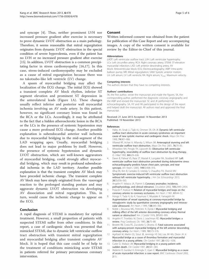
immediately after the coronary angiography showedsevere mitral regurgitation and systolic flow accelerationof the LVOT (Figure 4A). However, the LVOT pressuregradients were not measured due to the MR dopplercontamination. There was no regional wall motion abnor-mality or systolic anterior motion (SAM) of mitral leaflet.On the first day of the patient’s hospital stay, intravenous
furosemide was needed due to his newly-developedpulmonary edema. Because of the hypotension and dynamicLVOT obstruction, nitrate was not used. On the next day,the IABP was successfully removed without complication.In the follow-up echocardiography after the IABP weaning,the mitral regurgitation completely disappeared and the pres-sure gradient across the LVOT was restored to 8.95 mmHgduring the resting state. The pressure gradient across theLVOT was increased to 38.9 mmHg during the Valsalva state(Figure 4C). There was no left ventricular hypertrophy. Thepeak cardiac myoglobin and troponin-I at the time of thepatient’s admission were 12.6 ng/ml and 0.118 ng/ml, respec-tively. On the seventh day after admission, dobutamine stressechocardiography was performed to determine the degree ofthe dynamic LVOT obstruction between the resting andstressful states; however, there was no significant increase inthe pressure after the dobutamine infusion. Moreover, therewas no structural abnormality in the aortic valve or thesubaortic structure in the transesophageal echocardiography.On the basis of the evaluation, the patient was treatedmedically with diltiazem. The prescription of a β-blocker wasavoided due to the fact that the possibility of vasospasmcould not be completely excluded. In addition, the hydro-chlorothiazide was stopped to avoid aggravating the condi-tion of LVOT obstruction. Indeed, the patient remainedcomplaint-free during the first six months of follow-up.
DiscussionThis case is that of mimicked acute coronary syndromewith transient ST change, complete AV block and MR,
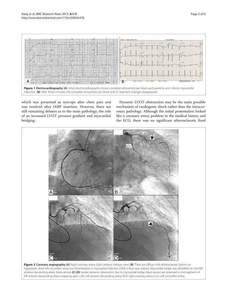
Figure 1 Electrocardiography (A) Initial electrocardiography shows complete atrioventricular block and posterior and inferior myocardialinfarction. (B) After three minutes, the complete atrioventricular block and ST-segment changes disappeared.
Kang et al. BMC Research Notes 2013, 6:478 Page 3 of 6http://www.biomedcentral.com/1756-0500/6/478
which was presented as syncope after chest pain andwas resolved after IABP insertion. However, there arestill remaining debates as to the main pathology, the roleof an increased LVOT pressure gradient and myocardialbridging.
Figure 2 Coronary angiography (A) Right coronary artery (right anterior obhypoplastic distal left circumflex artery but thrombolysis in myocardial infarctianterior descending artery (black errow) (C) (D) Severe dynamic obstruction dleft anterior descending artery-wrapping apex. LAD, left anterior descending a
Dynamic LVOT obstruction may be the main possiblemechanism of cardiogenic shock rather than the intracor-onary pathology. Although the initial presentation lookedlike a coronary artery problem in the medical history andthe ECG, there was no significant atherosclerotic fixed
lique view) (B) There are diffuse mild atherosclerotic lesions onon (TIMI) 3 flow was noticed. Myocardial bridge was identified on mid-leftue to myocardial bridge (black arrow) are observed in mid segment ofrtery; RCA, right coronary artery; LCx, Left circumflex artery.
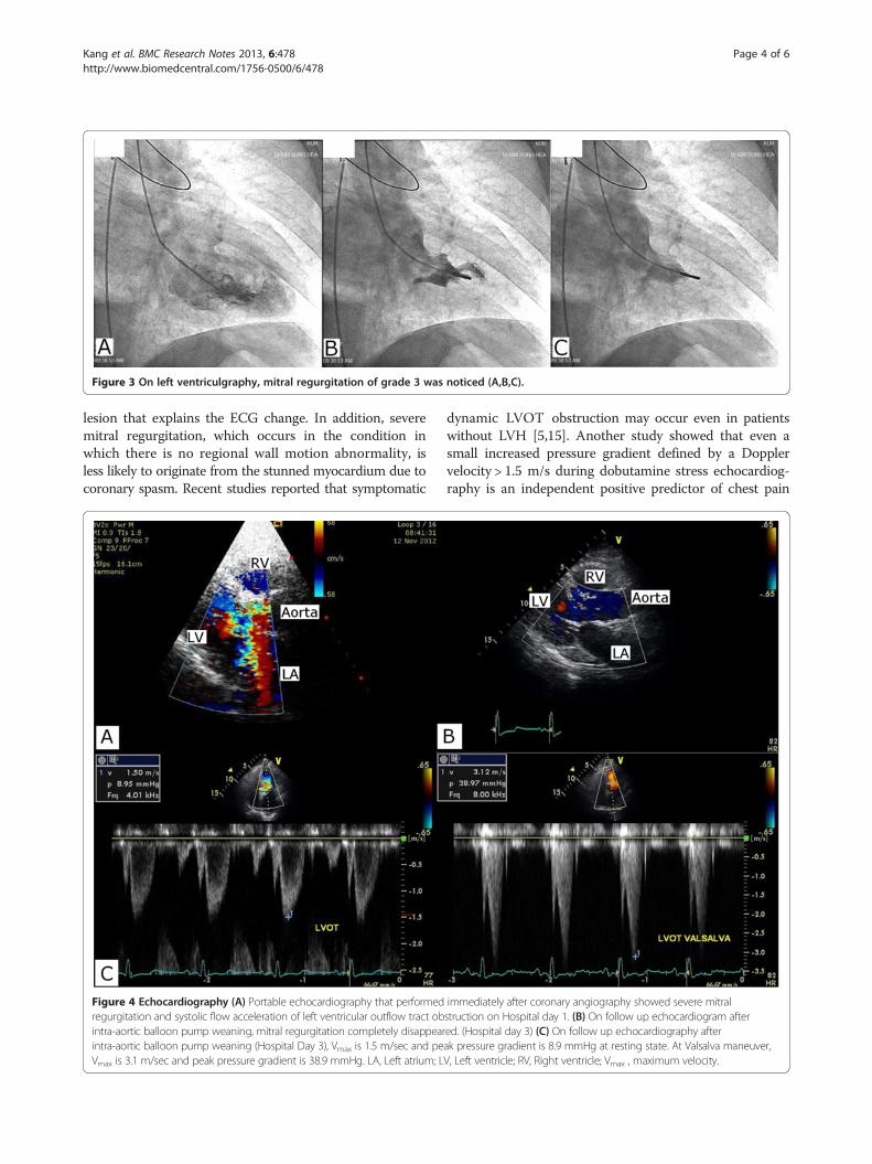
Figure 3 On left ventriculgraphy, mitral regurgitation of grade 3 was noticed (A,B,C).
Kang et al. BMC Research Notes 2013, 6:478 Page 4 of 6http://www.biomedcentral.com/1756-0500/6/478
lesion that explains the ECG change. In addition, severemitral regurgitation, which occurs in the condition inwhich there is no regional wall motion abnormality, isless likely to originate from the stunned myocardium due tocoronary spasm. Recent studies reported that symptomatic
Figure 4 Echocardiography (A) Portable echocardiography that performedregurgitation and systolic flow acceleration of left ventricular outflow tract obintra-aortic balloon pump weaning, mitral regurgitation completely disappearintra-aortic balloon pump weaning (Hospital Day 3), Vmax is 1.5 m/sec and peVmax is 3.1 m/sec and peak pressure gradient is 38.9 mmHg. LA, Left atrium; L
dynamic LVOT obstruction may occur even in patientswithout LVH [5,15]. Another study showed that even asmall increased pressure gradient defined by a Dopplervelocity > 1.5 m/s during dobutamine stress echocardiog-raphy is an independent positive predictor of chest pain
immediately after coronary angiography showed severe mitralstruction on Hospital day 1. (B) On follow up echocardiogram aftered. (Hospital day 3) (C) On follow up echocardiography afterak pressure gradient is 8.9 mmHg at resting state. At Valsalva maneuver,V, Left ventricle; RV, Right ventricle; Vmax , maximum velocity.
Kang et al. BMC Research Notes 2013, 6:478 Page 5 of 6http://www.biomedcentral.com/1756-0500/6/478
and syncope [4]. Thus, neither prominent LVH norincreased pressure gradient after exercise is necessaryto prove dynamic LVOT obstruction as a main pathology.Therefore, it seems reasonable that mitral regurgitationoriginates from dynamic LVOT obstruction in the specialcondition of severe hypovolemia, even if the patient hasno LVH or no increased pressure gradient after exercise[15]. In addition, LVOT obstruction is a common precipi-tating factor in stress cardiomyopathy [16,17]. In thiscase, stress-induced cardiomyopathy may be excludedas a cause of mitral regurgitation because there wasno takotsubo-like left ventricle (LV) change.A spasm of myocardial bridging may affect the
localization of the ECG change. The initial ECG showeda transient complete AV block rhythm, inferior STsegment elevation and prominent ST depression inthe anterolateral leads (Figure 1A). These changesusually reflect inferior and posterior wall myocardialischemia involving an AV node artery. In this patient,however, no significant coronary lesion was found inthe RCA or the LCx. Accordingly, it may be attributedto the fact that a hidden atherosclerotic lesion in the RCAor the LCx in the presence of systemic hypotension maycause a more profound ECG change. Another possibleexplanation is subendocardial anterior wall ischemiadue to myocardial bridging in the mid-segment of theLAD wrapping apex. Usually, myocardial bridgingdoes not lead to major problems by itself. However,the presence of systemic hypotension and dynamicLVOT obstruction, which are well-known aggravatorsof myocardial bridging, could strongly affect myocar-dial bridging, which may result in profound subendocar-dial ischemia in the LAD territory. Another possibleexplanation is that the transient complete AV block mayhave preceded ischemic change. The transient completeAV block may have simply originated from the vasovagalreaction to the prolonged standing posture and mayaggravate dynamic LVOT obstruction via developingAV dissociation and mitral regurgitation. This, inturn, would cause the ischemic change to appear onthe ECG.
ConclusionA rapid diagnosis of STEMI is mandatory for optimaltreatment. However, a small proportion of patients withsuspected STEMI suffer from other conditions. In thisreport, a case of cardiogenic shock was presented thatmimicked STEMI, due to dynamic left ventricular outflowtract obstruction with transient mitral regurgitationand myocardial bridging after transient complete AVblock. It is hoped that this case could be of help tothe treatment of conditions mimicking acute STEMIin patients referred for primary percutaneous coronaryintervention.
ConsentWritten informed consent was obtained from the patientfor publication of this Case Report and any accompanyingimages. A copy of the written consent is available forreview by the Editor-in-Chief of this journal.
AbbreviationsLVOT: Left ventricular outflow tract; LVH: Left ventricular hypertrophy;LCx: Left circumflex artery; RCA: Right coronary artery; STEMI: ST-elevationmyocardial infarction; LAD: Left anterior descending artery; AVblock: Atrioventricular block; ECG: Electrocardiography; IABP: Intra-aorticballoon pump; MR: Mitral regurgitation; SAM: Systolic anterior motion;LA: Left atrium; LV: Left ventricle; RA: Right atrium; Vmax: Maximum velocity.
Competing interestsThe authors declare that they have no competing interests.
Authors’ contributionsSK, the first author, wrote the manuscript and made the figures. SK, thecorresponding author, performed the diagnostic coronary angiography andthe IABP and revised the manuscript. SC and JK performed theechocardiography. SA, HY and HK participated in the design of the reportand helped draft the manuscript. All authors read and approved the finalmanuscript.
Received: 21 June 2013 Accepted: 14 November 2013Published: 19 November 2013
References1. Haley JH, Sinak LJ, Tajik AJ, Ommen SR, Oh JK: Dynamic left ventricular
outflow tract obstruction in acute coronary syndromes: an importantcause of new systolic murmur and cardiogenic shock. Mayo Clin Proc1999, 74:901–906.
2. Villareal RP, Achari A, Wilansky S, Wilson JM: Anteroapical stunning and leftventricular outflow tract obstruction. Mayo Clin Proc 2001, 76:79–83.
3. Wheeldon NH, Pringle TH, Lipworth BJ: Obstructive left ventricularhypertrophy, reversibility of outflow tract obstruction by drug therapy.Q J Med 1992, 84:629–636.
4. Dawn B, Paliwal VS, Raza ST, Mastali K, Longaker RA, Stoddard MF: Leftventricular outflow tract obstruction provoked during dobutamine stressechocardiography predicts future chest pain, syncope, and nearsyncope. Am Heart J 2005, 149(5):908–916.
5. Alhaj EK, Kim B, Cantales D, Uretsky S, Chaudhry FA, Sherrid MV:Symptomatic exercise-induced left ventricular outflow tract obstructionwithout left ventricular hypertrophy. J Am Soc Echocardiogr 2013,26(5):556–565.
6. Angelini P, Velasco JA, Flamm S: Coronary anomalies: incidence,pathophysiology, and clinical relevance. Circulation 2002, 105:2449–2454.
7. Polacek P, Kralove H: Relation of myocardial bridges and loops on thecoronary arteries to coronary occlusions. Am Heart J 1961, 61:44–52.
8. Hongo Y, Tada H, Ito K, Yasumura Y, Miyatake K, Yamagishi M:Augmentation of vessel squeezing at coronary-myocardial bridge bynitroglycerin: study by quantitative coronary angiography and intravas-cular ultrasound. Am Heart J 1999, 138:345–350.
9. Noble J, Bourassa MG, Petitclerc R, Dyrda I: Myocardial bridging andmilking effect of the left anterior descending coronary artery: Normalvariant or obstruction? Am J Cardiol 1976, 37:993–999.
10. Angelini P, Trivellato M, Donis J, Leachman RD: Myocardial bridges: areview. Prog Cardiovasc Dis 1983, 26:75–88.
11. Bestetti RB, Costa RS, Zucolotto S, Oliveira JS: Fatal outcome associatedwith autopsy-proven myocardial bridging of the left anterior descendingcoronary artery. Eur Heart J 1989, 10:573–576.
12. Agirbasli M, Martin GS, Stout JB, Jennings HS 3rd, Lea JW 4th, Dixon JH Jr:Myocardial bridge as a cause of thrombus formation and myocardialinfarction in a young athlete. Clin Cardiol 1997, 20:1032–1036.
13. Cutler D, Wallace JM: Myocardial bridging in a young patient withsudden death. Clin Cardiol 1997, 20:581–583.
14. Akdemir R, Gunduz H, Emiroglu Y, Uyan C: Myocardial bridging as a causeof acute myocardial infarction: a case report. BMC Cardiovasc Disord 2002,2:15.
Kang et al. BMC Research Notes 2013, 6:478 Page 6 of 6http://www.biomedcentral.com/1756-0500/6/478
15. Kim D, Mun JB, Kim EY, Moon J: Paradoxical heart failure precipitated byprofound dehydration: intraventricular dynamic obstruction andsignificant mitral regurgitation in a volume-depleted heart. Yonsei Med J2013, 54(4):1058–1061.
16. Brunetti ND, Ieva R, Rossi G, Barone N, De Gennaro L, Pellegrino PL, MavilioG, Cuculo A, Di Biase M: Ventricular outflow tract obstruction, systolicanterior motion and acute mitral regurgitation in Tako-Tsubo syndrome.Int J Cardiol 2008, 127(3):e152–e157.
17. El Mahmoud R, Mansencal N, Pilliére R, Leyer F, Abbou N, Michaud P, Nallet O,Digne F, Lacombe P, Cattan S, Dubourg O: Prevalence and characteristics ofleft ventricular outflow tract obstruction in Tako-Tsubo syndrome.Am Heart J 2008, 156(3):543–548.
doi:10.1186/1756-0500-6-478Cite this article as: Kang et al.: Cardiogenic shock accompanied bydynamic left ventricular outflow tract obstruction and myocardialbridging after transient complete atrioventricular block mimickingST-elevation myocardial infarction: a case report. BMC Research Notes2013 6:478.
Submit your next manuscript to BioMed Centraland take full advantage of:
• Convenient online submission
• Thorough peer review
• No space constraints or color figure charges
• Immediate publication on acceptance
• Inclusion in PubMed, CAS, Scopus and Google Scholar
• Research which is freely available for redistribution
Submit your manuscript at www.biomedcentral.com/submit