Embed Size (px)
Citation preview

CASE REPORT Open Access
The management of pneumothorax in patientswith anorexia nervosa: A case report and reviewof the literatureWalter L Biffl1,2*, Vignesh Narayanan3, Jennifer L Gaudiani3, Philip S Mehler2,3
Abstract
Of the many body systems adversely affected by severe anorexia nervosa (AN), the pulmonary system is relativelyspared. However, in the face of severe malnutrition of AN, the lung may undergo architectural changes thatadversely affect its integrity and healing capacity. We report herein a case of a pneumothorax in a patient withsevere AN, in which standard approaches to manage the pneumothorax were unsuccessful. Despite prolongedtube thoracostomy drainage, and subsequent thoracoscopic pleuredesis, the patient continued to have an air leakand non-resolution of her pneumothorax. We review the literature and discuss alternative approaches in thispatient population.
IntroductionAnorexia nervosa (AN) can be associated with a litanyof medical complications. A recent review cited adverseeffects on the gastrointestinal, cardiovascular, hematolo-gic, endocrine, renal, neurologic and dermatologic sys-tems [1]. The pulmonary system may be affected aswell. Although rare, there are descriptions of sponta-neous pneumothorax [2,3], spontaneous pneumomedias-tinum [4,5], and emphysema-like changes [6] in patientswith AN. We report herein a case of prolonged air leakfollowing a pneumothorax in a patient with AN, anddiscuss management strategies in light of potential pul-monary pathology unique to AN.
Case ReportA 26-year-old female with severe AN was admitted toour A.C.U.T.E. (Acute Comprehensive Urgent Treat-ment for Eating disorders) medical stabilization programfor monitored weight restoration to enable eventualtransfer to an inpatient eating disorder program. At thetime of hospitalization the patient was depressed andhad a body mass index (BMI) of 10.3 kg/m2. She dis-played hypothermia (34.2°C), a blood pressure of 72/40mmHg and heart rate of 36 bpm. Her skin was notable
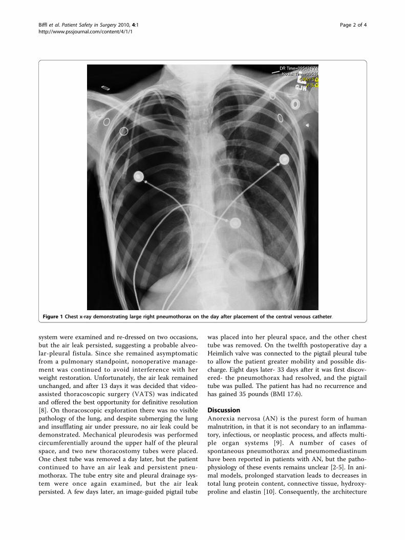
for prominent xerosis, and she had poor venous access.Laboratory testing revealed leukopenia and a markedelevation of her blood urea-nitrogen and serum amino-transferases (AST and ALT > 1,000 u/L). Her serumtotal protein and albumin levels were 6.9 and 4.4 g/dL,both within the normal range. She also quickly devel-oped refeeding hypophosphatemia [7]. Because of theneed for frequent blood draws and intravenous fluids totreat her marked dehydration and hypotension, a 7-French double-lumen Hohn catheter was inserted by aninterventional radiologist into her right internal jugularvein via an anterior cervical approach using ultrasoundlocalization and fluoroscopic guidance. The procedurewent smoothly and without apparent complication, anda post-procedure chest x-ray was unremarkable. How-ever, the next day an x-ray obtained for unrelated rea-sons demonstrated a sizable right pneumothorax (Figure1). Although the patient was asymptomatic, a 10-Frenchpigtail thoracostomy catheter was inserted at the bedsideand attached to 20 cm H2O suction to evacuate thepneumothorax and facilitate lung re-expansion.After one week, the patient appeared to have complete
re-expansion of the lung on x-ray, but the pneu-mothorax returned a day later. Moreover, she appearedto develop a spontaneous left pneumothorax. The leftpneumothorax resolved spontaneously, but she had apersistent right-sided pneumothorax and air leak fromthe tube. The tube entry site and pleural drainage
* Correspondence: [email protected] of Surgery, Denver Health Medical Center, Denver, Colorado,USA
Biffl et al. Patient Safety in Surgery 2010, 4:1http://www.pssjournal.com/content/4/1/1
© 2010 Biffl et al; licensee BioMed Central Ltd. This is an Open Access article distributed under the terms of the Creative CommonsAttribution License (http://creativecommons.org/licenses/by/2.0), which permits unrestricted use, distribution, and reproduction inany medium, provided the original work is properly cited.
system were examined and re-dressed on two occasions,but the air leak persisted, suggesting a probable alveo-lar-pleural fistula. Since she remained asymptomaticfrom a pulmonary standpoint, nonoperative manage-ment was continued to avoid interference with herweight restoration. Unfortunately, the air leak remainedunchanged, and after 13 days it was decided that video-assisted thoracoscopic surgery (VATS) was indicatedand offered the best opportunity for definitive resolution[8]. On thoracoscopic exploration there was no visiblepathology of the lung, and despite submerging the lungand insufflating air under pressure, no air leak could bedemonstrated. Mechanical pleurodesis was performedcircumferentially around the upper half of the pleuralspace, and two new thoracostomy tubes were placed.One chest tube was removed a day later, but the patientcontinued to have an air leak and persistent pneu-mothorax. The tube entry site and pleural drainage sys-tem were once again examined, but the air leakpersisted. A few days later, an image-guided pigtail tube
was placed into her pleural space, and the other chesttube was removed. On the twelfth postoperative day aHeimlich valve was connected to the pigtail pleural tubeto allow the patient greater mobility and possible dis-charge. Eight days later- 33 days after it was first discov-ered- the pneumothorax had resolved, and the pigtailtube was pulled. The patient has had no recurrence andhas gained 35 pounds (BMI 17.6).
DiscussionAnorexia nervosa (AN) is the purest form of humanmalnutrition, in that it is not secondary to an inflamma-tory, infectious, or neoplastic process, and affects multi-ple organ systems [9]. A number of cases ofspontaneous pneumothorax and pneumomediastinumhave been reported in patients with AN, but the patho-physiology of these events remains unclear [2-5]. In ani-mal models, prolonged starvation leads to decreases intotal lung protein content, connective tissue, hydroxy-proline and elastin [10]. Consequently, the architecture
Figure 1 Chest x-ray demonstrating large right pneumothorax on the day after placement of the central venous catheter.
Biffl et al. Patient Safety in Surgery 2010, 4:1http://www.pssjournal.com/content/4/1/1
Page 2 of 4

may be weakened and may be more susceptible to injury[3].Patients with severe AN require frequent blood draws
during the early stages of their refeeding program, aswell as a need for intravenous fluids, blood productsand occasionally parenteral nutrition [11]. However,venous access for these therapies may be quite limitedin patients who are severely malnourished. In addition,the discomfort and annoyance to the patient of dailyvenipuncture may interfere with the therapeutic treat-ment plan and medical stabilization. Therefore, in termsof the management of this patient, the central venouscatheter was considered a necessary intervention, andappropriate precautions were used to minimize the riskof complications. A relatively high anterior approach tothe internal jugular vein was selected, and ultrasoundwas used to guide the insertion.In the current case, although the immediate post-pro-
cedure film did not demonstrate a pneumothorax, thetemporal association suggests this was likely an iatro-genic, and not a spontaneous, pneumothorax. Further-more, on thoracoscopic examination, the right lungappeared normal, without evidence of apical bullae orany other significant pathology. On the other hand, theprolonged air leak suggests that she had some underly-ing abnormality. Malnutrition is a well-known impedi-ment to normal wound healing. This patient hadnormal serum protein and albumin levels at the time ofadmission. However, malnutrition was assumed basedon her overall condition and pattern of weight loss.Indeed, we have found that conventional markers ofnutritional status such as serum albumin may not corre-late with the severity of AN, and could be paradoxicallynormal even in very advanced stages of this disease [12].Moreover, as Coxson and colleagues [6] described,patients with AN may develop architectural changes anddecreased surfactant production. This could result in alung that heals poorly and will not seal itself. The factthat no abnormality- specifically, no hole- was found inthe lung at the time of thoracoscopic examination, doesnot necessarily mean that a hole did not exist. Indeed,the fact that the air leak continued suggests that a per-foration was missed. It is possible that the lack ofinflammation or exudate at the site of injury made itmore difficult to detect. It is unlikely that further ima-ging (e.g., CT scan) would have directed our efforts, asthere was no gross pathology present on visual inspec-tion of the lung. Similarly, in the absence of visiblepathology, lung resection would have been difficult tojustify- particularly in the face of compromised woundhealing.Tube thoracostomy allows re-expansion and resolu-
tion of pneumothoraces, but it may take variableamounts of time for air leaks to seal. Persistent air leak
is accepted as an indication for VATS [8]. In this case,VATS was deferred based on concern that post-proce-dural pain and the need for narcotic pain medicationswould impede her feeding progress. In general, the lowmorbidity and typical course (i.e., eating a regular dietsoon after the procedure) of VATS would make this aminor concern, but patients with AN can be very sensi-tive to changes in their course, and may be easilyderailed from their nutritional plan. In retrospect, theprocedure was of dubious benefit. Although mosthealthy individuals do well with removal of one chesttube in the early postoperative period, it may be prudentto leave two tubes in place in the patient with AN.Furthermore, it is possible that higher levels of suctionwould have been successful in promoting lung expan-sion and sealing following the pleuredesis procedure.This should be considered in future cases. An alterna-tive strategy, as proposed by Cerfolio [13], would havebeen to switch from suction or water seal drainage to aHeimlich valve earlier in the course. In fact, in consulta-tion with a pulmonologist, it was decided that thiswould be a reasonable step toward patient discharge.We suggest that this strategy is safe and allows fullmobility and earlier discharge.
ConclusionsIn sum, the malnutrition of AN is associated with pul-monary changes that may predispose to spontaneouspneumothorax or persistent air leak. Whether thesechanges are reversible with refeeding, and how long ittakes to reestablish normal pulmonary physiology arenot currently known. However, a prolonged alveolar-pleural fistula may be anticipated in these patients dur-ing the severe stage of their AN. Based on our experi-ence, this problem may be managed effectively with aHeimlich valve to expedite the patient’s transition to thenext stage of their recovery from AN.
Abbreviations ListALT: Alanine aminotransferase; AN: Anorexia nervosa;AST: Aspartate aminotransferase; BMI: Body massindex; VATS: Video-assisted thoracoscopic surgery
ConsentWritten informed consent was obtained from the patientfor publication of this case report and accompanyingimages. A copy of the written consent is available forreview by the Editor-in-Chief of this Journal.
AcknowledgementsThe patient gave permission for the publication of this case report.
Biffl et al. Patient Safety in Surgery 2010, 4:1http://www.pssjournal.com/content/4/1/1
Page 3 of 4
Author details1Department of Surgery, Denver Health Medical Center, Denver, Colorado,USA. 2Department of Patient Safety and Quality, Denver Health MedicalCenter, Denver, Colorado, USA. 3Department of Medicine, Denver HealthMedical Center, Denver, Colorado, USA.
Authors’ contributionsWLB, VN, JLG, and PSM have all made substantial contributions to thedrafting of the manuscript and critical revision, and all have given finalapproval of the version to be published.
Competing interestsThe authors declare that they have no competing interests.
Received: 28 September 2009Accepted: 1 February 2010 Published: 1 February 2010
References1. Goldstein MA, Herzog DB, Misra M, Sagar P: A 19-year-old man with
weight loss and abdominal pain. N Engl J Med 2008, 359:1272-1283.2. Adson DE, Crow SJ, Mitchell JE: Spontaneous pneumothorax in anorexia
nervosa. Psychosomatics 1998, 39:162-164.3. Corless JA, Delaney JC, Page RD: Simultaneous bilateral spontaneous
pneumothoraces in a young woman with anorexia nervosa. Int J EatDisord 2001, 30:110-112.
4. Fergusson RJ, Shaw TRD, Turnbull CM: Spontaneous pneumomediastinum:a complication of anorexia nervosa?. Postgrad Med J 1985, 61:815-817.
5. van Veelen I, Hogeman PHG, van Elburg, Nielsen-Abbring FW,Heggelman BGF, Mahieu HF: Pneumomediastinum: a rare complication ofanorexia nervosa in children and adolescents. A case study and reviewof the literature. Eur J Pediatr 2008, 167:171-174.
6. Coxson HO, Chan IHT, Mayo JR, Hlynsky J, Nakano Y, Birmingham CL: Earlyemphysema in patients with anorexia nervosa. Am J Respir Crit Care Med2004, 170:748-752.
7. Crook MA, Hally V, Panteli JV: The importance of the refeeding syndrome.Nutrition 2001, 17:632-637.
8. Ng CSH, Lee TW, Wan S, Yim APC: Video assisted thoracic surgery in themanagement of spontaneous pneumothorax: the current status.Postgrad Med J 2006, 82:179-185.
9. Attea E, Walsh BT: Behavioral management for anorexia nervosa. N Engl JMed 2009, 360:500-506.
10. Sahebjami H, Macgee J: Effects of starvation on lung mechanics andbiochemistry in young and old rats. J Appl Physiol 1985, 58:778-784.
11. Mehler PS, Kolpak S, Padilla R: Anorexia nervosa and the use of totalparenteral nutrition. Curr Nutr Food Sci 2005, 1:97-104.
12. Narayanan V, Gaudiani JL, Mehler PS: Serum albumin levels may notcorrelate with weight status in severe anorexia nervosa. Eating Disorders2009, 17:322-326.
13. Cerfolio RJ: Recent advances in the treatment of air leaks. Curr Opin PulmMed 2005, 11:319-323.
doi:10.1186/1754-9493-4-1Cite this article as: Biffl et al.: The management of pneumothorax inpatients with anorexia nervosa: A case report and review of theliterature. Patient Safety in Surgery 2010 4:1.
Submit your next manuscript to BioMed Centraland take full advantage of:
• Convenient online submission
• Thorough peer review
• No space constraints or color figure charges
• Immediate publication on acceptance
• Inclusion in PubMed, CAS, Scopus and Google Scholar
• Research which is freely available for redistribution
Submit your manuscript at www.biomedcentral.com/submit
Biffl et al. Patient Safety in Surgery 2010, 4:1http://www.pssjournal.com/content/4/1/1
Page 4 of 4


![CASE REPORT Open Access Tension pneumothorax … proven in a retrospective study with diaphragmatic ... presentation of a tension pneumothorax [3,7]. In this case the tension pneumothorax](https://img.pdfslide.net/doc/110x75/5ae6a4b67f8b9ae1578df685/case-report-open-access-tension-pneumothorax-proven-in-a-retrospective-study.jpg)


![Journal of Clinical & Experimental Cardiology...ISSN:2155-9880 JCEC, an open access journal [1]. Pneumothorax may be detected during the procedure or within 24h after implantation](https://img.pdfslide.net/doc/110x75/5e67a16a6cb9fe6dd548fa6a/journal-of-clinical-experimental-cardiology-issn2155-9880-jcec-an-open.jpg)