Embed Size (px)
Citation preview
Alteration of Cytokine Profiles Inhibits Efficacy of Silver Nanoparticle-basedNeutralization of arenavirusesNathan Elrod1, Jeff Brady2,3, Harold Rathburn1 and Janice L. Speshock1*
1Tarleton State University, Department of Biological Sciences, Stephenville, TX 76402, USA2Tarleton State University, Department of Wildlife, Sustainability, and Ecosystem Sciences, Stephenville, TX 76402, USA3Texas A&M AgriLife Research and Extension Center, Stephenville, TX 76401, USA*Corresponding author: Janice L. Speshock, Tarleton State University, Department of Biological Sciences, 1333. Washington Street, Box T-0100, Stephenville, TX76402, USA, Tel: (254) 968-9341; E-mail: [email protected]
Received date: March 27, 2016; Accepted date: April 14, 2017; Published date: April 19, 2017
Copyright: © 2017 Elrod N. This is an open-access article distributed under the terms of the Creative Commons Attribution License, which permits unrestricted use,distribution, and reproduction in any medium, provided the original author and source are credited.
Abstract
Background: Arenaviruses are important pathogens that can cause hemorrhagic fever or meningoencephalitis.There is not an effective treatment targeting arenaviruses, which necessitates a search for therapies against thisvirus family, as well as others. Silver nanoparticles (Ag-NPs) have been shown to have effective antiviral activity incell culture models against arenaviruses, and therefore it was hypothesized that they may make an effectivetherapeutic against viral meningitis. However, the silver nanoparticles interfered with normal cytokine profilesproduced in response to the virus infection, which led to exacerbated pathology.
Methods: Mice were infected with Tacaribe virus, a mouse-adapted arenavirus that causes lethal encephalitis.The virus was left untreated or treated with 10 nm Ag-NPs, which were demonstrated previously to inhibit virusreplication in cell culture. Virus replication, brain pathology, and cytokine profiles (pro-inflammatory interleukins andtype I interferons), as well as over morbidity and mortality were assessed in the mice with and without treatment.
Results: Although the viral loads appeared to be reduced in the Ag-NP treated mice, mortality was observed inall virus-infected animals, regardless of whether or not they received nanoparticle therapy. The Ag-NP treatmentsalso impacted the innate immune response against the virus infection and unregulated the production ofinterleukin-1 beta, which likely contributed to the observed mortality in the mice.
Conclusions: This study confirms why it is important to assess promising in vitro results using in vivo models.Biological barriers can impact the efficacy of therapeutics in mammals. Although Ag-NPs may still have success asan antiviral therapeutic in other areas of the body that are less constricted, in sensitive areas such as the brain, thenanoparticles can cause very adverse reactions.
Keywords: Silver nanoparticles; Immunotoxicology; Inflammation;Interferons; Arenavirus; Tacaribe virus
IntroductionThe Arenaviridae family of 18 species of viruses is divided into New
World and Old World clades [1]. These viruses have an ambisenseRNA genome with two structural segments, the S segment and the Lsegment, each encoding viral proteins [1]. The New World, or TacaribeSerocomplex, clade consists of Tacaribe Virus (TCRV), as well as thehemorrhagic fever viruses Junin and Machupo [1,2]. At an amino acidlevel TCRV is a 67-78% match to the highly pathogenic Junin in the 4main viral proteins (NP, GPC, L, and Z) [3]. TCRV is unique fromother arenaviruses in that it fails to suppress host type I and IIinterferon (IFN), and consequently, does not cause viral hemorrhagicfever in humans making it safe for study at the biosafety level 2 (BSL-2)[4,5]. TCRV is also unusual compared to other members ofArenaviridae as it has been isolated in bats as opposed to the strict useof rodent hosts in the other arenaviruses in its clade, however it can bemouse-adapted quite easily for use in rodent models [6]. In BALB/cmice of all ages inoculated intra-cranially (IC), following serialpassage, TCRV can manifest clinical symptoms (ruffled fur, hind limbparalysis, lethargy) in 9 to 11 days followed by death 1 to 3 days after
symptoms manifest [7,8]. Newborn BALB/c mice andimmunocompromised mice lacking IFN-α/β and IFN-γ receptorsinoculated intra-peritoneally (IP) also showed the same clinicalsymptoms followed by death in the same time-frame [9,10]. Mouse-adaptation of TCRV causes mortality by lethal meningoencephalitis, orlesions in both the meninges and brain tissue caused by inflammation,via over-activation of the cellular immune response, which is moresimilar to the pathology observed with the Old World humanarenavirus, Lymphocytic Choriomeningitis Virus (LCMV) [7-11].There are a lack of therapies and preventative measures to combathemorrhagic fever and meningitis infections caused by arenaviruses[12]. An effective treatment to arenavirus infection would require thecombination of an agent that will limit both virus replication and theproinflammatory cellular response, which contributes to the swellingobserved in viral meningoencephalitis [13].
Nanomaterials have been shown to have numerous physical,biological, and pharmaceutical properties, including antiviral [14].Silver nanoparticles (Ag-NPs) in the 10 to 20 nm size range, have beenshown to prevent viral replication of Human Immunodeficiency Virustype 1 [15], Monkeypox virus [16], adenovirus serotype 3 [17], andTCRV using cell culture models [18]. It is hypothesized that the Ag-NPs inhibit viral infection by binding to the viral glycoproteins to
Toxicology: Open Access Elrod et al., Toxicol Open Access 2017, 3:2DOI: 10.4172/2476-2067.1000124
Research Article OMICS International
Toxicol Open Access, an open access journalISSN:2476-2067
Volume 3 • Issue 2 • 1000124
prevent association of the virion with the host cell receptor, or as in thecase of TCRV, to stimulate the uptake of an inert virus particle [15-18].TCRV was shown to associate with the Ag-NPs, but instead of blockingbinding, as observed with most viruses, the association of Ag-NPs andTCRV increased the binding efficiency and uptake of the virions intoVero cells, but the virus did not replicate once it was internalized, thusdemonstrating virus inactivation [18]. It was therefore hypothesizedthat similar inhibitory effects could be demonstrated using mouse-adapted TCRV with Ag-NP treatment in Balb/c mice. Juvenile micewere inoculated IC with TCRV treated with Ag-NPs, and the resultingmorbidity and mortality of the virus was observed in Ag-NP andmock-treated infections.
Materials and Methods
Mouse adaptation of TCRVAll procedures were performed under an approved protocol by the
Tarleton State University Institutional Animal Care and UsageCommittee to ensure laboratory animal health and safety. The viruswas mouse adapted following three serial IC passages in 7 week oldICR (CD-1) mice (Harlan Laboratories, Indianapolis, IN) to increasevirulence using a modified protocol [8]. Briefly, thirty microliters ofTCRV (VR-1556; ATCC, Manassas, VA) was given IC to 3 mice. Micewere observed daily and were sacrificed via lethal injection of sodiumpentobarbital (150 µL IP) when mice demonstrated clinical symptomsof severe inactivity or inappetence, or had lost more than 25% of theirinitial body weight, which occurred in two of the three mice at day 6post-infection. Those two mice were sacrificed and the brain tissueswere harvested and homogenized in 1 ml of TNE buffer (0.01 M Tris-HCl, 0.15 M NaCl, 1 mM EDTA, buffered at pH 7.4). Thehomogenized brains from the 2 mice showing signs of illness werecombined in equal 100 μL proportions to create a total volume of 200μL of homogenate which was combined with 200 μL of Fetal BovineSerum (FBS; Atlanta Biologicals, Norcross, GA) and 600 μL of TNE tocreate serial passage 1 (TCRVp1). This process was repeated with twoadditional serial passages into the brain to generate a stock that waslethal to 100% of mice (TCRVp3), which was determined using a viralplaque assay to be 6.5 x 10⁵ plaque forming units (pfu)/mL. Thisinoculum was stored at -80°C until further experimentation.
Viral attenuation with Ag-NPsBiopure silver nanospheres at a diameter of 10 ± 2 nm were
obtained from nanoComposix Inc (San Diego, CA). The nanoparticleswere synthesized with a 0.01% polyvinylpyrrolidone (PVP) coating toincrease biological stability and decrease toxicity [19]. PVP coating isused to keep the nanoparticles from agglomeration duringmanufacture and reduces cytotoxic effects in vitro [19,20]. The abilityof Ag-NPs to neutralize TCRV was performed in a similarexperimental design as an in vitro neutralization assay in Vero cells[18]. The lethal dose at 50% (LD50) for the virus via IC inoculation wasdetermined to be 280 pfu/mL, therefore a 1000-fold dilution ofTCRVp3 (650 pfu/mL) was used to assess Ag-NP neutralization. Thisconcentration of TCRV was incubated with 50 μg/mL of Ag-NP, 25 μLof 0.01% PVP, which reflected the amount of PVP in the Ag-NPsample at 50 μg/mL, or 25 μL TNE. These viral suspensions were thenincubated at room temperature in a rotating mixer for 1 hour to allowfor interaction. The dosage of 50 μg/mL nanoparticle was used as it hadbeen determined to have a completely inhibitory effect on Tacaribevirus in Vero cells [18].
These attenuated preparations were then given at 30 μL IC to groupsof ICR mice (n=4) at 7 weeks old. Morbidity and mortality between theAg-NP and control groups were recorded for comparison. In addition,brain tissue was excised post mortem for RNA extraction andhistopathological examination. One mouse per group’s brain waspreserved in 4% paraformaldehyde and sent for histological processingand slide preparation with hematoxylin and eosin staining. Theremaining mice brains were homogenized in 1 mL TNE each and 50μLof homogenate was used for mRNA extraction.
Messenger RNA ExtractionRNA was extracted from the mouse brains using 50 μL of brain
homogenate and the GET Total RNA kit, with optional DNasetreatment, from G-biosciences (St. Louis, MO) as per themanufacturer’s instructions. The mRNA was quantified using theQuant-iT Ribogreen RNA assay kit (ThermoFisher Scientific,Waltham, MA) in a BioTek Flx800 multiwell fluorimeter (Winooski,VT). All RNA samples were normalized to 500 nanograms (ng) ofRNA.
Quantitative reverse transcriptase polymerase chain reaction(qRT-PCR)
One microliter of each RNA sample was added to 2.5 μL of Sybrgreen, 1 μL DNase/RNase-free water, and 0.5 μL of reversetranscriptase from the qScript One-Step SYBR Green qRT-PCR kit(Quanta Biosciences, Beverly, MA) and 0.5 μL of primer mix (0.2 μMof each primer). Each RNA sample was prepared in triplicate with eachprimer set listed in Table 1 and loaded into a 384-well PCR plate foranalysis in the Roche 480 Light Cycler (Basel, Switzerland) todetermine the threshold cycle of each sample. An initial 10 minuteincubation at 50°C allowed for the conversion of the RNA tocomplementary DNA using reverse transcriptase, which was thenfollowed by the following cycles for DNA amplification: 1 minutedenature at 95°C; 45 cycles of 10 seconds at 95°C, 30 seconds at 60°Cwith a quantification fluorescence read, and 30 seconds at 72°C;followed by a melt curve of 10 seconds at 95°C, 30 seconds at 40°Cwith continuous fluorescence readings to 95°C; and a final cool downto 40°C. The threshold cycles were used to determine the fold changeusing the delta-delta CT method [21].
Gene Forward Primer Reverse Primer
TCRV S[18]
TGTGTTGGCTGGCAGAT AGGAGAGTGAACAAAGACAT
IFNα [22] GGACTTTGGATTCCCGCAGGAGAAG
GCTGCATCAGACAGCCTTGCAGGTC
IFNβ [22] AACCTCACCTACAGGGCGGACTTCA
TCCCACGTCAATCTTTCCTCTTGCTT
IL-1β [23] CAACCAACAAGTGATATTCTCCATG
GATCCACAGTGTGGACGTGCA
IL-6 [23] GAGGATACCACTCCCAACAGACC
AAGTGCATCATCGTTGTTCATACA
TNFα [23] CATCTTCTCAAAATTCGAGTGACAA
TGGGCGTAGACAAGGTACAACCC
L19 [24] ATGAGTATGCTCAGGCTACAG
TGACGGGAGTTGGCATTG
Table 1: Primer sequences for qRT-PCR.
Citation: Elrod N, Brady J, Rathburn H, Speshock JL (2017) Alteration of Cytokine Profiles Inhibits Efficacy of Silver Nanoparticle-basedNeutralization of arenaviruses. Toxicol Open Access 3: 124. doi:10.4172/2476-2067.1000124
Page 2 of 6
Toxicol Open Access, an open access journalISSN:2476-2067
Volume 3 • Issue 2 • 1000124
HistologyFormalin-fixed brains were shipped to Tarleton State University
Department of Medical Laboratory Sciences (Fort Worth, TX) forhistological processing and slide preparation. The slides were stainedwith hematoxylin and eosin for viewing of histopathology. The slideswere viewed on a Zeiss Jenalumar Contrast Microscope (Oberkochen,Germany) and imaged using a WESCO 291CU digital camera(Pittsburgh, PA).
Statistical analysisAll qRT-PCR analyses were compared between control and
treatment groups using a one-way Analysis of Variance (ANOVA) withsignificance determination at p< 0.05. Histopathic effect was reportedby visual observation.
Results
Ag-NP neutralization of TCRVAlthough mice that received an inoculation of Ag-NP-treated
TCRV lived longer than mice that received a mock attenuation ofTCRV (12 days versus 6 days), only one of the Ag-NP treated micesurvived the exposure Table 2. This compared to 0 mice in the mock-treatment (TNE) or PVP treatment Table 2.
Attenuation Treatment Number of mice that survived (n=4)
TNE+TCRV 0
PVP+TCRV 0
Ag-NP+TCRV 1
Table 2: Survivability assessment of TCRV-infected mice followingattenuation treatments (n=4).
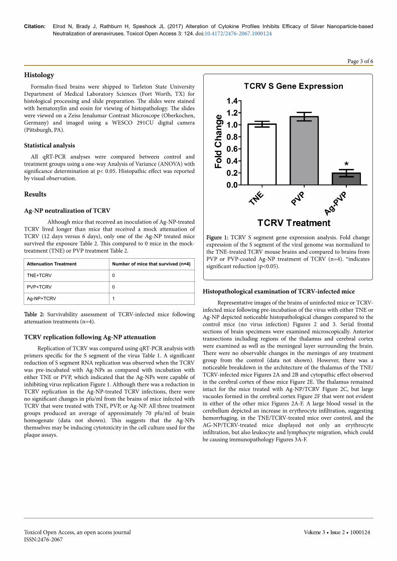
TCRV replication following Ag-NP attenuationReplication of TCRV was compared using qRT-PCR analysis with
primers specific for the S segment of the virus Table 1. A significantreduction of S segment RNA replication was observed when the TCRVwas pre-incubated with Ag-NPs as compared with incubation witheither TNE or PVP, which indicated that the Ag-NPs were capable ofinhibiting virus replication Figure 1. Although there was a reduction inTCRV replication in the Ag-NP-treated TCRV infections, there wereno significant changes in pfu/ml from the brains of mice infected withTCRV that were treated with TNE, PVP, or Ag-NP. All three treatmentgroups produced an average of approximately 70 pfu/ml of brainhomogenate (data not shown). This suggests that the Ag-NPsthemselves may be inducing cytotoxicity in the cell culture used for theplaque assays.
Figure 1: TCRV S segment gene expression analysis. Fold changeexpression of the S segment of the viral genome was normalized tothe TNE-treated TCRV mouse brains and compared to brains fromPVP or PVP-coated Ag-NP treatment of TCRV (n=4). *indicatessignificant reduction (p<0.05).
Histopathological examination of TCRV-infected miceRepresentative images of the brains of uninfected mice or TCRV-
infected mice following pre-incubation of the virus with either TNE orAg-NP depicted noticeable histopathological changes compared to thecontrol mice (no virus infection) Figures 2 and 3. Serial frontalsections of brain specimens were examined microscopically. Anteriortransections including regions of the thalamus and cerebral cortexwere examined as well as the meningeal layer surrounding the brain.There were no observable changes in the meninges of any treatmentgroup from the control (data not shown). However, there was anoticeable breakdown in the architecture of the thalamus of the TNE/TCRV-infected mice Figures 2A and 2B and cytopathic effect observedin the cerebral cortex of these mice Figure 2E. The thalamus remainedintact for the mice treated with Ag-NP/TCRV Figure 2C, but largevacuoles formed in the cerebral cortex Figure 2F that were not evidentin either of the other mice Figures 2A-F. A large blood vessel in thecerebellum depicted an increase in erythrocyte infiltration, suggestinghemorrhaging, in the TNE/TCRV-treated mice over control, and theAG-NP/TCRV-treated mice displayed not only an erythrocyteinfiltration, but also leukocyte and lymphocyte migration, which couldbe causing immunopathology Figures 3A-F.
Citation: Elrod N, Brady J, Rathburn H, Speshock JL (2017) Alteration of Cytokine Profiles Inhibits Efficacy of Silver Nanoparticle-basedNeutralization of arenaviruses. Toxicol Open Access 3: 124. doi:10.4172/2476-2067.1000124
Page 3 of 6
Toxicol Open Access, an open access journalISSN:2476-2067
Volume 3 • Issue 2 • 1000124
Figure 2: Histological examination of brains from TCRV-infectedmice. Frontal cross sections from the center of the brain includingthe thalamus-cerebral cortex border from the brains of mice thatwere infected with TCRV that had been pre-treated with TNE (aand d), PVP (b and e), or Ag-NPs (c and f). The top 3 images (a-c)are at 6.3x magnification and the bottom 3 images (d-f) representthe lower left quadrant of the 6.3x image magnified to 50x.
Figure 3: Histological examination of brain vasculature fromTCRV-infected mice. Frontal cross sections from the center of thebrain including a blood vessel from the cerebellum of brains of micethat were infected with TCRV that had been pre-treated with TNE(a and d), PVP (b and e), or Ag-NPs (c and f). The top 3 images (a-c) are at 6.3x magnification and the bottom 3 images (d-f) representthe lower left quadrant of the 6.3x image magnified to 50x.
Immune responses in the brains of TCRV-infected miceSince there was a reduction in TCRV replication following Ag-NP
treatment, but an increase in observable histopathology, it washypothesized that the immune system was contributing to thepathology. Type I interferon (IFN) responses were assessed since theyhave been shown to contribute greatly to the recovery from arenavirusinfections. A slight decline in IFN beta and a significant reduction inIFN alpha were observed following Ag-NP treatment Figure 4, whichshould have led to an increase in virus replication, not a decrease,suggesting further that the Ag-NPs themselves might be contributingto the pathology and mortality observed, by suppressing immunefunction. It was also hypothesized that the Ag-NPs were stimulating aninflammatory response, which would cause swelling of the brain, and
thereby significant morbidity and mortality. Therefore the geneexpression changes were assessed for the pro-inflammatory cytokinesinterleukin-1 beta (IL-1β), interleukin-6 (IL-6), and tumor necrosisfactor alpha (TNF-α). There were no differences observed in theexpression of IL-1β or TNF-α with any of the treatment groupscompared to control, and the IL-6 TCRV infection with Ag-NPtreatment actually had a reduction in expression Figure 5. These dataindicate that there was no induction of inflammation contributing tothe pathology of the mice brains, and that the Ag-NPs may suppressimmune response in infected animals.
Figure 4: Type I interferon gene expression analysis. Fold changeexpression of the host type I interferons (alpha and beta) werenormalized to the TNE-treated TCRV mouse brains and comparedto brains from PVP or PVP-coated Ag-NP treatment of TCRV(n=4). *indicates significant change in gene expression (p<0.05).
DiscussionAttenuation with silver nanoparticles did impact the amount of
innate immune response by the host Figures 4 and 5, which likelycontributed to the observed pathology since there was also a reductionin TCRV S segment expression, following pre-incubation with Ag-NPs,compared to controls Figure 1. The inactivation of the virus was notsufficient to prevent death in the majority of the mice tested Table 1,which leads one to hypothesize that the Ag-NPs may be causing celldeath of the neurons in addition to the virus through a direct orindirect (inflammation) mechanism. An in vitro cell culture model didnot demonstrate any discernable cell death from the virus-nanoparticleagglomerate [18], but the cells used for this study were kidney cells,which tend to be more stable than neurons [18,20]. Also, the in vitrostudy utilized Ag-NPs that were uncoated or polysaccharide-coated,and the Biopure PVP coating may have interfered with the TCRVinteraction with silver. The first attempt in vivo was using the uncoated10 nm Ag-NPs from the same manufacturer as the cell culture trials,but there was no decline in mortality and a great deal of inflammationwas observed (data not shown). Therefore a more biocompatiblepolymer coating was selected for the Ag-NPs for the in vivo study, todecrease side-effects from the nanoparticle. These PVP-coated Ag-NPsdo exhibit some mild in vivo toxicity and inflammation [25], but didnot cause mortality alone in an open system [25]. However, thesensitivity of neurons to external stimuli and the enclosed nature of the
Citation: Elrod N, Brady J, Rathburn H, Speshock JL (2017) Alteration of Cytokine Profiles Inhibits Efficacy of Silver Nanoparticle-basedNeutralization of arenaviruses. Toxicol Open Access 3: 124. doi:10.4172/2476-2067.1000124
Page 4 of 6
Toxicol Open Access, an open access journalISSN:2476-2067
Volume 3 • Issue 2 • 1000124
mouse brain may have exacerbated the toxicity of the nanoparticles.Mouse neuroblastoma cells have been shown to be much moresensitive to nanoparticle toxicity than African Green Monkey kidneycells (Vero cells) using cell culture toxicity assays [18,20]. In previousstudies of TCRV, it was demonstrated that the Purkinje cells of thecerebellum appear to be the major targets of the virus, which may bemore vulnerable to increases in inflammation and decreases in theanti-viral protection of the interferons [8]. The histology obtained fromthis study showed most histological changes around the regions of thethalamus and the cerebellum, confirming this target area for the virus.Since more histopathological effects occurred in the nanoparticle-treated virus infection, it is possible that the virus is targeting thePurkinje cells allowing for increased uptake of the Ag-NPs, which inturn are causing cytotoxicity.
Figure 5: Inflammatory gene expression analysis. Fold changeexpression of the pro-inflammatory cytokine genes werenormalized to the TNE-treated TCRV mouse brains and comparedto brains from PVP or PVP-coated Ag-NP treatment of TCRV(n=4). *indicates significant reduction (p<0.05).
This study illustrates the potential conflict in the results betweenan in vitro study, where the virus was completely inactivated, and an invivo study, where there is still mortality from the virus infection,although it is hypothesized that the Ag-NPs are causing the mortality.Cell culture models lack the diversity of cell populations andcomplexity of living organisms, and therefore can be misleading attimes. Neurons are generally not utilized in vitro for virus studies dueto their known instability against external stimuli [19]. Either thereplication-deficient virions, the Ag-NPs, or both, are capable ofdestroying these sensitive cells in an in vivo model of infection. SinceXiang et al. demonstrated efficacy of Ag-NPs in vivo against influenzavirus [26], there still is the possibility of using Ag-NPs as a broad-spectrum antiviral agent, but anatomical location of treatment mayneed to be considered prior to use, especially in more sensitive areaslike the brain.
Declarations
Animal Ethics ApprovalAll procedures were performed under an approved protocol
(AUP_Speshock_2014) by the Tarleton State University InstitutionalAnimal Care and Usage Committee to ensure laboratory animal healthand safety. Animals were housed according to guidelines outlined in anapproved standard operating procedures (SOP_0026) document.
Competing InterestsThe authors have no competing interests to declare.
AcknowledgementsThis project was funded through Tarleton State University’s Office of
Student Research and Creative Activities through a Student ResearchGrant awarded to Mr. Elrod. There was no external funding source forthis research. We would like to thank Dr. Brooke Dubansky fromTarleton State University Medical Laboratory Sciences for histologicalprocessing and staining.
References1. Buchmeier MJ, Bowen MD, Peters CJ (2013) Arenaviridae: Fields
Virology. 6thedn Lippincott, Williams & Wilkins, Philadelphia, pp:1283-1303.
2. Charrel RN, de Lamballerie X (2003) Arenaviruses other than Lassavirus. Antiviral Res 57: 89-100.
3. Bolken TC, Laquerre S, Zhang Y, Bailey TR, Pevear DC, et al. (2006)Identification and characterization of potent small molecule inhibitor ofhemorrhagic fever New World arenaviruses. Antiviral Res 69: 86-97.
4. Bray M (2005) Pathogenesis of viral hemorrhagic fever. Curr OpinImmunol 17: 399-403.
5. Martinez-Sobrido L, Giannakas P, Cubitt B, García-Sastre A, Carlos de laTorre J (2007) Differential inhibition of type I interferon induction byarenavirus nucleoproteins. J Virol 81: 12696-12703.
6. Downs WG, Anderson CR, Spence L, Aitken TH, Greenhall AH (1963)Tacaribe virus, a new agent isolated from artibeus bats and mosquitoes inTrinidad, West Indies. Am J Trop Med Hyg 12: 640-646.
7. Borden EC, Nathanson N (1974) Tacaribe Virus-Infection of Mouse -Immunopathologic Disease Model. Laboratory Investigation 30: 465-473.
8. Rosato RR, Elwell MR, Eddy GA (1978) Virulence alterations of tacaribevirus infection in adult mice: lethal model for encephalitis. Arch Virol 58:137-147.
9. Gowen BB, Wong MH, Larson D, Ye W, Jung KH, et al. (2010)Development of a new Tacaribe arenavirus infection model and its use toexplore antiviral activity of a novel aristeromycin analog. Plos One 5: 11.
10. Borden EC, Murphy FA, Nathanson N, Monath TPC (1971) Effect ofantilymphocyte serum on Tacaribe virus infection in infant mice. InfectImmun 3: 466–471.
11. Shresta S, Sharar KL, Prigozhin DM, Beatty PR, Harris E (2006) Murinemodel for dengue virus-induced lethal disease with increased vascularpermeability. J Virol 80: 10208-10217.
12. Leyssen P, De Clercq E, Neyts J (2008) Molecular strategies to inhibit thereplication of RNA viruses. Antiviral Res 78: 9-25.
13. Sefing EJ, Wong MH, Larson DP, Hurst BL, Van Wettere AJ, et al. (2010)Vascular leak ensues a vigorous proinflammatory cytokine response toTacaribe arenavirus infection in AG129 mice. Virology Journal 10: 221.
14. Sondi I, Salopek-Sondi B (2007) Silver nanoparticles as antimicrobialagent: a case study on E. coli as a model for Gram-negative bacteria. JColloid Interface Sci 275: 177-182.
Citation: Elrod N, Brady J, Rathburn H, Speshock JL (2017) Alteration of Cytokine Profiles Inhibits Efficacy of Silver Nanoparticle-basedNeutralization of arenaviruses. Toxicol Open Access 3: 124. doi:10.4172/2476-2067.1000124
Page 5 of 6
Toxicol Open Access, an open access journalISSN:2476-2067
Volume 3 • Issue 2 • 1000124
15. Elechiguerra JL, Burt JL, Morones JR, Camacho-Bragado A, Gao X, et al.(2005) Interaction of silver nanoparticles with HIV-1. JNanobiotechnology 3: 6.
16. Rogers JV, Parkinson CV, Choi YW, Speshock JL, Hussain SM (2008) Apreliminary assessment of silver nanoparticle inhibition of monkeypoxvirus plaque formation. Nanoscale Res Lett 3: 129-133.
17. Chen N, Zheng Y, Yin J, Li X, Zheng C (2013) Inhibitory effects of silvernanoparticles against adenovirus type 3 in vitro. J Virol Methods 193:470-477.
18. Speshock JL, Murdock RC, Braydich-Stolle LK, Schrand AM, Hussain SM(2013) Interaction of silver nanoparticles with Tacaribe virus. JNanobiotechnology 8: 19.
19. Kittler S, Greulich C, Gebauer JS, Diendorf J, Treuel L, et al. (2010) Theinfluence of proteins on the dispersibility and cell-biological activity ofsilver nanoparticles. J Mater Chem 20: 512-518.
20. Schrand AM, Braydich-Stolle LK, Schlager JJ, Dai L, Hussain SM (2008)Can silver nanoparticles be useful as potential biological labels?Nanotechnol 19: 235104.
21. Livak KJ, Schittgen TD (2001) Analysis of relative gene expression datausing real-time quantitative PCR and the 2-ΔΔCT method. Methods 25:402–408.
22. Liu G, Friggeri A, Yang YP, Park YJ, Tsuruta Y, et al. (2009) miR-147, amicroRNA that is induced upon Toll-like receptor stimulation, regulatesmurine macrophage inflammatory responses. PNAS 106: 15819-15824.
23. Overbergh L, Giulietti A, Valckx D, Decallonne R, Bouillon R, et al.(2003) The use of real-time reverse transcriptase PCR for thequantification of cytokine gene expression. J Biomol Tech 14: 33-43.
24. Speshock JL, Doyon-Reale N, Rabah R, Neely MN, Roberts PC (2007)Filamentous influenza A virus infection predisposes mice to fatalsepticemia following superinfection with Streptococcus pneumoniaeserotype 3. Infect Immun 75: 3102-3111.
25. Speshock JL, Elrod N, Sadoski DK, Maurer E, Braydich-Stolle LK, et al.(2016) Differential organ toxicity in the adult zebra fish following exposreto acute sub-lethal dose of 10 nm silver nanoparticles. Frontiers inNanosci and Nanotechnol 2: 114-120.
26. Xiang D, Zheng Y, Duan W, Li X, Yin J, et al. (2013) Inhibition of A/Human/Hubei/3/2005 (H3N2) influenza virus infection by silvernanoparticles in vitro and in vivo. Int J Nanomed 8: 4103-4114.
Citation: Elrod N, Brady J, Rathburn H, Speshock JL (2017) Alteration of Cytokine Profiles Inhibits Efficacy of Silver Nanoparticle-basedNeutralization of arenaviruses. Toxicol Open Access 3: 124. doi:10.4172/2476-2067.1000124
Page 6 of 6
Toxicol Open Access, an open access journalISSN:2476-2067
Volume 3 • Issue 2 • 1000124