Embed Size (px)
Citation preview
Int J Clin Exp Pathol 2016;9(2):2459-2465www.ijcep.com /ISSN:1936-2625/IJCEP0019775
Case Report Pleomorphic adenoma of the breast: a report of two cases and a literature review
Yunan Han1, Qingfu Zhang2, Shawn Xiang Li3, Liang Feng1, Lei Zhang1, Zhan Li1, Xueshan Qiu2, Feng Jin1, Bo Chen1
1Department of Breast Surgery, The First Hospital of China Medical University, Shenyang, China; 2Department of Pathology, College of Basic Medical Sciences and The First Hospital of China Medical University, Shenyang, China;
3Department of International Education College, China Medical University, Shenyang, China
Received November 14, 2015; Accepted January 10, 2016; Epub February 1, 2016; Published February 15, 2016
Abstract: Pleomorphic adenoma of the breast (PAB) is an extremely rare tumor in the breast. Herein, we reported two Asian female patients diagnosed as PAB and reviewed the relevant literature briefly. The two cases were both diag-nosed as fibroadenoma in breast ultrasonography before surgery. Case one is a 28-year-old woman presented with a seven year history of a slowly-growing painless mass in left breast, which was about 1.46×0.93 cm in size. Case two is a 47-year-old female who had a complaint of left breast mass three months ago, which was 2.31×1.37 cm in size. The two tumors both located under areola. The two patients both underwent lumpectomy. Microscopically, they were both comprised of mixture of epithelial cells and myoepithelial cells embedded in myxochondroid matrix background. Immunohistochemically, the two cases displayed positive cytokeratin 7 (CK7) for epithelial cells, posi-tive smooth muscle actin (SMA) and S-100 for myoepithelial cells. PAS staining was positive in case two. According to the pathologic histology and immunohistochemical findings, the two cases were diagnosed as PAB. However, the pathogenesis of PAB has been unclear and further studies remain to be researched.
Keywords: Pleomorphic adenoma, fibroadenoma, breast
Introduction
Pleomorphic adenoma (PA) is a common tumor occurred in salivary glands. The occurrence of PA located in breast is extremely rare and PAB was mostly found in the mammary subareola [1] of postmenopausal female [2]. PA has a characteristic mixture of epithelial components and myoepithelial components embedded in myxochondroid matrix [3]. To our knowledge, only 77 cases of PAB [4-7] have been reported in the literature since the first reported case of PAB by French scholar Lecène in 1906 [8]. Indeed, the breast is a rare location of the PA and the rarity causes the confusion and diffi-culty in pathological diagnosis among other breast neoplasms, such as mucinous carcino-ma [9] and metaplastic carcinoma [10]. Herein, we reported two cases of PAB and reviewed rel-evant literature briefly in order to avoid misdiag-nosis in this rare location.
Case report
Case 1
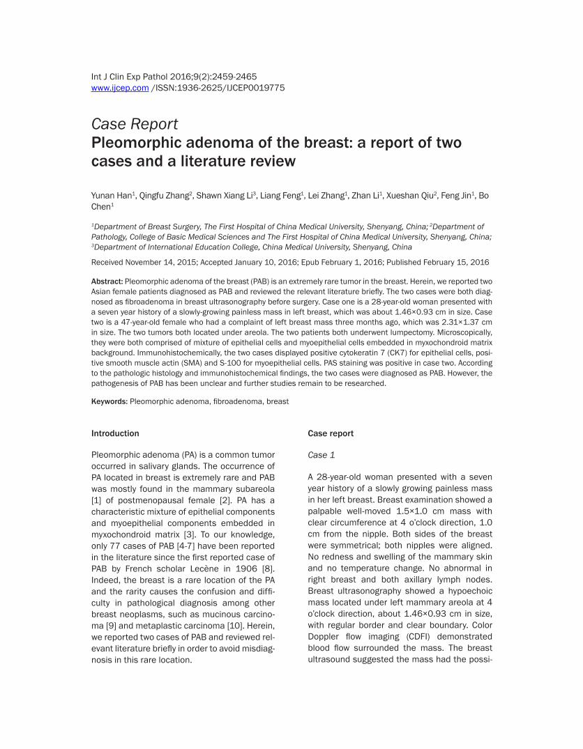
A 28-year-old woman presented with a seven year history of a slowly growing painless mass in her left breast. Breast examination showed a palpable well-moved 1.5×1.0 cm mass with clear circumference at 4 o’clock direction, 1.0 cm from the nipple. Both sides of the breast were symmetrical; both nipples were aligned. No redness and swelling of the mammary skin and no temperature change. No abnormal in right breast and both axillary lymph nodes. Breast ultrasonography showed a hypoechoic mass located under left mammary areola at 4 o’clock direction, about 1.46×0.93 cm in size, with regular border and clear boundary. Color Doppler flow imaging (CDFI) demonstrated blood flow surrounded the mass. The breast ultrasound suggested the mass had the possi-
Pleomorphic adenoma of the breast
2460 Int J Clin Exp Pathol 2016;9(2):2459-2465
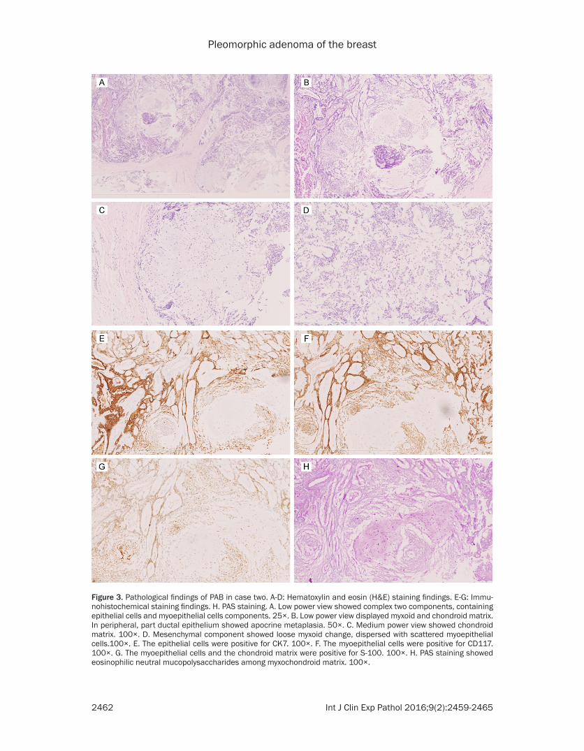
Acid-Schiff (PAS) staining showed eosinophilic neutral mucopolysaccharides among myxo-chondroid matrix (Figure 3H).
Discussion
PA mainly occurs in the salivary gland, yet it also can be found in sella turcica [11], lacrimal gland [12], larynx [13], skin [14], vulva [15], lungs [16], and possibly in the kidney [17]. The breast is a modified sweat gland which shares similar embryological ectodermal layer with the salivary glands [3]. Hence, although breast is an extremely uncommon location, similar tumors and their pathological patterns can affect these locations.
According to the 77 cases of PAB have been reported in English literature, PAB most com-monly occurs in women, however it can also occur in man and only four cases of PAB in men have been reported [18-21]. The age of the PAB patients ranges from 19 to 85 years old [5, 22]. The tumor size ranges from 0.6 to 20 cm in diameter, most were around 2 cm in diameter [3, 20]. PAB always presents with a solidary pal-pable mass in the subareola area. In our cases, the mass was considered as fibroadenoma of the breast through clinical and imaging exami-nations preoperatively. Some authors had reported the preoperative diagnosis was carci-
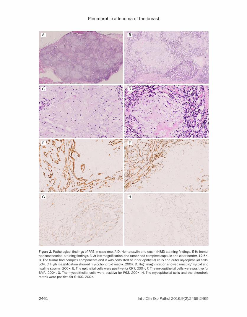
bility of fibroadenoma of the left breast (BI-RADS 4A) (Figure 1). Then the patient underwent lumpectomy for left mammary mass. In gross examination, the lesion was a well-capsular mass, measuring 1.5×1.0 cm in diameter, showing silver-white at cut surface with toughness texture. Microscopically, the tumor was comprised of epithelial cells and myoepithelial cells embedded in myxochon-droid matrix, with complete capsule and clear border (Figure 2A-D). Immunohistochemical (IHC) staining displayed the epithelial cells were positive for cytokeratin 7 (CK7) (Figure 2E), the myoepithelial cells were positive for smooth muscle actin (SMA) (Figure 2F), P63 (Figure 2G) and S-100 (Figure 2H). According to the his-tological patterns and IHC findings, the tumor was diagnosed as PAB.
Case 2
A 47-year-old female had a complaint of left breast mass three months ago. At beginning the lesion was in bean-size below the left nip-ple, and then it grew to peanut-size. Breast ultrasonography showed a hypoechoic mass in subareola location, measuring 2.31×1.37 cm. CDFI demonstrated no blood flow signal. The breast ultrasound suggested the mass had the possibility of fibroadenoma of the left breast. Then the patient underwent left breast local
excision surgery at the local hospital. She was referred to our hospital for consultation in pathology and manage-ment. We reviewed the patho-logical slides of the patient and confirmed it to be PAB. Microscopically, the tumor was comprised of complex two components, containing epithelial cells and myoepi-thelial cells components (Fi- gure 3A). Stroma structures showed chondromyxoid ma- trix, loose myxoid change, dis-persed with scattered myoep-ithelial cells (Figure 3B-D). Immunohistochemically, the epithelial cells were positive for CK7 (Figure 3E), and the myoepithelial cells were posi-tive for CD117 (Figure 3F) and S-100 (Figure 3G). Periodic
Figure 1. Breast ultrasonography of case 1 showed a left breast hypoechoic mass about 1.46×0.93 cm in size, with regular border and clear boundary.
Pleomorphic adenoma of the breast
2461 Int J Clin Exp Pathol 2016;9(2):2459-2465
Figure 2. Pathological findings of PAB in case one. A-D: Hematoxylin and eosin (H&E) staining findings. E-H: Immu-nohistochemical staining findings. A. At low magnification, the tumor had complete capsule and clear border. 12.5×. B. The tumor had complex components and it was consisted of inner epithelial cells and outer myoepithelial cells. 50×. C. High magnification showed myxochondroid matrix. 200×. D. High magnification showed mucoid/myxoid and hyaline stroma. 200×. E. The epithelial cells were positive for CK7. 200×. F. The myoepithelial cells were positive for SMA. 200×. G. The myoepithelial cells were positive for P63. 200×. H. The myoepithelial cells and the chondroid matrix were positive for S-100. 200×.
Pleomorphic adenoma of the breast
2462 Int J Clin Exp Pathol 2016;9(2):2459-2465
Figure 3. Pathological findings of PAB in case two. A-D: Hematoxylin and eosin (H&E) staining findings. E-G: Immu-nohistochemical staining findings. H. PAS staining. A. Low power view showed complex two components, containing epithelial cells and myoepithelial cells components. 25×. B. Low power view displayed myxoid and chondroid matrix. In peripheral, part ductal epithelium showed apocrine metaplasia. 50×. C. Medium power view showed chondroid matrix. 100×. D. Mesenchymal component showed loose myxoid change, dispersed with scattered myoepithelial cells.100×. E. The epithelial cells were positive for CK7. 100×. F. The myoepithelial cells were positive for CD117. 100×. G. The myoepithelial cells and the chondroid matrix were positive for S-100. 100×. H. PAS staining showed eosinophilic neutral mucopolysaccharides among myxochondroid matrix. 100×.
Pleomorphic adenoma of the breast
2463 Int J Clin Exp Pathol 2016;9(2):2459-2465
noma through both clinical and radiographic findings [9]. Diagnosis of PAB through preoper-ative clinical and imaging examinations is very challenging. Even through the preoperative biopsy, misdiagnosis can be found, such as mucinous carcinoma [9] and metaplastic carci-noma [10], due to limited tissue samples. The final diagnosis of PAB was made after opera-tion and through the histopathological and IHC findings of paraffin sections and IHC stains.
Therapy of the PAB was surgical excision through reviewing the previous reports. The local excision of tumor margin with a narrow (2 to 5 mm) rim of normal breast tissue was rec-ommended for PAB [20]. Though PA generally has indolent benign behavior, the local recur-rence of PAB can also find in two cases [23, 24] and even one case recurred for the second time following surgery [24]. The local recur-rence of PA in salivary glands had relationship with the extent of surgery, the pseudopodia outside the pseudopodia or fingerlike tumor extensions under microscopy [25], and multifo-cality of tumor [24]. Due to several multifocal PAB had been observed [22] and rare reports occurred malignant transformation of PAB [26], patients diagnosed as PAB should be informed of the risk of recurrence and John BJ et. al sug-gested that the patients do yearly examinations and follow-up at least for five years [24].
Three cases of malignant PAB (i.e., carcinoma ex PAB) have been reported [27]; however, none had metastasized to axillary lymph nodes or distant sites. The histological features of malignant PAB were depended on tumor infil-trative growth patterns, marked cytological atypia, high mitotic rate, presence of atypia, necrosis and high Mib-1 index [27]. No clinical criteria for diagnosis of malignant PAB had been presented.
The tendency of most PAB located in juxta-are-olar region suggested PAB might originate from the large mammary ducts in subareola area [28]. The histogenesis of PAB remains contro-versial, whether PAB derived from epithelial cells, or myoepithelial cells, or both of them remains unclear. Many reports suggested myo-epithelial cell proliferation as a key factor in oncogenesis [29, 30]. The abundant myoepi-thelial cells in large mammary ducts around the areolar region might explain the frequent occur-rence in this area. Nartia et al. postulated that
PAB originated from multipotent ductal cells, which differentiated into the myoepithelial cells [1]. Further studies on histogenesis of PAB are needed.
PA of the salivary glands harbor chromosomal translocations, involving 8q12 (with the target gene PLAG1) [31], 12q15 (with the target gene HMGI-C) [32], and 6p21 (with the target gene HMGIY) [33]. In PAB, Sato et al. reported IHC-positive staining of HMGI-C and HMGIY and they suggested the tumor might show abnor-mal expression of HMGI-C and HMGIY proteins [34]. Some further studies on chromosomal rearrangements in PAB and exact mechanisms remain to be evaluated.
In conclusion, we reported two cases of PAB. Diagnosis of PAB through clinical examinations, imaging findings and preoperative biopsy was difficult and the tumor may be misdiagnosed as fibroadenoma or malignant breast tumor. Clinical doctors and pathologists should pay more attention to the diagnosis of PAB.
Acknowledgements
This work was supported by grants from the Natural Science Foundation of Liaoning Province of China (No: L2015598). We greatly appreciate Shu-han Wang, at Cornell University, who helped us search for references and give us endless support.
Disclosure of conflict of interest
None.
Address correspondence to: Dr. Bo Chen, Depart- ment of Breast Surgery, The First Hospital of China Medical University, Shenyang 110001, China. E-mail: [email protected]
References
[1] Narita T and Matsuda K. Pleomorphic adeno-ma of the breast: case report and review of the literature. Pathol Int 1995; 45: 441-447.
[2] Rakha EA, Aleskandarany MA, Samaka RM, Hodi Z, Lee AH and Ellis IO. Pleomorphic ade-noma-like tumour of the breast. Histopathology 2016; 68: 405-10.
[3] Reid-Nicholson M, Bleiweiss I, Pace B, Azueta V and Jaffer S. Pleomorphic adenoma of the breast. A case report and distinction from mu-cinous carcinoma. Arch Pathol Lab Med 2003; 127: 474-477.
Pleomorphic adenoma of the breast
2464 Int J Clin Exp Pathol 2016;9(2):2459-2465
[4] Khamechian T, Alizargar J and Mazoochi T. Reporting a Rare Case of Pleomorphic Adenoma of the Breast. Case Rep Med 2014; 2014: 1-4.
[5] Leekha N, Muralee M, Mathews A, Preethi TR and Ahamed MI. Pleomorphic Adenoma of Breast-A Case Report and Review of Literature. Indian J Surg Oncol 2014; 5: 152-154.
[6] Ginter PS, Scognamiglio T, Tauchi-Nishi P, Antonio LB and Hoda SA. Pleomorphic adeno-ma of breast: a radiological and pathological study of a common tumor in an uncommon lo-cation. Case Rep Pathol 2015; 2015: 1-3.
[7] Kelten C, Boyaci C, Trabulus DC and Sirin S. Benign mixed tumour of the breast and breast skin, two cases with diagnostic difficulties. BMJ Case Rep 2015; 2015.
[8] Lecène AL. Observation d’un cas de tumeur “mixte” du sein. Revue de Chirurgie 1906; 33: 434-468.
[9] Iyengar P, Cody HS 3rd and Brogi E. Pleomorphic adenoma of the breast: case report and review of the literature. Diagn Cytopathol 2005; 33: 416-420.
[10] Djakovic A, Engel JB, Geisinger E, Honig A, Tschammler A and Dietl J. Pleomorphic adeno-ma of the breast initially misdiagnosed as metaplastic carcinoma in preoperative stereo-tactic biopsy: a case report and review of the literature. Eur J Gynaecol Oncol 2011; 32: 427-430.
[11] Takahashi S, Mikami S, Akiyama T and Kawase T. Intrasellar salivary gland-like pleomorphic adenoma: case report. Neurosurgery 2011; 68: E562-565; discussion E566.
[12] Gupta A and Khandelwal A. Lacrimal gland pleomorphic adenoma: an inconceivable diag-nosis in a child. BMJ Case Rep 2013; 2013.
[13] Dubey SP, Banerjee S, Ghosh LM and Roy S. Benign pleomorphic adenoma of the larynx: report of a case and review and analysis of 20 additional cases in the literature. Ear Nose Throat J 1997; 76: 548-550, 552, 554-547.
[14] Limaiem F, Bouslama S, Haddad I, Ben Slama S, Bouraoui S, Lahmar A and Mzabi-Regaya S. Chondroid syringoma: report of four cases. Skinmed 2015; 13: 104-106.
[15] Su A, Apple SK and Moatamed NA. Pleomorphic adenoma of the vulva, clinical reminder of a rare occurrence. Rare Tumors 2012; 4: e16.
[16] Moran CA, Suster S, Askin FB and Koss MN. Benign and malignant salivary gland-type mixed tumors of the lung. Clinicopathologic and immunohistochemical study of eight cas-es. Cancer 1994; 73: 2481-2490.
[17] Pacchioni D, Volante M, Casetta G, Sapino A, Marchio C and Bussolati G. Myxoid renal tumor with myoepithelial differentiation mimicking a salivary gland pleomorphic adenoma: descrip-
tion of a case. Am J Surg Pathol 2007; 31: 632-636.
[18] Molland JG, Morgan GJ, Walker DM and Lin BP. Pleomorphic adenoma of the parotid and breast in a male patient. Pathology 2005; 37: 263-265.
[19] Makek M and von Hochstetter AR. Pleomorphic adenoma of the human breast. J Surg Oncol 1980; 14: 281-286.
[20] Diaz NM, McDivitt RW and Wick MR. Pleomorphic adenoma of the breast: a clinico-pathologic and immunohistochemical study of 10 cases. Hum Pathol 1991; 22: 1206-1214.
[21] Simha MR, Doctor VM and Udwadia TE. Mixed tumour of salivary gland type of the male breast. Indian J Cancer 1992; 29: 14-17.
[22] Moran CA, Suster S and Carter D. Benign mixed tumors (pleomorphic adenomas) of the breast. Am J Surg Pathol 1990; 14: 913-921.
[23] Soreide JA, Anda O, Eriksen L, Holter J and Kjellevold KH. Pleomorphic adenoma of the human breast with local recurrence. Cancer 1988; 61: 997-1001.
[24] John BJ, Griffiths C and Ebbs SR. Pleomorphic adenoma of the breast should be excised with a cuff of normal tissue. Breast J 2007; 13: 418-420.
[25] Henriksson G, Westrin KM, Carlsoo B and Silfversward C. Recurrent primary pleomorphic adenomas of salivary gland origin: intrasurgi-cal rupture, histopathologic features, and pseudopodia. Cancer 1998; 82: 617-620.
[26] Hayes MM, Lesack D, Girardet C, Del Vecchio M and Eusebi V. Carcinoma ex-pleomorphic ad-enoma of the breast. Report of three cases suggesting a relationship to metaplastic carci-noma of matrix-producing type. Virchows Arch 2005; 446: 142-149.
[27] Hayes MM, Lesack D, Girardet C, Del Vecchio M and Eusebi V. Carcinoma ex-pleomorphic ad-enoma of the breast. Report of three cases suggesting a relationship to metaplastic carci-noma of matrix-producing type. Virchows Archiv 2004; 446: 142-149.
[28] van der Walt JD and Rohlova B. Pleomorphic adenoma of the human breast. A report of a benign tumour closely mimicking a carcinoma clinically. Clin Oncol 1982; 8: 361-365.
[29] Ballance WA, Ro JY, el-Naggar AK, Grignon DJ, Ayala AG and Romsdahl MG. Pleomorphic ad-enoma (benign mixed tumor) of the breast. An immunohistochemical, flow cytometric, and ul-trastructural study and review of the literature. Am J Clin Pathol 1990; 93: 795-801.
[30] Review of the literature. J Surg Oncol 1987; 36: 58-63.
[31] Voz ML, Agten NS, Van de Ven WJ and Kas K. PLAG1, the main translocation target in pleo-morphic adenoma of the salivary glands, is a
Pleomorphic adenoma of the breast
2465 Int J Clin Exp Pathol 2016;9(2):2459-2465
positive regulator of IGF-II. Cancer Res 2000; 60: 106-113.
[32] Geurts JM, Schoenmakers EF, Roijer E, Stenman G and Van de Ven WJ. Expression of reciprocal hybrid transcripts of HMGIC and FHIT in a pleomorphic adenoma of the parotid gland. Cancer Res 1997; 57: 13-17.
[33] Rohen C, Rogalla P, Meyer-Bolte K, Bartnitzke S, Chilla R and Bullerdiek J. Pleomorphic adenomas of the salivary glands: absence of HMGIY rearrangements. Cancer Genet Cytogenet 1999; 111: 178-181.
[34] Sato K, Ueda Y, Shimasaki M, Ozaki M, Nitta N, Chada K, Ishikawa Y and Katsuda S. Pleomorphic adenoma (benign mixed tumor) of the breast: A case report and review of the literature. Pathol Res Pract 2005; 201: 333-339.


![Ductal Adenocarcinoma Ex Pleomorphic Adenoma of the ... · lesions [2, 5]. Carcinoma ex pleomorphic adenoma (Ca ex PA) is a rare transformation of a benign primary PA to a malignant](https://img.pdfslide.net/doc/110x75/60bd399bb7acaf776f026cd1/ductal-adenocarcinoma-ex-pleomorphic-adenoma-of-the-lesions-2-5-carcinoma.jpg)


![[PAPER] Pleomorphic Adenoma Print.docx](https://img.pdfslide.net/doc/110x75/56d6bd9b1a28ab30168ea546/paper-pleomorphic-adenoma-printdocx.jpg)