Embed Size (px)
Citation preview
Hindawi Publishing CorporationCase Reports in Obstetrics and GynecologyVolume 2013, Article ID 483689, 3 pageshttp://dx.doi.org/10.1155/2013/483689
Case ReportPrimary Mesenteric Smooth Muscle Tumor: An Entity withUnpredictable Biologic Behavior
Ioannis Kalogiannidis,1 Thomas Stavrakis,1 Ioannis Amplianitis,2 Sophia Grammenou,1
Georgios Mavromatidis,1 and David Rousso1
1 3rd Department of Obstetrics & Gynecology, Aristotle University of Thessaloniki, Konstantinoupoleos 49,54642 Thessaloniki, Greece
2 Department of Pathological Anatomy, Hippokration General Hospital, Konstantinoupoleos 49, 54642 Thessaloniki, Greece
Correspondence should be addressed toThomas Stavrakis; [email protected]
Received 18 June 2013; Accepted 24 July 2013
Academic Editors: K. Dafopoulos and H.-C. Lai
Copyright © 2013 Ioannis Kalogiannidis et al. This is an open access article distributed under the Creative Commons AttributionLicense, which permits unrestricted use, distribution, and reproduction in any medium, provided the original work is properlycited.
Smooth muscle tumors of the mesentery are rare lesions with unpredictable, usually malignant, biologic behavior irrespective oftheir histologic appearance. Such case is presented here.We present a case of a large smoothmuscle tumor located in the mesenteryof a 48 years old patient. The histopathologic features of the surgically excised tumor were that of a benign-appearing smoothmuscle tumor, either a primary mesenteric smooth muscle tumor of unknown biologic behavior or a parasitic leiomyoma. Thepatient was discharged 4 days after from the hospital without any early postoperative complication. Close followup was furtherdecided. Nine months after her primary therapy, our patient is alive and with no evidence of recurrent disease. Increased awarenessmust be considered for large mesenteric smooth muscle tumors, because even when they present indolent histologic features, theyusually behave aggressively.
1. Introduction
Solid primary tumors of mesenteric origin are quite rare.Among them, gastrointestinal stromal tumors (GISTs) andsmooth muscle tumors seem to be the most commonneoplasms [1–3]. Regarding the latter tumors, especiallythose of large size, the prediction of the biologic behaviorbased on histologic grounds is not efficient. Most of thelarge mesenteric smooth muscle tumors behave aggressivelyirrespective of their histologic appearance [4–6].
We present a case of a 48 years old female patient with alarge smooth muscle tumor located in the mesentery with asynchronous uterine leiomyoma. Diagnostic and therapeuticapproach, postoperative dilemmas, and review of the litera-ture are presented as well.
2. Case Presentation
A 48 years old woman, mother of two children with normalmenstruation and insignificant prior history, presented with
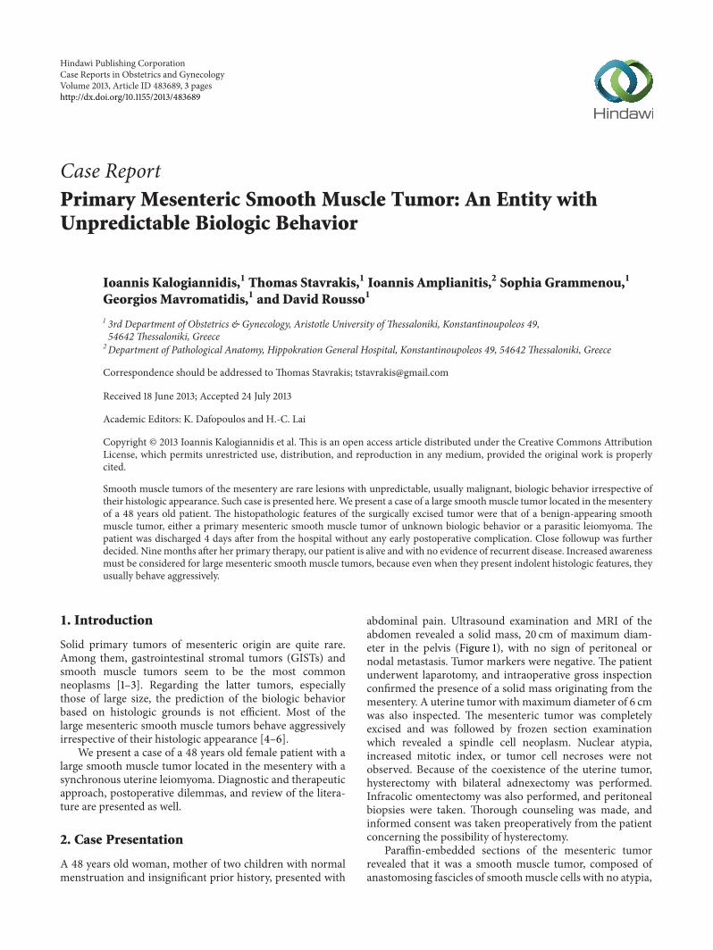
abdominal pain. Ultrasound examination and MRI of theabdomen revealed a solid mass, 20 cm of maximum diam-eter in the pelvis (Figure 1), with no sign of peritoneal ornodal metastasis. Tumor markers were negative. The patientunderwent laparotomy, and intraoperative gross inspectionconfirmed the presence of a solid mass originating from themesentery. A uterine tumor with maximum diameter of 6 cmwas also inspected. The mesenteric tumor was completelyexcised and was followed by frozen section examinationwhich revealed a spindle cell neoplasm. Nuclear atypia,increased mitotic index, or tumor cell necroses were notobserved. Because of the coexistence of the uterine tumor,hysterectomy with bilateral adnexectomy was performed.Infracolic omentectomy was also performed, and peritonealbiopsies were taken. Thorough counseling was made, andinformed consent was taken preoperatively from the patientconcerning the possibility of hysterectomy.
Paraffin-embedded sections of the mesenteric tumorrevealed that it was a smooth muscle tumor, composed ofanastomosing fascicles of smoothmuscle cells with no atypia,

2 Case Reports in Obstetrics and Gynecology
Figure 1: Magnetic resonance imaging (MRI) of the abdomenrevealing a solid mass 20 cm of maximum diameter.
Figure 2: Mesenteric tumor stained with hematoxylin.
mitosis, or tumor cell necrosis. Immunohistochemically, thesmooth muscle cells were positive for SMA and desmin andnegative for CD117 (Figures 2 and 3). Peritoneal biopsies andomentum were disease free. The uterine tumor was a typicalbenign leiomyoma.
Thepatient was discharged four days later.Thepostopera-tive course was uneventful. Because of the dilemma regardingthe biologic behavior of the mesenteric tumor, long-termclose followup was decided. Nine months after surgery, norecurrent disease has been noted.
3. Discussion
Primary solid tumors of the mesentery are usually of mes-enchymal nature [1, 3]. Most commonly, they are smoothmuscle tumors or GISTs [3]. Fibromatosis (desmoid tumor),well-differentiated liposarcoma, malignant fibrous histiocy-toma, and peripheral nerve sheath tumors also occur in thelocation [7, 8].
Figure 3: Mesenteric tumor stained with (smooth muscle actin)SMA.
Regarding the primary, mesenteric smooth muscletumors, their biologic behavior seems to be unpredictable,because these tumors, when large, usually behave in amalignant fashion, even in the absence of nuclear atypia,tumor cell necrosis, or increased mitotic count [4]. Thisis in contrast with their uterine counterparts. The uterinesmooth muscle tumors with low mitotic count, none-to-mild nuclear atypia, and no tumor cell necrosis arecharacterized as leiomyomas and behave in a benign fashion[1, 4, 5, 9].
In our case, the histologic examination of the mesen-teric tumor showed that it was a smooth muscle tumorwith no atypia, no tumor cell necrosis, and no increasedmitotic count. If the mesenteric tumor is considered aprimary mesenteric smooth muscle tumor, despite the blandhistopathologic characteristics, the large size of the tumorindicated that it will behave in a malignant fashion [5, 6, 10].
If the mesenteric tumor is not considered a primarytumor of the mesentery, other entities, mainly the parasiticleiomyoma, should enter the differential diagnosis [5]. Thepatient had a synchronous uterine leiomyoma, and thepossibility of a second leiomyoma detached form a subserosallocation and attached to themesentery could not be excluded.In such a case, the indolent histologic features of the neo-plasm assure the benign biologic behavior [5, 11, 12].
Another entity entering the differential diagnosis couldbe the benign metastasizing leiomyoma, but most of thewomen suffering from this mysterious condition presentlesions in the lungs and have a smooth muscle tumor excisedand inadequately studied in the past [12].
In conclusion, primary solid mesenteric tumors con-stitute a histological heterogeneous group of neoplasms.Histologic examination can reveal the histogenetic nature ofa primary solid mesenteric tumor, more often such as GIST,smooth muscle tumor, or desmoid tumor. In the case ofthe primary mesenteric smooth muscle tumor, the histologicfeatures, namely, the lack of cytologic atypia, mitoses, andtumor cell necrosis do not correlate with the prognosis,because when large, they usually behave in a malignant fash-ion. On the other hand, a diagnosis of parasitic leiomyomashould be made with great caution. In any case, we believe
Case Reports in Obstetrics and Gynecology 3
that mesenteric smooth muscle tumors, either primary orparasitic, regardless of the histologic characteristics, shouldhave close follow up, because of the serious possibility ofmalignant behavior, even in the absence of histologic criteriaof malignancy.
Conflict of Interests
The authors declare that they have no conflict of interests.
Authors, Contribution
All authors contributed equally.
References
[1] C. Dufay, A. Abdelli, V. Le Pennec, and L. Chiche, “Mesenterictumors: diagnosis and treatment,” Journal of Vascular Surgery,vol. 149, no. 4, pp. e239–e251, 2012.
[2] N. P. Gupta, M. Aron, and S. Sood, “Pedimculated mesentericleiomyoma masquerading as a retrovesical mass lesion: adiagnostic dilemma,” British Journal of Urology, vol. 82, no. 1,pp. 134–135, 1998.
[3] S. I. Schwartz and F. C. Brunicardi, Schwartz’s Principlesof Surgery, McGraw-Hill; Medical Publication Division, NewYork, NY, USA, 9th edition, 2010.
[4] K. Yannopoulos and A. P. Stout, “Primary solid tumors of themesentery,” Cancer, vol. 16, pp. 914–927, 1963.
[5] N. Fasih, A. K. P. Shanbhogue, D. B. Macdonald et al., “Leiomy-omas beyond the uterus: unusual locations, rare manifesta-tions,” Radiographics, vol. 28, no. 7, pp. 1931–1948, 2008.
[6] H. Hashimoto, M. Tsuneyoshi, and M. Enjoji, “Malignantsmooth muscle tumors of the retroperitoneum and mesentary:a clinicopathologic analysis of 44 cases,” Journal of SurgicalOncology, vol. 28, no. 3, pp. 177–186, 1985.
[7] L. Montagliani and V. Duverger, “Desmoid tumors,” Journal deChirurgie, vol. 145, no. 1, pp. 20–26, 2008.
[8] S. L. Singla, K. N. Rattan, and N. Kaushik, “Mesenteric leiomy-oma in infancy,” Indian Journal of Pediatrics, vol. 67, no. 11, pp.857–858, 2000.
[9] S. Roy, V. Saroha, and D. Jain, “Highly cellular leiomyomamimics a malignant small round-cell tumor: a diagnosticdilemma on frozen sections,” Taiwanese Journal of Obstetricsand Gynecology, vol. 49, no. 2, pp. 203–205, 2010.
[10] J. Rosai and L. V. Ackerman, RoSai and Ackerman’s SurgicalPathology, Mosby, Edinburgh, UK, 9th edition, 2004.
[11] M. R. Hendrickson and R. L. Kempson, “Surgical pathology ofthe uterine corpus,”Major Problems in Pathology, vol. 12, pp. 1–580, 1979.
[12] L. Tietze, K. Gunther, A. Horbe et al., “Benign metastasizingleiomyoma: a cytogenetically balanced but clonal disease,”Human Pathology, vol. 31, no. 1, pp. 126–128, 2000.

Submit your manuscripts athttp://www.hindawi.com
Stem CellsInternational
Hindawi Publishing Corporationhttp://www.hindawi.com Volume 2014
Hindawi Publishing Corporationhttp://www.hindawi.com Volume 2014
MEDIATORSINFLAMMATION
of
Hindawi Publishing Corporationhttp://www.hindawi.com Volume 2014
Behavioural Neurology
EndocrinologyInternational Journal of
Hindawi Publishing Corporationhttp://www.hindawi.com Volume 2014
Hindawi Publishing Corporationhttp://www.hindawi.com Volume 2014
Disease Markers
Hindawi Publishing Corporationhttp://www.hindawi.com Volume 2014
BioMed Research International
OncologyJournal of
Hindawi Publishing Corporationhttp://www.hindawi.com Volume 2014
Hindawi Publishing Corporationhttp://www.hindawi.com Volume 2014
Oxidative Medicine and Cellular Longevity
Hindawi Publishing Corporationhttp://www.hindawi.com Volume 2014
PPAR Research
The Scientific World JournalHindawi Publishing Corporation http://www.hindawi.com Volume 2014
Immunology ResearchHindawi Publishing Corporationhttp://www.hindawi.com Volume 2014
Journal of
ObesityJournal of
Hindawi Publishing Corporationhttp://www.hindawi.com Volume 2014
Hindawi Publishing Corporationhttp://www.hindawi.com Volume 2014
Computational and Mathematical Methods in Medicine
OphthalmologyJournal of
Hindawi Publishing Corporationhttp://www.hindawi.com Volume 2014
Diabetes ResearchJournal of
Hindawi Publishing Corporationhttp://www.hindawi.com Volume 2014
Hindawi Publishing Corporationhttp://www.hindawi.com Volume 2014
Research and TreatmentAIDS
Hindawi Publishing Corporationhttp://www.hindawi.com Volume 2014
Gastroenterology Research and Practice
Hindawi Publishing Corporationhttp://www.hindawi.com Volume 2014
Parkinson’s Disease
Evidence-Based Complementary and Alternative Medicine
Volume 2014Hindawi Publishing Corporationhttp://www.hindawi.com