Embed Size (px)
Citation preview

253
benign tumors is very important. Radiologic images of heman-giopericytoma typically shows a diffusely enhancing tumor with a broad-based meningeal attachment, which is also observed in meningioma13). A few characteristics of hemangiopericytoma were known15). However, they are not definite and are also ob-served in aggressive meningioma. Here, we describe a patient with an unexpected intradural hemangiopericytoma in the cer-vical spine, and we describe an unexpected hemangiopericyto-ma case with large bleeding and management of the tumor.
CASE REPORT
A 21-year-old man visited complaining of neck pain and a tingling sensation in both hands. The intensity of neck pain had progressed over 1 year. The tingling sensation in the hands had developed 1 month prior and had worsened. Motor function and reflexes did not decrease. His magnetic resonance (MR)
INTRODUCTION
Hemangiopericytoma is a highly vascularized, aggressive neoplasm that is classified as a mesenchymal nonmeningotheli-al tumor with uncertain malignant potential or borderline ma-lignancy. This tumor develops in the pericytes of soft tissue and is usually located in subcutaneous tissue and skeletal muscle. Hemangiopericytomas of the central nervous system are rare, comprising 2% to 4% of all meningeal tumors7,8). Hemangio-pericytoma rarely occurs in the spine, with only approximately 60 previously reported cases of which 10 were located in the in-tradural extramedullary (IDEM) region1-3,5,9,10,12,14).
Hemangiopericytomas sometimes look like aggressive hem-angiomas such as arteriovenous shunting or congested spinal vessels5). Local recurrence and metastasis of hemangiopericyto-ma were reported to 60% and 23%, respectively11). Therefore, differential diagnosis between hemangiopericytoma and other
Spinal Hemangiopericytoma Which Needed Intraoperative Embolization due to Unexpected Bleeding
Chang-Hyun Lee, M.D., Ki-Jeong Kim, M.D., Ph.D., Tae-Ahn Jahng, M.D., Hyun-Jib Kim, M.D.
Department of Neurosurgery, Spine Center, Seoul National University Bundang Hospital, Seoul National University College of Medicine, Seongnam, Korea
Spinal intradural hemangiopericytoma is a very rare tumor and can be characterized by massive bleeding during surgeries, frequent recurrence, and metastasis. However, definite radiologic differential points of hemangiopericytoma are not known. We describe an unexpected hemangiopericy-toma case with large bleeding and management of the tumor. A 21-year-old man visited complaining of progressive neck pain and tingling sensa-tion in both hands. Magnetic resonance imaging of his spine revealed C1-2 ventral intradural mass. When the dura was opened, the intradural tu-mor was placed behind spinal accessary nerves. The tumor was partially exposed only after some accessary nerves had been cut. When internal debulking was performing, unexpected bleeding was noted and it was difficult to control because of narrow surgical field and hypervascularity. In-traoperative spinal angiography and embolization were performed. The tumor was completely removed after embolization. Pathological diagnosis was consistent with hemangiopericytoma. When surgeons meet a flesh-red tumor that bleeds unexpectedly during surgery, hemangiopericytoma may be considered. When feeder control is hard due to reciprocal location of spinal cord, the tumor, and feeders, intraoperative angiography and embolization may be a possible option.
Key Words : Hemangiopericytoma · Intradural · Spine · Surgery · Angiography.
Case Report
• Received : May 7, 2013 • Revised : July 28, 2013 • Accepted : September 8, 2013• Address for reprints : Ki-Jeong Kim, M.D., Ph.D. Department of Neurosurgery, Spine Center, Seoul National University Bundang Hospital, Seoul National University College of Medicine, 82 Gumi-ro 173beon-gil, Bundang-gu, Seongnam 463-707, Korea Tel : +82-31-787-7166, Fax : +82-31-787-4097, E-mail : [email protected] • This is an Open Access article distributed under the terms of the Creative Commons Attribution Non-Commercial License (http://creativecommons.org/licenses/by-nc/3.0) which permits unrestricted non-commercial use, distribution, and reproduction in any medium, provided the original work is properly cited.
J Korean Neurosurg Soc 54 : 253-256, 2013
http://dx.doi.org/10.3340/jkns.2013.54.3.253
Copyright © 2013 The Korean Neurosurgical Society
Print ISSN 2005-3711 On-line ISSN 1598-7876www.jkns.or.kr
254
J Korean Neurosurg Soc 54 | September 2013
cluding brain, abdomen, and pelvis. Our presumptive diagnosis at preoperative state was meningioma, but schwannoma, and neurofibroma were also considered.
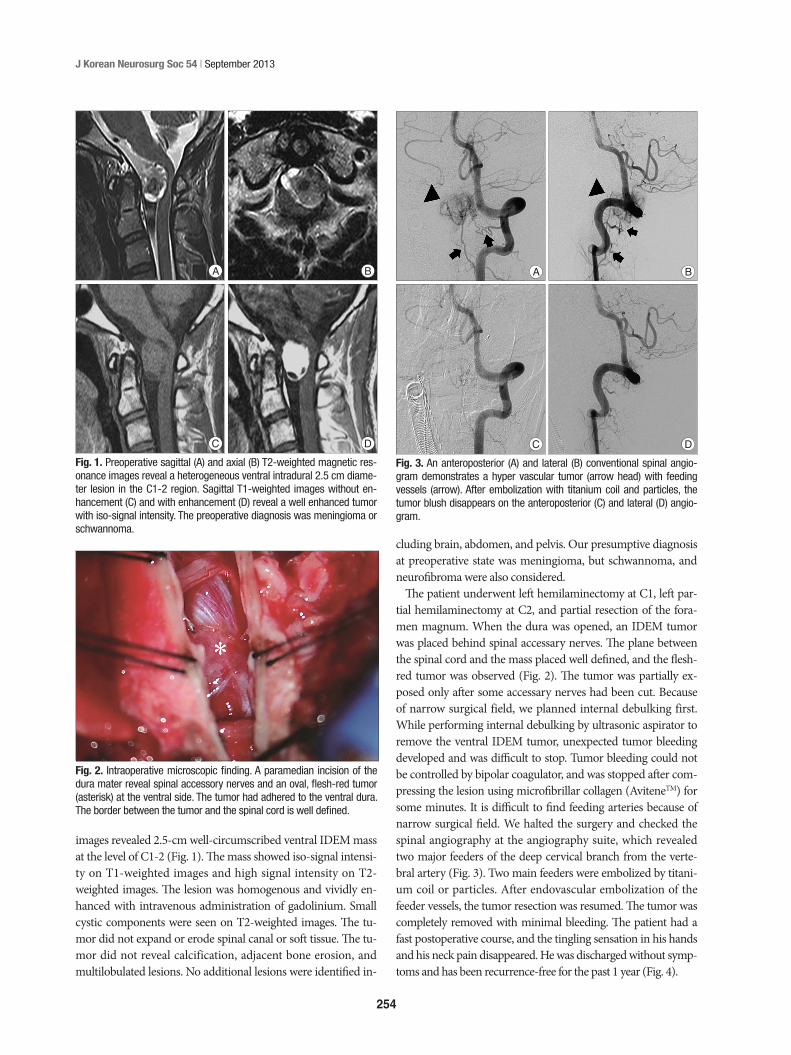
The patient underwent left hemilaminectomy at C1, left par-tial hemilaminectomy at C2, and partial resection of the fora-men magnum. When the dura was opened, an IDEM tumor was placed behind spinal accessary nerves. The plane between the spinal cord and the mass placed well defined, and the flesh-red tumor was observed (Fig. 2). The tumor was partially ex-posed only after some accessary nerves had been cut. Because of narrow surgical field, we planned internal debulking first. While performing internal debulking by ultrasonic aspirator to remove the ventral IDEM tumor, unexpected tumor bleeding developed and was difficult to stop. Tumor bleeding could not be controlled by bipolar coagulator, and was stopped after com-pressing the lesion using microfibrillar collagen (AviteneTM) for some minutes. It is difficult to find feeding arteries because of narrow surgical field. We halted the surgery and checked the spinal angiography at the angiography suite, which revealed two major feeders of the deep cervical branch from the verte-bral artery (Fig. 3). Two main feeders were embolized by titani-um coil or particles. After endovascular embolization of the feeder vessels, the tumor resection was resumed. The tumor was completely removed with minimal bleeding. The patient had a fast postoperative course, and the tingling sensation in his hands and his neck pain disappeared. He was discharged without symp-toms and has been recurrence-free for the past 1 year (Fig. 4).
images revealed 2.5-cm well-circumscribed ventral IDEM mass at the level of C1-2 (Fig. 1). The mass showed iso-signal intensi-ty on T1-weighted images and high signal intensity on T2-weighted images. The lesion was homogenous and vividly en-hanced with intravenous administration of gadolinium. Small cystic components were seen on T2-weighted images. The tu-mor did not expand or erode spinal canal or soft tissue. The tu-mor did not reveal calcification, adjacent bone erosion, and multilobulated lesions. No additional lesions were identified in-
Fig. 1. Preoperative sagittal (A) and axial (B) T2-weighted magnetic res-onance images reveal a heterogeneous ventral intradural 2.5 cm diame-ter lesion in the C1-2 region. Sagittal T1-weighted images without en-hancement (C) and with enhancement (D) reveal a well enhanced tumor with iso-signal intensity. The preoperative diagnosis was meningioma or schwannoma.
Fig. 3. An anteroposterior (A) and lateral (B) conventional spinal angio-gram demonstrates a hyper vascular tumor (arrow head) with feeding vessels (arrow). After embolization with titanium coil and particles, the tumor blush disappears on the anteroposterior (C) and lateral (D) angio-gram.
D D
B B
C C
A A
Fig. 2. Intraoperative microscopic finding. A paramedian incision of the dura mater reveal spinal accessory nerves and an oval, flesh-red tumor (asterisk) at the ventral side. The tumor had adhered to the ventral dura. The border between the tumor and the spinal cord is well defined.

255
Intraoperative Embolization of Spinal Hemangiopericytom | CH Lee, et al.
mor have to be controlled first. When feeder control is hard due to reciprocal location of spinal cord, the tumor, and feeders, in-traoperative angiography and embolization may be a possible option.
In intracranial hemangiopericytoma patients, adjuvant radio-therapy or radiosurgery was usually recommended6,16). In spinal hemangiopericytoma patients, the evidence of adjuvant radio-therapy or radiosurgery was not yet clear1). In 10 previously re-ported cases of IDEM spinal hemangiopericytoma, 1 of 2 pa-tients who had undergone radiotherapy recurred at 2 years after treatment. Two of 7 patients who had not undergone ra-diotherapy showed tumor recurrence. The data on radiation for
Pathologic findingsA gross specimen revealed a nodular mass of soft tissue mea-
suring 2×2 cm. The cut surface of the mass was reddish yellow. Hematoxylin and eosin staining showed a highly cellular neo-plasm growing in patternless sheets and with high mitotic activi-ty. There was proliferation of the tumor cells with oval nuclei, and the tumor was surrounded by abundant blood vessels resulting in the “staghorn” appearance typical of hemangiopericytoma (Fig. 5). After immunostaining, the tumor cells were negative for CD34 and epithelial membrane antigen. On the basis of these findings, we made a final diagnosis of hemangiopericytoma.
DISCUSSION
Hemangiopericytoma is distinguished from meningioma by multilobulated le-sion, expanding/eroding spinal canal, large soft tissue component, or no calci-fication15). Ross et al.15) addressed that malignant, invasive meningioma may be very hard to distinguish from heman-giopericytoma. However, hemangio-pericytomas need to be distinguished from other tumors because they some-times bleed substantially, recur frequent-ly, and metastases. In this case, the tu-mor showed no lobulation, no bony erosion, and no soft tissue invasion on MR images like meningioma. Hence, our presumptive diagnosis, meningioma or schwannoma, was wrong. Hemangio-pericytoma has a chance of missing di-agnosis by radiologic studies.
In the review of previous reports, 7 of 10 papers reported hemangiopericyto-mas were presented as red mass in surgi-cal field in Table 1. One tumor was de-scribed as having the gross appearance of a meningioma10), and the others were not described3,5). Although hemangio-pericytoma could not be distinguished by radiologic studies in preoperative state, hemangiopericytoma seems to be distinguished by the gross feature (red color) on surgical field, which means a hypervascular tumor. Surgeons had bet-ter take extra precaution and suspect hemangiopericytoma when they watch a flesh-red mass during surgeries of IDEM tumors. Hemangiopericytoma sometimes makes large bleeding, al-though not in all. In case of unexpected large bleeding, feeding vessels of the tu-
Fig. 4. (A) Postcontrast sagittal T1-weighted image 6 months after the surgical resection demon-strates no recurred or residual tumor. Axial (B) and sagittal (C) T2-weighted images reveals that the spinal cord shifted anteriorly without the tumor. The cord is slightly diminished in caliber.
A B C
Fig. 5. (A) The tumor was somewhat firm and composed of cystic and nodular lesions. (B) Hematoxylin and eosin staining (original magnification, ×100) shows an irregular array of oval nu-clei. The tumor is surrounded by many blood vessels resulting in the “staghorn” appearance typical of hemangiopericytoma. Upon immunostaining of the specimen, tumor cells are negative for CD34 (C) and EMA (D). EMA : epithelial membrane antigen.
C
A
D
B

256
J Korean Neurosurg Soc 54 | September 2013
AJNR Am J Neuroradiol 30 : 152-154, 20096. Guthrie BL, Ebersold MJ, Scheithauer BW, Shaw EG : Meningeal
hemangiopericytoma : histopathological features, treatment, and long-term follow-up of 44 cases. Neurosurgery 25 : 514-522, 1989
7. Jang JH, Rhim SC, Lee DG, Roh SW : Multiple spinal metastases of hemangiopericytoma : case report. J Korean Neurosurg Soc 32 : 380-383, 2002
8. Jääskeläinen J, Servo A, Haltia M, Wahlström T, Valtonen S : Intracrani-al hemangiopericytoma : radiology, surgery, radiotherapy, and outcome in 21 patients. Surg Neurol 23 : 227-236, 1985
9. Kashiwazaki D, Hida K, Yano S, Seki T, Iwasaki Y : Subpial hemangio-pericytoma with marked extramedullary growth : case report. Neuro-surgery 61 : E1336-E1337; discussion E1337, 2007
10. Kruse F Jr : Hemangiopericytoma of the meniges (angioblastic menin-gioma of Cushing and Eisenhardt). Clinico-pathologic aspects and fol-low-up studies in 8 cases. Neurology 11 : 771-777, 1961
11. Mena H, Ribas JL, Pezeshkpour GH, Cowan DN, Parisi JE : Hemangio-pericytoma of the central nervous system : a review of 94 cases. Hum Pathol 22 : 84-91, 1991
12. Moscovici S, Ramirez-DeNoriega F, Fellig Y, Rosenthal G, Cohen JE, It-shayek E : Intradural extramedullary hemangiopericytoma of the tho-racic spine infiltrating a nerve root : a case report and literature review. Spine (Phila Pa 1976) 36 : E1534-E1539, 2011
13. Osborne DR, Dubois P, Drayer B, Sage M, Burger P, Heinz ER : Primary intracranial meningeal and spinal hemangiopericytoma : radiologic manifestations. AJNR Am J Neuroradiol 2 : 69-74, 1981
14. Pitlyk PJ, Dockery MB, Miller RH : Hemangiopericytoma of the spinal cord : report of three cases. Neurology 15 : 649-653, 1965
15. Ross JS, Brant-Zawadzki M, Moore KR, Crim J, Chen MZ, Katzman GL : Diagnostic imaging : Spine, ed 1. Utah : Amirsys, 2004, ppIV.1.82-85
16. Soyuer S, Chang EL, Selek U, McCutcheon IE, Maor MH : Intracranial meningeal hemangiopericytoma : the role of radiotherapy : report of 29 cases and review of the literature. Cancer 100 : 1491-1497, 2004
spinal intradural lesions remains unclear because of few cases. In addition, neither radiotherapy nor radiosurgery has demon-strated protection against neuroaxis4). Therefore, we recom-mend closely following patients with a hemangiopericytoma clinically with serial MR imaging.
CONCLUSION Intradural spinal hemangiopericytoma is rare but need to be
distinguished from other benign tumors because it is aggres-sive. When surgeons meet a flesh-red tumor that bleeds unex-pectedly during surgery, hemangiopericytoma may be consid-ered. When feeder control is hard due to reciprocal location of spinal cord, the tumor, and feeders, intraoperative angiography and embolization may be a possible option.
References 1. Ackerman PD, Khaldi A, Shea JF : Intradural hemangiopericytoma of
the thoracic spine : a case report. Spine J 11 : e9-e14, 20112. Betchen S, Schwartz A, Black C, Post K : Intradural hemangiopericyto-
ma of the lumbar spine : case report. Neurosurgery 50 : 654-657, 20023. Ciappetta P, Celli P, Palma L, Mariottini A : Intraspinal hemangioperi-
cytomas. Report of two cases and review of the literature. Spine (Phila Pa 1976) 10 : 27-31, 1985
4. Dufour H, Métellus P, Fuentes S, Murracciole X, Régis J, Figarella-Branger D, et al. : Meningeal hemangiopericytoma : a retrospective study of 21 patients with special review of postoperative external radio-therapy. Neurosurgery 48 : 756-762; discussion 762-763, 2001
5. Fitzpatrick D, Mahajan J, Lewkowitz M, Black K, Setton A, Woldenberg R : Intradural hemangiopericytoma of the lumbar spine : a rare entity.
Table 1. Previously reported papers of spinal hemangioblastoma
Year Author(s) Sex/age Location Gross finding Treatment Radio-therapy Recurrence Follow-up
1961 Kruse10) M/53 C3 Mimicked meningioma Partial resection† Yes At 2 yr 3 yr
1965 Pitlyk et al.14) M/60 C4 “Grayish red” Total resection No NA NAF/49 C3 “Meat-red” Total resection No None 10 yrM/39 T8 “Meat-red” Total resection* No At 9 yr 18 yr
1985 Ciappetta et al.3) M/48 C3-C5 NA Total resection No At 6 yr 6 yr2002 Betchen et al.2) M/31 L4 “Red” Total resection No None 6 mo2007 Kashiwazaki et al.9) M/31 T4-T5 “Red” Total resection† No None 3 yr2009 Fitzpatrick et al.5) M/54 L4-L5 NA Embolization, total resection Yes NA NA2011 Ackerman et al.1) M/58 T9-T11 “Reddish tan” Total resection† No None None2011 Moscovici et al.12) M/20 T9-T10 “Bluish” Total resection No None 2 yr2012 Present case M/21 C1-C2 Flesh red Embolization, total resection† No None 1 yr
*The tumor was reported to have been a meningioma initially and was considered to have been totally removed, †The tumor was hyper vascular, and substantial bleed-ing was described. NA : not available, yr : years, mo : months

![Metastatic Intracranial Hemangiopericytoma to the Spinal ... · 128 INTRODUCTION Intracranial hemangiopericytoma (HPC) is a rare tumor with malignant features [1], of which incidence](https://img.pdfslide.net/doc/110x75/5d4d5a7488c993a90e8bc971/metastatic-intracranial-hemangiopericytoma-to-the-spinal-128-introduction.jpg)